. 2024 May 1;81(5):525-533.
doi: 10.1001/jamaneurol.2024.0126.

Magnetic Resonance Imaging Characteristics of LGI1-Antibody and CASPR2-Antibody Encephalitis

Affiliations

Magnetic Resonance Imaging Characteristics of LGI1-Antibody and CASPR2-Antibody Encephalitis

Mark J Kelly et al. JAMA Neurol. .

Abstract

Importance: Rapid and accurate diagnosis of autoimmune encephalitis encourages prompt initiation of immunotherapy toward improved patient outcomes. However, clinical features alone may not sufficiently narrow the differential diagnosis, and awaiting autoantibody results can delay immunotherapy.

Objective: To identify simple magnetic resonance imaging (MRI) characteristics that accurately distinguish 2 common forms of autoimmune encephalitis, LGI1- and CASPR2-antibody encephalitis (LGI1/CASPR2-Ab-E), from 2 major differential diagnoses, viral encephalitis (VE) and Creutzfeldt-Jakob disease (CJD).

Design, setting, and participants: This cross-sectional study involved a retrospective, blinded analysis of the first available brain MRIs (taken 2000-2022) from 192 patients at Oxford University Hospitals in the UK and Mayo Clinic in the US. These patients had LGI1/CASPR2-Ab-E, VE, or CJD as evaluated by 2 neuroradiologists (discovery cohort; n = 87); findings were validated in an independent cohort by 3 neurologists (n = 105). Groups were statistically compared with contingency tables. Data were analyzed in 2023.

Main outcomes and measures: MRI findings including T2 or fluid-attenuated inversion recovery (FLAIR) hyperintensities, swelling or volume loss, presence of gadolinium contrast enhancement, and diffusion-weighted imaging changes. Correlations with clinical features.

Results: Among 192 participants with MRIs reviewed, 71 were female (37%) and 121 were male (63%); the median age was 66 years (range, 19-92 years). By comparison with VE and CJD, in LGI1/CASPR2-Ab-E, T2 and/or FLAIR hyperintensities were less likely to extend outside the temporal lobe (3/42 patients [7%] vs 17/18 patients [94%] with VE; P < .001, and 3/4 patients [75%] with CJD; P = .005), less frequently exhibited swelling (12/55 [22%] with LGI1/CASPR2-Ab-E vs 13/22 [59%] with VE; P = .003), and showed no diffusion restriction (0 patients vs 16/22 [73%] with VE and 8/10 [80%] with CJD; both P < .001) and rare contrast enhancement (1/20 [5%] vs 7/17 [41%] with VE; P = .01). These findings were validated in an independent cohort and generated an area under the curve of 0.97, sensitivity of 90%, and specificity of 95% among cases with T2/FLAIR hyperintensity in the hippocampus and/or amygdala.

Conclusions and relevance: In this study, T2 and/or FLAIR hyperintensities confined to the temporal lobes, without diffusion restriction or contrast enhancement, robustly distinguished LGI1/CASPR2-Ab-E from key differential diagnoses. These observations should assist clinical decision-making toward expediting immunotherapy. Their generalizability to other forms of autoimmune encephalitis and VE should be examined in future studies.


Conflict of interest statement

Conflict of Interest Disclosures: Dr Kelly reported grants from Wellcome Trust via the Irish Clinical Academic Training (ICAT) Programme during the conduct of the study. Dr Binks reported funding from a National Institute for Health Research Clinical Lectureship during the conduct of the study; travel expenses from ECTRIMS outside the submitted work; and having had a patent for PCT/GB2019/051257 pending, “Diagnostic Strategy to improve specificity of CASPR2 antibody detection.” Dr Ramanathan reported grants from the National Health and Medical Research Council, Royal Australasian College of Physicians Research Establishment Fellowship, Brain Foundation, and University of Sydney; speaker honoraria from Biogen, Novartis, Alexion, and Limbic Neurology; and consultant fees from UCB for serving on a steering committee of a phase 3 trial outside the submitted work. Dr Handel reported grants from the Medical Research Council (MRC) and UCB-Pharma during the conduct of the study. Dr Dubey reported grants and/or consulting fees from UCB, Arialys, DOD, and David J. Tomassoni ALS Research outside the submitted work and having had patents pending for LUZP4-IgG, cavin-4-IgG, and SKOR2 IgG as markers of neurological autoimmunity and patents licensed for KLHL11 IgG. Dr Day reported grants from the National Institutes of Health (NIH/NIA) (K23AG064029) during the conduct of the study and in-kind support from Amgen Pharmaceuticals for a clinical trial in anti-NMDAR encephalitis (ExTINGUISH) and personal income related to consulting for Arialys Pharmaceuticals regarding development of a clinical trial for the treatment of autoimmune encephalitis. Dr Flanagan reported research support from UCB; serving on advisory boards for Alexion, Roche, and Horizon Therapeutics; royalties from UpToDate outside the submitted work; and having had a patent for Dach1-IgG pending (Mayo Clinic) as a marker of neurologic autoimmunity. Dr Irani reported grants from Wellcome and UK Research and Innovation/MRC during the conduct of the study; personal fees from UCB, MedImmun, Roche, Janssen, Cerebral Therapeutics, and ADC Therapeutics; grants from Brain, CSL Behring, and Ono Pharma outside the submitted work; and having had a patent for WO/2010/046716 with royalties paid from Euroimmun, a patent for WO2019211633 issued, and a patent for WO202189788A1 pending. No other disclosures were reported.

Figures

Figure 1.
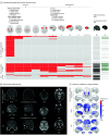
Figure 1.. Regional T2 Magnetic Resonance Imaging (MRI) Hyperintensities, Diffusion Restriction, and Contrast Uptake
A, T2 hyperintensities across brain regions (red), the presence of diffusion restriction (dark gray), and contrast enhancement (green) in LGI1/CASPR2-antibody encephalitis (LGI1/CASPR2-Ab-E, n = 55), viral encephalitis (VE, n = 22), and Creutzfeldt-Jakob disease (CJD, n = 10). Light gray indicates absence; white indicates not performed. C, Raw data for frequencies are available in eTable 2 in Supplement 1. ADC indicates apparent diffusion coefficient; AE, autoimmune encephalitis; DWI, diffusion-weighted imaging; FLAIR, fluid-attenuated inversion recovery; HSV1, herpes simplex virus 1.
Figure 2.
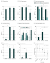
Figure 2.. Magnetic Resonance Imaging (MRI) Features and Distinctions Between LGI1- and CASPR2-Antibody Autoimmune Encephalitis (AE), Viral Encephalitis (VE), and Creutzfeldt-Jakob Disease (CJD)
MRI abnormalities (A) included T2 hyperintensities (B) in almost all LGI1/CASPR2-antibody AE and VE cases and a smaller but high proportion of CJD cases. Fisher exact test was used for data in panels A-H. I, History of seizures was the only clinical feature associated with T2 and/or fluid-attenuated inversion recovery (FLAIR) hyperintensities in the hippocampus on multivariable regression analysis. Data expressed as log of odds ratios from univariable Fisher exact tests. Error bars indicate 95% CI. ERC indicates entorhinal cortex; HAJ, hippocampus-amygdala junction. aP < .01. bP < .05. cP < .001.
Figure 3.
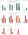
Figure 3.. Subgroup Analyses in Combined Discovery and Validation Cohorts of LGI1-Antibody Encephalitis vs CASPR2-Antibody Encephalitis and LGI1/CASPR2-Antibody Autoimmune Encephalitis (AE) vs Viral Encephalitis (VE) Subtypes
LGI1-antibody AE demonstrated higher rates of abnormality than CASPR2-antibody AE but similar rates of T2/FLAIR hyperintensities, temporal lobe involvement, hippocampus/amygdala confinement, and extratemporal extension. Compared with HSV1 VE, other forms of VE demonstrated fewer abnormalities, T2/FLAIR hyperintensities, hippocampus/amygdala swelling, diffusion restriction, and contrast enhancement. Error bars indicate 95% CI. FLAIR indicates fluid-attenuated inversion recovery; HSV1, herpes simplex virus 1. aP < .01. bP < .001. cP < .05.
Figure 4.
Figure 4.. Receiver Operating Characteristic (ROC) Curves Reporting the Predictive Value of Diffusion Restriction, Contrast Enhancement, and Extratemporal T2 and/or Fluid-Attenuated Inversion Recovery (FLAIR) Hyperintensity
Distinguishing LGI1/CASPR2-antibody autoimmune encephalitis (AE) from non-AE cases across both cohorts separately and combined. All cases entered into this analysis demonstrated T2 and/or FLAIR hyperintensity of the hippocampus and/or amygdala. Absence of all 3 of these features was predictive of LGI1/CASPR2-antibody AE with an area under the curve (AUC) of 0.97, sensitivity of 90%, and specificity of 95% among cases with hippocampus/amygdala T2/FLAIR hyperintensity. For similar analyses including all cases (including scans without hippocampus/amygdala changes and normal scans) and comparing LGI1/CASPR1-antibody AE against herpes simplex virus 1 cases only, see eFigure 4 in Supplement 1.

References

    1. Irani SR, Alexander S, Waters P, et al. . Antibodies to Kv1 potassium channel-complex proteins leucine-rich, glioma inactivated 1 protein and contactin-associated protein-2 in limbic encephalitis, Morvan’s syndrome and acquired neuromyotonia. Brain. 2010;133(9):2734-2748. doi:10.1093/brain/awq213 - DOI - PMC - PubMed
    1. Day GS. Rapidly progressive dementia. Continuum (Minneap Minn). 2022;28(3):901-936. - PMC - PubMed
    1. Thompson J, Bi M, Murchison AG, et al. ; Faciobrachial Dystonic Seizures Study Group . The importance of early immunotherapy in patients with faciobrachial dystonic seizures. Brain. 2018;141(2):348-356. doi:10.1093/brain/awx323 - DOI - PMC - PubMed
    1. Binks SNM, Veldsman M, Easton A, et al. . Residual fatigue and cognitive deficits in patients after leucine-rich glioma-inactivated 1 antibody encephalitis. JAMA Neurol. 2021;78(5):617-619. doi:10.1001/jamaneurol.2021.0477 - DOI - PMC - PubMed
    1. Flanagan EP, Kotsenas AL, Britton JW, et al. . Basal ganglia T1 hyperintensity in LGI1-autoantibody faciobrachial dystonic seizures. Neurol Neuroimmunol Neuroinflamm. 2015;2(6):e161. doi:10.1212/NXI.0000000000000161 - DOI - PMC - PubMed

Publication types

MeSH terms

Supplementary concepts