. 2024 Mar 18;16(6):5123-5148.
doi: 10.18632/aging.205630. Epub 2024 Mar 18.

The comprehensive landscape of prognosis, immunity, and function of the GLI family by pan-cancer and single-cell analysis

Affiliations

The comprehensive landscape of prognosis, immunity, and function of the GLI family by pan-cancer and single-cell analysis

Yinteng Wu et al. Aging (Albany NY). .

Abstract

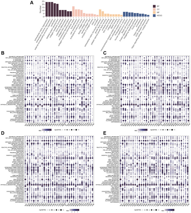
The Hedgehog (Hh) signaling pathway has been implicated in the pathogenesis of various cancers. However, the roles of the downstream GLI family (GLI1, GLI2, and GLI3) in tumorigenesis remain elusive. This study aimed to unravel the genetic alterations of GLI1/2/3 in cancer and their association with the immune microenvironment and related signaling pathways. Firstly, we evaluated the expression profiles of GLI1/2/3 in different cancer types, analyzed their prognostic and predictive values, and assessed their correlation with tumor-infiltrating immune cells. Secondly, we explored the relationships between GLI1/2/3 and genetic mutations, epigenetic modifications, and clinically relevant drugs. Finally, we performed enrichment analysis to decipher the underlying mechanisms of GLI1/2/3 in cancer initiation and progression. Our results revealed that the expression levels of GLI1/2/3 were positively correlated in most cancer tissues, suggesting a cooperative role of these factors in tumorigenesis. We also identified tissue-specific expression patterns of GLI1/2/3, which may reflect the distinct functions of these factors in different cell types. Furthermore, GLI1/2/3 expression displayed significant associations with poor prognosis in several cancers, indicating their potential as prognostic biomarkers and therapeutic targets. Importantly, we found that GLI1/2/3 modulated the immune microenvironment by regulating the recruitment, activation, and polarization of cancer-associated fibroblasts, endothelial cells, and macrophages. Additionally, functional enrichment analyses indicated that GLI1/2/3 are involved in the regulation of epithelial-mesenchymal transition (EMT). Together, our findings shed new light on the roles of GLI1/2/3 in tumorigenesis and provide a potential basis for the development of novel therapeutic strategies targeting GLI-mediated signaling pathways in cancer.

Keywords: GLI; function; immune; pan-cancer; prognosis; single-cell.


Conflict of interest statement

CONFLICTS OF INTEREST: The authors declare no conflicts of interest related to this study.

Figures

Figure 1
Figure 1
Correlation between GLI1, GLI2, and GLI3. Correlation of (A) GLI1 and GLI2, (B) GLI1 and GLI3, (C) GLI2 and GLI3 in normal tissues. Correlation of (D) GLI1 and GLI2, (E) GLI1 and GLI3, (F) GLI2 and GLI3 in tumor cell lines. Correlation of (G) GLI1 and GLI2, (H) GLI1 and GLI3, (I) GLI2 and GLI3 in tumor tissues.
Figure 2
Figure 2
Expression profiles of GLI1, GLI2, and GLI3. Expression levels of GLI1, GLI2, and GLI3 in normal tissues (AC), tumor cell lines (DF), and tumor tissues (GI) using data from the GTEx database. Combined TCGA and GTEx data of GLI1 (J), GLI2 (K), and GLI3 (L) expression differences between tumor and normal tissues (*p < 0.05, **p < 0.01, and ***p < 0.001; Abbreviation: ns: no significance). Differential expression of GLI1 (M), GLI2 (N), and GLI3 (O) between paired tumors and normal tissues using data from the TGGA database (*p < 0.05, **p < 0.01, and ***p < 0.001; Abbreviation: ns: no significance).
Figure 3
Figure 3
Protein expression of GLI1 in normal and tumor tissues. Immunohistochemical staining of normal and tumor tissues in the HPA database. (A) Breast normal, BRCA. (B) Cervix normal, CESC. (C) Lung normal, LUNG. (D) Oral mucosa normal, HNSC. (E) Ovary normal, OV. (F) Prostate normal, PRAD. (G) Rectum normal, READ. (H) Pancreas normal, PAAD.
Figure 4
Figure 4
Protein subcellular localization the immunofluorescence images of (A) GLI1, (B) GLI2, and (C) GLI3 protein.
Figure 5
Figure 5
Expression of GLI1, GLI2, and GLI3 with OS, PFI, and DSS in tumor patients. Forest plots of hazard ratios of GLI1, GLI2, and GLI3 in OS (AC), PFI (DF), and DSS (GI).
Figure 6
Figure 6
Kaplan–Meier OS curves for patients stratified by different expression levels of GLI1 in seventeen cancer types.
Figure 7
Figure 7
Correlation between GLI1 (A), GLI2 (B), and GLI3 (C) expression with clinical stage in cancer patients.
Figure 8
Figure 8
Mutation signature mapping of GLI1, GLI2, and GLI3 genes in the cBioPortal database. (A) An overview of the genomic alternations of GLI1, GLI2, and GLI3 occurred in pan-cancer. The mutation frequency and corresponding mutation types of GLI1 (B), GLI2 (C), and GLI3 (D) in different cancers. Mutation sites of GLI1 (E), GLI2 (F), and GLI3 (G). Kaplan-Meier plot showing the comparison of OS (H), DSS (I), and PFS (J) in cases with/without GLI1 gene alterations in the tumor.
Figure 9
Figure 9
Correlation analysis of GLI1, GLI2, and GLI3 with CNV, methylation, and mutation frequency. (A) Type of genetic variation. (B) CNV expression of GLI1, GLI2, and GLI3 in human pan-cancer. (C) Correlation between CNV expression of GLI1, GLI2, and GLI3 and their mRNA expression. (D) Correlation between GLI1, GLI2, and GLI3 and methylation in various tumors. (E) Correlations between GLI1, GLI2, and GLI3 mRNA expression and methylation in various tumors. (F) The mutation frequency of GLI1, GLI2, and GLI3 in various tumors. (G) SNV oncoplot. An oncoplot showing the mutation distribution of GLI1, GLI2, and GLI3 and a classification of SNV types.
Figure 10
Figure 10
Infiltration of immune cells in tumors. Correlation of GLI1 (A), GLI2 (B), and GLI3 (C) with tumor-infiltrating immune cells according to different algorithms. (D) Correlation of GLI1, GLI2, and GLI3 with different species of immune cell subtypes. (E) Relationship between GLI1, GLI2, and GLI3 and three scores such as ESTIMATEScore, ImmuneScore, and StromalScore in different cancer types.
Figure 11
Figure 11
Coexpression of GLI1 (A), GLI2 (B), and GLI3 (C) with immune-related genes (X means P > 0.05).
Figure 12
Figure 12
Correlation analysis of GLI1 (A), GLI2 (B), and GLI3 (C) expression with DNA methyltransferases (Red represents DNMAT1, blue represents DNMT2, green represents DNMT3A, and purple represents DNMT3B). Correlation analysis of GLI1 (D), GLI2 (E), and GLI3 (F) expression with MMRs genes in human pan-cancer (*P < 0.05, **P < 0.01, ***P < 0.001).
Figure 13
Figure 13
Relationship of GLI1, GLI2, and GLI3 with TMB, MSI, and drug sensitivity. The Relationship of GLI1, GLI2, and GLI3 expression with TMB (AC) and MSI (DF). Drug sensitivity analysis in CTRP database (G) and GDSC database (H).
Figure 14
Figure 14
Enrichment Analysis. (A) GO and KEGG of similar genes. GSEA of GLI1 (B), GLI2 (C), GLI3 (D), and GLI1/2/3 gene sets (E).
Figure 15
Figure 15
Single-cell analysis. The expression distribution of GLI1 (A), GLI2 (B), and GLI3 (C) at the single-cell level. The correlation between the gene set of GLI1/2/3 and cancer-related functional states (D). The correlation between the gene set of GLI1/2/3 and cancer-related functional states in RB (E). The expression distribution of the gene set of GLI1/2/3 with t-SNE plot in RB is displayed (F). T-SNE describes the distribution of cells, where each point represents a single cell, and the color of the point indicates the expression level of the gene list in that cell.

Similar articles

Cited by

References

    1. Chen DS, Mellman I. Oncology meets immunology: the cancer-immunity cycle. Immunity. 2013; 39:1–10. 10.1016/j.immuni.2013.07.012 - DOI - PubMed
    1. Okazaki T, Chikuma S, Iwai Y, Fagarasan S, Honjo T. A rheostat for immune responses: the unique properties of PD-1 and their advantages for clinical application. Nat Immunol. 2013; 14:1212–8. 10.1038/ni.2762 - DOI - PubMed
    1. Pardoll D, Allison J. Cancer immunotherapy: breaking the barriers to harvest the crop. Nat Med. 2004; 10:887–92. 10.1038/nm0904-887 - DOI - PubMed
    1. Wei SC, Duffy CR, Allison JP. Fundamental Mechanisms of Immune Checkpoint Blockade Therapy. Cancer Discov. 2018; 8:1069–86. 10.1158/2159-8290.CD-18-0367 - DOI - PubMed
    1. Desbois M, Wang Y. Cancer-associated fibroblasts: Key players in shaping the tumor immune microenvironment. Immunol Rev. 2021; 302:241–58. 10.1111/imr.12982 - DOI - PubMed

Publication types