. 2024 Apr;300(4):107174.
doi: 10.1016/j.jbc.2024.107174. Epub 2024 Mar 16.

Truncation of the constant domain drives amyloid formation by immunoglobulin light chains

Affiliations

Truncation of the constant domain drives amyloid formation by immunoglobulin light chains

Francesca Lavatelli et al. J Biol Chem. 2024 Apr.

Abstract

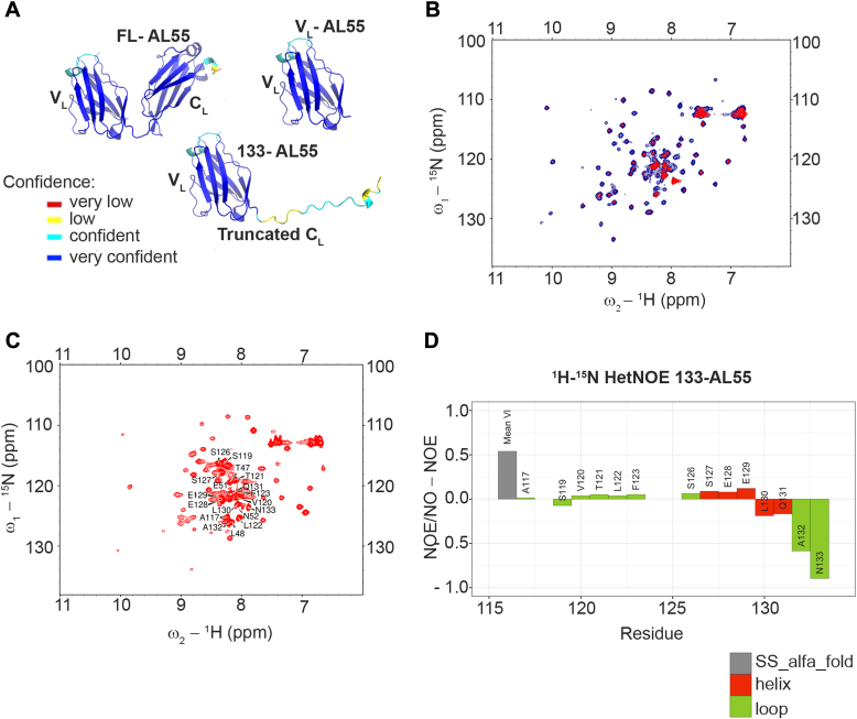
AL amyloidosis is a life-threatening disease caused by deposition of immunoglobulin light chains. While the mechanisms underlying light chains amyloidogenesis in vivo remain unclear, several studies have highlighted the role that tissue environment and structural amyloidogenicity of individual light chains have in the disease pathogenesis. AL natural deposits contain both full-length light chains and fragments encompassing the variable domain (VL) as well as different length segments of the constant region (CL), thus highlighting the relevance that proteolysis may have in the fibrillogenesis pathway. Here, we investigate the role of major truncated species of the disease-associated AL55 light chain that were previously identified in natural deposits. Specifically, we study structure, molecular dynamics, thermal stability, and capacity to form fibrils of a fragment containing both the VL and part of the CL (133-AL55), in comparison with the full-length protein and its variable domain alone, under shear stress and physiological conditions. Whereas the full-length light chain forms exclusively amorphous aggregates, both fragments generate fibrils, although, with different kinetics, aggregate structure, and interplay with the unfragmented protein. More specifically, the VL-CL 133-AL55 fragment entirely converts into amyloid fibrils microscopically and spectroscopically similar to their ex vivo counterpart and increases the amorphous aggregation of full-length AL55. Overall, our data support the idea that light chain structure and proteolysis are both relevant for amyloidogenesis in vivo and provide a novel biocompatible model of light chain fibrillogenesis suitable for future mechanistic studies.

Keywords: AL amyloidosis; amyloidogenesis; cardiomyopathy; constant domain; immunoglobulin light chains; proteolysis.


Conflict of interest statement

Conflict of interest The authors declare that they have no conflicts of interest with the contents of this article.

Figures

Figure 1
Figure 1
Sequence and conformational characterization of AL-55–related species.A, scheme of the three AL55 proteoforms under investigation with V (variable), J (joining), and C (constant) regions. Full-length sequence of AL55 shown in the lower part of panel A with highlighted sequences of 133-AL55 (in bold) and VL-AL55 (underlined), respectively. B, far-UV CD spectra of the three AL55 species at pH 7.4, 10 °C, and 85 °C. C, secondary structure content of the three proteoforms based on far-UV CD spectra collected at 10 °C. D, thermal stability monitored by CD signal at 203 nm. After heating up to 85 °C (blue lines), samples were cooled down at 10 °C before repeating a second heating treatment (gray lines) to assess the reversibility of the temperature-induced conformational changes. E, temperature of the midpoint (Tmp). Values of Tmp for each species were determined from the corresponding curves reported in E. All data shown in B–E are expressed as mean and SD of three independent measurements. VL, light chain’s variable domain.
Figure 2
Figure 2
Structural evaluation of AL55 species.A, structures predicted using AlphaFold2. Residues in the 3D structures are colored according to their pLDDT value (AlphaFold per residue estimate of model confidence) with the following code: red pLDDT <50; yellow 50 ≤pLDDT <70; cyan 70 ≤pLDDT <90; blue pLDDT ≥ 90. B, conformational stability of 133-AL55 at 37 °C monitored by solution NMR. Superposition of the 2D [1H-15N] HSQC spectra of uniformly [13C, 15N] labeled 133-AL55 at 0.5 mM in PBS pH 7.4/D2O, 95/5, vol/vol, recorded immediately after dissolution (blue trace) and after 6 h at 37 °C (red trace). C, 2D [1H-15N] HSQC spectrum of uniformly [13C, 15N] labeled 133-AL55 at 0.5 mM in PBS pH 7.4/D2O, 95/5, vol/vol, recorded at 15 °C: the 19 assigned peaks (over 97 detectable peaks) are labeled. D, backbone dynamics of 133-AL55 investigated by 1H-15N heteronuclear NOE, at 15 °C on the same sample used for backbone resonances assignment. The 1H-15N NOE values are plotted versus sequential residue number for the 14 assigned amino acids belonging to the CL domain and compared with the average of the remaining 59 peaks. CL, light chain’s constant domain; HSQC, heteronuclear single quantum coherence.
Figure 3
Figure 3
Fibrillogenesis of full-length AL55, 133-AL55, and VL-AL55.A, ThT emission fluorescence monitored during incubation of each protein at 20 μM concentration, in the microplate assay format, using agitation speed at 100 rpm and (B), at 900 rpm. Shown VL-AL55 (blue), 133-AL55 (green), full-length AL55 (red). C–E, representative TEM images of aggregates of VL-AL55, 133-AL55, and full-length AL55, respectively. Samples were withdrawn after 48 h of incubation and directly applied onto the carbon grid (negative staining; the scale bars represent 200 nm). TEM, transmission electron microscopy; ThT, Thioflavin T; VL, light chain’s variable domain.
Figure 4
Figure 4
Fibrillogenesis of 133-AL55 or VL-AL55 in mixture with full-length AL55.A, ThT emission fluorescence of full-length AL55 incubated with equimolar concentration of soluble VL-AL55 monitored with time (orange). ThT signal of VL-AL55 is shown in blue. B, ThT emission fluorescence of full-length AL55 incubated with equimolar concentration of soluble 133-AL55 monitored with time (black). ThT signal of 133-AL55 alone is shown in green. T50 values are significantly different (respectively, 7.6 h for 133-AL55 alone versus 12.6 h for 133-AL55 in the presence of the full length; p = 0.002; Student’s t test). All incubations were carried out using 20 μM protein concentrations in microplates under agitation at 900 rpm. Curves shown as mean and SD of three independent experiments. ThT, Thioflavin T; VL, light chain’s variable domain.
Figure 5
Figure 5
Fibrillogenesis of [13C]full-length AL55, VL-AL55, and 133-AL55 studied by FTIR spectroscopy. A–C, second derivatives of the FTIR absorption spectra of [13C]full-length AL55 (A), VL-AL55 (B), and 133-AL55 (C) collected at different incubation times. At 24, 48, 72, and 144 h of incubation at 37 °C, 20 μl aliquots of each sample were withdrawn and the FTIR spectra of supernatants (SN, middle spectra in panel A) and pellets (bottom spectra in all panels) were acquired. Representative spectra with the main peak positions are shown. VL, light chain’s variable domain.
Figure 6
Figure 6
Fibrillogenesis of 133-AL55 or VL-AL55 in mixture with [13C]full-length AL55 by isotope-edited FTIR.A, second derivatives of the absorption spectra of samples containing both [13C]full-length AL55 and unlabeled VL-AL55 at different times of incubation at 37 °C. Representative spectra with main peak positions assigned to [13C]full-length AL55 (red numbers) and VL-AL55 (blue numbers) are reported. B, second derivatives of the absorption spectra of samples containing both [13C]full-length AL55 and unlabeled 133-AL55 at different times of incubation at 37 °C. Representative spectra with main peak positions assigned to [13C] full-length AL55 (red numbers) and 133-AL55 (green numbers) are shown. At 24, 48, 72, and 144 h of incubation, FTIR spectra of supernatants and pellets were also acquired and shown in A and B panels. C, time course aggregation monitored with the ratio of the IR peak assigned to the native β-sheets of each variant to the tyrosine peak, based on the second derivative spectra. The ratio was calculated for the following samples: [13C] full-length AL55 (intensity at ⁓1596 cm−1/intensity at ⁓1480 cm−1) alone (red) and in the presence of VL-AL55 (magenta) or 133-AL55 (orange); VL-AL55 (intensity at ⁓1638 cm−1/intensity at ⁓1516 cm−1) alone (blue) and in the presence of [13C] full-length AL55 (light blue); 133-AL55 (intensity at ⁓1638 cm−1/intensity at ⁓1516 cm−1) alone (blue) and in the presence of [13C] full-length AL55 (light blue). D, intensity ratios for 133-AL55 alone (green bar) and in the presence of [13C] full-length AL55 (light green bar) are shown as mean and SD of three independent experiments. ∗p <  0.05, Student’s t test. VL, light chain’s variable domain.
Figure 7
Figure 7
Electrophoretic and TEM analyses of aggregates from the fibrillogenesis of 133-AL55 or VL-AL55 in mixture with full-length AL55.A and B, SDS 4 to 20% PAGE analysis of supernatants and pellets from the coincubation of full-length AL55 with VL-AL55 and 133-AL55, respectively. Samples were withdrawn after 48 h of incubation; insoluble aggregates were separated by centrifugation, as detailed in Methods. Mixtures at time 0 (T0h) are shown for comparison. C and D, TEM images of aggregates from the cofibrillogenesis of full-length AL55 with VL-AL55 and 133-AL55, respectively. Samples were withdrawn after 48 h of incubation and directly applied onto the carbon grid (negative staining; the scale bars represent 200 nm). NR, nonreducing conditions; P, pellet; S, supernatant; R, reducing conditions; TEM, transmission electron microscopy; VL, light chain’s variable domain.

References

    1. Buxbaum J.N., Dispenzieri A., Eisenberg D.S., Fändrich M., Merlini G., Saraiva M.J.M., et al. Amyloid nomenclature 2022: update, novel proteins, and recommendations by the international society of amyloidosis (ISA) nomenclature committee. Amyloid. 2022;29:213–219. - PubMed
    1. Merlini G., Dispenzieri A., Sanchorawala V., Schönland S.O., Palladini G., Hawkins P.N., et al. Systemic immunoglobulin light chain amyloidosis. Nat. Rev. Dis. Primers. 2018;4:38. - PubMed
    1. Bellotti V., Mangione P., Merlini G. Review: immunoglobulin light chain amyloidosis--the archetype of structural and pathogenic variability. J. Struct. Biol. 2000;130:280–289. - PubMed
    1. Kourelis T.V., Dasari S., Theis J.D., Ramirez-Alvarado M., Kurtin P.J., Gertz M.A., et al. Clarifying immunoglobulin gene usage in systemic and localized immunoglobulin light-chain amyloidosis by mass spectrometry. Blood. 2017;129:299–306. - PubMed
    1. Kyle R.A., Greipp P.R. “Idiopathic” Bence Jones proteinuria: long-term follow-up in seven patients. N. Engl. J. Med. 1982;306:564–567. - PubMed

Publication types

MeSH terms