. 2024 Mar 18;7(6):e202302525.
doi: 10.26508/lsa.202302525. Print 2024 Jun.

A genetic screen to uncover mechanisms underlying lipid transfer protein function at membrane contact sites

Affiliations

A genetic screen to uncover mechanisms underlying lipid transfer protein function at membrane contact sites

Shirish Mishra et al. Life Sci Alliance. .

Abstract

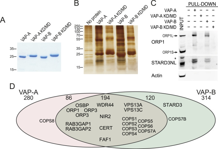
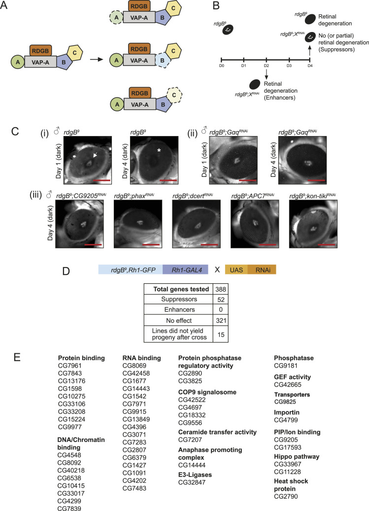
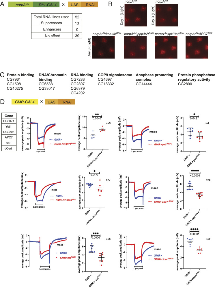
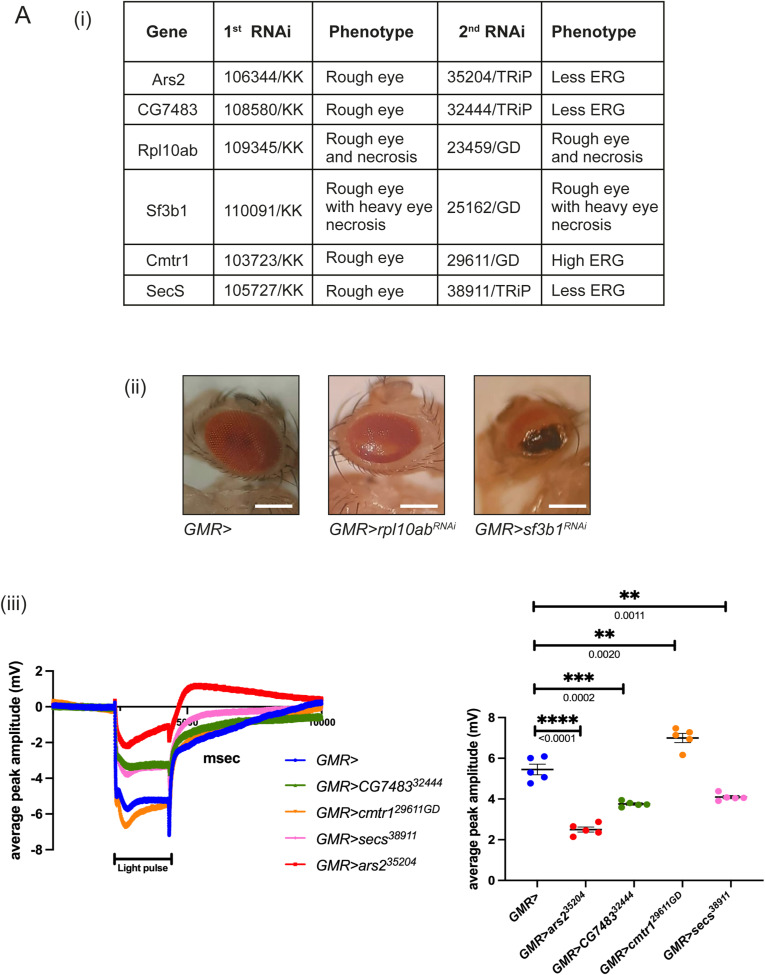
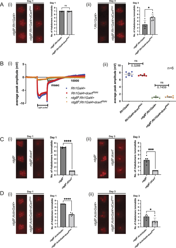
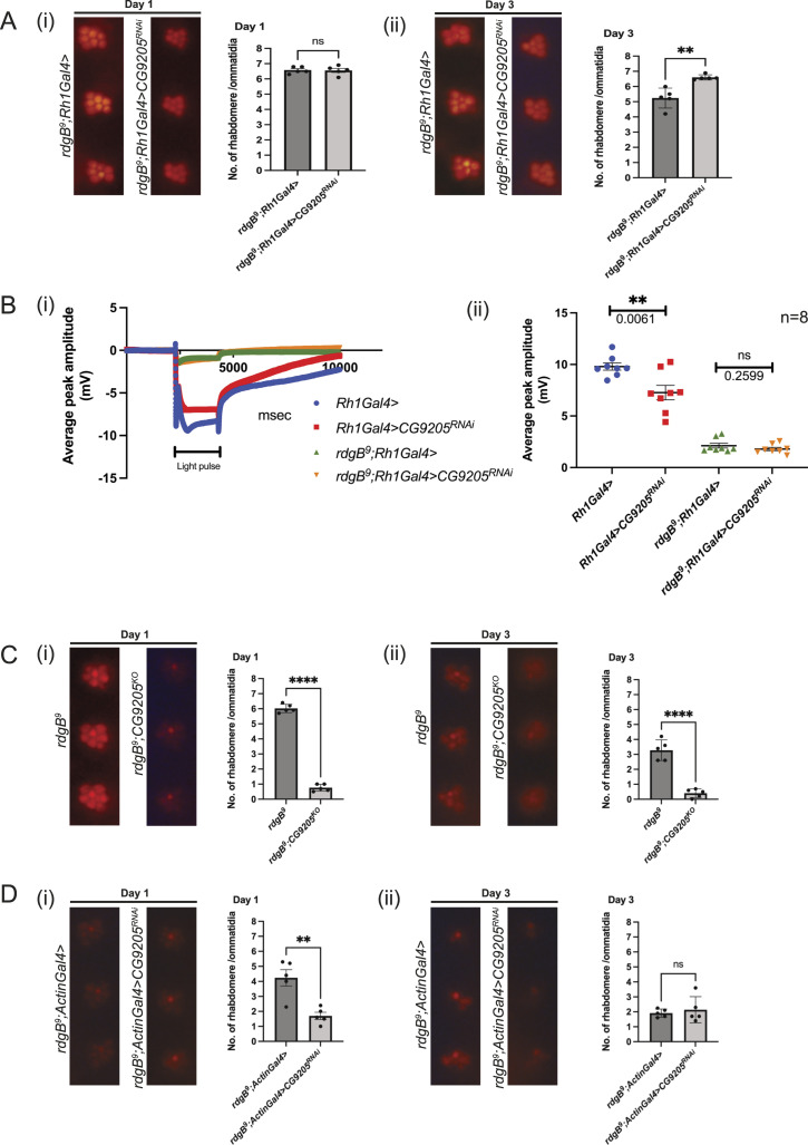
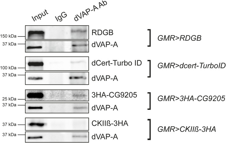
Lipid transfer proteins mediate the transfer of lipids between organelle membranes, and the loss of function of these proteins has been linked to neurodegeneration. However, the mechanism by which loss of lipid transfer activity leads to neurodegeneration is not understood. In Drosophila photoreceptors, depletion of retinal degeneration B (RDGB), a phosphatidylinositol transfer protein, leads to defective phototransduction and retinal degeneration, but the mechanism by which loss of this activity leads to retinal degeneration is not understood. RDGB is localized to membrane contact sites through the interaction of its FFAT motif with the ER integral protein VAP. To identify regulators of RDGB function in vivo, we depleted more than 300 VAP-interacting proteins and identified a set of 52 suppressors of rdgB The molecular identity of these suppressors indicates a role of novel lipids in regulating RDGB function and of transcriptional and ubiquitination processes in mediating retinal degeneration in rdgB9 The human homologs of several of these molecules have been implicated in neurodevelopmental diseases underscoring the importance of VAP-mediated processes in these disorders.


Conflict of interest statement

The authors declare that they have no conflict of interest.

Figures

Figure 1.
Figure 1.. Identification of VAP-A and VAP-B binding partners.
(A) Coomassie Blue staining of the recombinant WT and KD/MD mutant MSP domains of VAP-A and VAP-B after SDS–PAGE. (B) Silver nitrate staining of proteins pulled down using WT MSP domains of VAP-A and VAP-B, and the KD/MD mutant MSP domains, after SDS–PAGE. (C) Western blot analysis of proteins pulled down using the WT and mutant MSP domain of VAP-A and VAP-B. The input and pull-down fractions correspond to HeLa cell total protein extract and bound proteins, respectively. *: non-specific band. (D) Venn diagram of proteins pulled down by VAP-A and VAP-B (and not by mutant VAP-A and VAP-B). A total of 401 proteins were pulled down with either VAP-A or VAP-B. 194 proteins were pulled down with both VAP-A and VAP-B.
Figure 2.
Figure 2.. Strategy of the genetic screen and hits found.
(A) Cartoon depicting classes of VAP interactors used in the present genetic screen. Three classes of genetic interactors of rdgB are shown based on the likely molecular mechanism: loss of A, a direct physical interactor of VAP-A; loss of B, a direct interactor of VAP-A that also interacts with C, a protein required for RDGB function; and loss of C, a protein required for rdgB function but only interacts with VAP-A via B. Depletion of a specific VAP interactor is depicted with a dotted line. Fly homologs were filtered using DIOPT in FlyBase (http://flybase.org/). (B) Genetic scheme used to find either enhancers or suppressors of the retinal degeneration phenotype of rdgB9. (C) Pseudopupil imaging: (i) rdgB9 showed retinal degeneration by day 4 in dark when checked via deep pseudopupil imaging (depicted by *). (ii) The degeneration was partially suppressed when levels of Gαq were down-regulated in rdgB9 on day 4. (iii) Selected hits that showed suppression of retinal degeneration in rdgB9 on day 4 (scale bar 225 μm). (D) Table showing the full list of genes used in the screen and the number of suppressor genes identified. (E) Positive hits (suppressor genes) are divided into different categories depending on their cellular functions. n = 5 flies/RNAi line.
Figure 3.
Figure 3.. Genetic screen using norpAp24.
(A) Scheme used to test for genetic interaction of each of the 52 su(rdgB) with norpAp24 under illumination conditions (constant light 2000 Lux). (B) norpAp24 flies degenerate by day 3 under light conditions, and examples of su(RDGB) candidates that suppressed the norpAp24 retinal degeneration phenotype. n = 5 flies/RNAi line. (C) Complete list of 13 genes with their cellular functions that suppressed the norpAp24 phenotype. ERG screen. (D) Of 52 candidates, five su(RDGB) showed reduced (CG9205, Yeti, Apc7, Set, and dCert) and one (CG3071) showed higher ERG phenotype (traces and quantification shown) when down-regulated in an otherwise WT background. The number of flies used for the experimental set is mentioned along with the quantification. Scatter plots with the mean ± SEM are shown. Statistical tests: unpaired t test.
Figure S1.
Figure S1.. Six su(rdgB) affecting either eye development or physiology.
(A) Second category of su(rdgB) was variable in either showing a rough eye phenotype in the first RNAi line and ERG defects in the second independent RNAi line. (ii) rpl10Ab and sf3b1 are the only candidates that consistently showed a rough eye phenotype in two independent RNAi lines (scale bar 225 μm). (iii) ERG traces and quantifications of the rest of the su(rdgB) with their respective RNAi line mentioned. The number of flies used for the experimental set is mentioned along with the quantification. Scatter plots with the mean + SEM are shown. Statistical tests: unpaired t test.
Figure 4.
Figure 4.. Spatial and temporal down-regulation of dCert in rdgB9.
(A) Suppression of retinal degeneration when dCert RNAi line (35579/TRiP, BDRC) was expressed using Rh1 promoter. After eclosion, flies were kept in the dark and assayed on either day 1 or 3: (i) on day 1, there was no appreciable difference in two genotypes and rhabdomeres were intact; and (ii) on day 3, down-regulation of dCert in rdgB9 suppressed the retinal degeneration observed in rdgB9 control. (B) When subjected to ERG analysis, down-regulation of dCert using Rh1-GAL4 in the background of rdgB9 did not suppress the ERG phenotype: (i) ERG trace and (ii) quantification. n = 6 flies. Scatter plots with the mean ± SEM are shown. Statistical tests: unpaired t test. (C) Double mutant of rdgB9;dcert1 showed enhancement of retinal degeneration: (i, ii) by day 1 alone, double mutant has severely enhanced retinal degeneration phenotype when compared to rdgB9. (D) Enhancement of retinal degeneration when dCert (35579/TRiP, BDRC) was down-regulated with a whole-body Actin-Gal4 promoter in the rdgB9 background: (i) on day 1, rhabdomere loss is significant in the experimental files compared with control that worsens by day 3 and phenocopies the retinal degeneration present in the double mutant. For optical neutralization experiments, scoring was done by quantifying 10 ommatidia/fly head, n = 5 fly heads.
Figure 5.
Figure 5.. Spatial and temporal down-regulation of CG9205 in rdgB9.
(A) Suppression of retinal degeneration when CG9205 RNAi line (29079/GD, VDRC) was expressed using Rh1 promoter. After eclosion, flies were kept in the dark and assayed on either day 1 or 3: (i) on day 1, there was no appreciable difference in two genotypes and rhabdomeres were intact; and (ii) on day 3, down-regulation of CG9205 in rdgB9 suppressed the retinal degeneration observed in rdgB9 control. (B) When subjected to ERG analysis, down-regulation of CG9205 using Rh1-GAL4 in the background of rdgB9 did not suppress the ERG phenotype, whereas down-regulation of CG9205 using Rh1-GAL4 in an otherwise WT background shows reduced ERG amplitude: (i) ERG trace and (ii) quantification. n = 8 flies. scatter plots with the mean ± SEM are shown. Statistical tests: unpaired t test. (C) Double mutant of rdgB9;CG9205KO showed enhancement of retinal degeneration: (i, ii) by day 1 alone, double mutant has severely enhanced retinal degeneration phenotype when compared to rdgB9. (D) Enhancement of retinal degeneration when CG9205 (29079/GD, VDRC) was down-regulated with a whole-body Actin-Gal4 promoter in the rdgB9 background: (i) on day 1, rhabdomere loss is significant in the experimental files compared with control that remains the same on day 3 and phenocopies the retinal degeneration present in the double mutant. For optical neutralization experiments, scoring was done by quantifying 10 ommatidia/fly head, n = 5 fly heads.
Figure S2.
Figure S2.. Representative immunoblots showing co-immunoprecipitation of RDGB (used as a control) and dCert and CG9205 after pulling with dVAP-A antibody in fly heads.
UAS constructs of RDGB, dCert::TurboID, and 3HA::CG9205 were expressed under GMR promoter. The experiment was repeated twice.

References

    1. Alpy F, Stoeckel M-E, Dierich A, Escola J-M, Wendling C, Chenard M-P, Vanier MT, Gruenberg J, Tomasetto C, Rio M-C (2001) The steroidogenic acute regulatory protein homolog MLN64, a late endosomal cholesterol-binding protein. J Biol Chem 276: 4261–4269. 10.1074/jbc.M006279200 - DOI - PubMed
    1. Alpy F, Latchumanan VK, Kedinger V, Janoshazi A, Thiele C, Wendling C, Rio M-C, Tomasetto C (2005) Functional characterization of the MENTAL domain. J Biol Chem 280: 17945–17952. 10.1074/jbc.M500723200 - DOI - PubMed
    1. Alpy F, Rousseau A, Schwab Y, Legueux F, Stoll I, Wendling C, Spiegelhalter C, Kessler P, Mathelin C, Rio M-C, et al. (2013) STARD3 or STARD3NL and VAP form a novel molecular tether between late endosomes and the ER. J Cell Sci 126: 5500–5512. 10.1242/jcs.139295 - DOI - PubMed
    1. Bonini NM, Fortini ME (2003) Human neurodegenerative disease modeling using Drosophila. Annu Rev Neurosci 26: 627–656. 10.1146/annurev.neuro.26.041002.131425 - DOI - PubMed
    1. Cabukusta B, Berlin I, van Elsland DM, Forkink I, Spits M, de Jong AWM, Akkermans JJLL, Wijdeven RHM, Janssen GMC, van Veelen PA, et al. (2020) Human VAPome analysis reveals MOSPD1 and MOSPD3 as membrane contact site proteins interacting with FFAT-related FFNT motifs. Cell Rep 33: 108475. 10.1016/j.celrep.2020.108475 - DOI - PubMed

LinkOut - more resources