. 2024 Mar;13(3):e12417.
doi: 10.1002/jev2.12417.

Y-box binding protein 1 in small extracellular vesicles reduces mesenchymal stem cell differentiation to osteoblasts-implications for acute myeloid leukaemia

Affiliations

Y-box binding protein 1 in small extracellular vesicles reduces mesenchymal stem cell differentiation to osteoblasts-implications for acute myeloid leukaemia

Venkatesh Kumar Chetty et al. J Extracell Vesicles. 2024 Mar.

Abstract

Small extracellular vesicles (sEVs) released by acute myeloid leukaemia (AML) cells have been reported to influence the trilineage differentiation of bone marrow-derived mesenchymal stem cells (BM-MSCs). However, it remains elusive which biological cargo from AML-sEVs is responsible for this effect. In this study, sEVs were isolated from cell-conditioned media and blood plasma using size-exclusion chromatography and ultrafiltration and characterized according to MISEV2018 guidelines. Our results demonstrated that AML-sEVs increased the proliferation of BM-MSCs. Conversely, key proteins that are important for normal haematopoiesis were downregulated in BM-MSCs. Additionally, we revealed that AML-sEVs significantly reduced the differentiation of BM-MSCs to osteoblasts without affecting adipogenic or chondrogenic differentiation. Next, LC-MS/MS proteomics elucidated that various proteins, including Y-box-binding protein 1 (YBX1), were upregulated in both AML-sEVs and BM-MSCs treated with AML-sEVs. Clinically relevant, we found that YBX1 is considerably upregulated in most paediatric AML patient-derived sEVs compared to healthy controls. Interestingly, sEVs isolated after the downregulation of YBX1 in AML cells remarkably rescued the osteoblastic differentiation of BM-MSCs. Altogether, our data demonstrate for the first time that YBX1 containing AML-sEVs is one of the key players that disrupt the normal function of bone marrow microenvironment by reducing the osteogenic differentiation of BM-MSCs.

Keywords: Y-box binding protein 1; acute myeloid leukaemia; bone marrow microenvironment; mesenchymal stem cells; osteoblasts; small extracellular vesicles.


Conflict of interest statement

The authors report no conflict of interest.

Figures

FIGURE 1
FIGURE 1
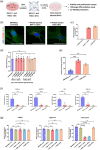
Isolation and characterization of leukaemia sEVs. (a) Steps involved in the sEVs isolation using tangential flow filtration, size‐exclusion chromatography and ultrafiltration, collectively named as TSU. (b) TEM imaging of sEVs derived from MV4‐11 and K562 cell lines. Scale bars‐ 100 nm. (c) Graph showing the particle size distribution of MV4‐11 and K562 sEVs determined by NTA. (d) Mean values of particle count evaluated using NTA (left y‐axis, bar plot), and free protein concentration determined using micro‐BCA (right y‐axis, dot plot). Data correspond to the mean ± S.E.M. obtained from three independent experiments. (e) Detection of canonical EV markers (syntenin, TSG101 and Hsp70) and EV negative marker (calnexin) using Western blot. The black outer line box implies the samples that were run together.
FIGURE 2
FIGURE 2
MV4‐11 and K562 sEVs influence BM‐MSCs differentiation. (a) Schematic workflow of bone marrow‐derived mesenchymal stem cells (BM‐MSCs) treatment with MV4‐11 and K562 sEVs. Small EVs were incubated with BM‐MSCs with a ratio of 50:1 (sEV particle number/recipient cells). Analysis of PKH26‐labelled MV4‐11 and K562 sEVs transferred into BM‐MSCs after 24 h using (b) confocal microscopy and (c) flow cytometry. Representative confocal images showing cell nuclei (DAPI, blue), cell membrane (WGA‐Alexa488, green) and PKH26‐labelled leukaemia sEVs (PKH26, red). Scale bars‐ 10 µm. (d) MTT assay of BM‐MSCs treated with different MV4‐11 and K562 sEVs concentrations for 24 h. (e) Colony‐forming unit (CFU) assay performed on BM‐MSCs treated with leukaemia sEVs for three weeks. (f) Relative mRNA expression of various genes important for normal hematopoietic function in bone marrow microenvironment. (g) Trilineage differentiation of BM‐MSCs treated with leukaemia and healthy control sEVs. Graphs showing the influence of adipogenic (FABP4), chondrogenic (aggrecan) and osteogenic (osteocalcin) differentiation of BM‐MSCs due to the sEVs treatment. Data shown in c‐g are mean ± S.E.M. obtained from three independent experiments, and statistical significance is calculated in reference to either untreated BM‐MSCs or BM‐MSCs treated with healthy control sEVs (****p < 0.0001, ***p < 0.001, **p < 0.01, and ns‐ non‐significant).
FIGURE 3
FIGURE 3
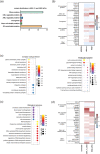
Identification of AML‐derived sEV protein targets that influence osteogenic differentiation of BM‐MSCs. (a) Different classes of proteins enriched in both MV4‐11 and K562 sEVs (CML‐chronic myeloid leukemia). (b) Heat map showing the different classes of proteins contained in leukaemia sEVs. (c) Gene ontology (GO) enrichment analysis of the proteome of MV4‐11 and K562 sEVs. BM‐MSCs were treated with leukaemia sEVs with a ratio of 50:1 (50 sEV particles per cell). (d) Heat map illustrating the different classes of proteins contained in BM‐MSCs treated with leukaemia sEVs.
FIGURE 4
FIGURE 4
Upregulation of YBX1 protein cargo inside AML‐derived sEVs. (a) Western blot analysis of YBX1 in various AML and non‐AML samples. C1‐C15 and P1‐P15 indicates sEVs isolated from peripheral blood plasma of 15 paediatric healthy controls and 15 paediatric AML patients, respectively. (b) Quantification of YBX1 expression using ELISA (N = 15 for paediatric healthy control and AML patient sEVs). (c) Table showing the level of YBX1 (ng/mL) on various paediatric AML patients sEVs. Evaluation of YBX1 expression on BM‐MSCs treated with AML and non‐AML sEVs. sEVs were incubated with BM‐MSCs in the ratio of 50:1 (50 sEV particles per cell). (d) Immunoblotting of GAPDH and YBX1 on untreated BM‐MSCs and BM‐MSCs treated with different sEVs. Quantification of YBX1 relative to corresponding GAPDH signal. (e) Relative YBX1‐mRNA level on untreated BM‐MSCs and BM‐MSCs treated with different sEVs using qRT‐PCR. (f) Demonstration of the location of YBX1 cargo in leukaemia sEVs by incubation with Proteinase K (Prot K) and Triton X. Electroporation‐based downregulation of YBX1 in MV4‐11 cells. (g) Determination of YBX1 relative mRNA and protein level on MV4‐11 cells transfected with si‐YBX1 using qRT‐PCR, ELISA and western blot. Data illustrated in b, e, f and g for the cell line and their corresponding sEVs are mean ± S.E.M. of n = 3 experiments. All p values were calculated using one‐way ANOVA with Dunnett's multiple comparisons test except the ELISA analysis in f in which p‐value was calculated using an unpaired t‐test (****p < 0.0001, ***p < 0.001, **p < 0.01, *p < 0.05 and ns‐ non‐significant).
FIGURE 5
FIGURE 5
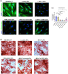
Osteogenic differentiation analysis of BM‐MSCs. Small EVs were incubated with BM‐MSCs in the ratio of 50:1 (50 sEV particles per cell). (a) Representative confocal images showing the osteoblastic differentiation of BM‐MSCs treated with AML and non‐AML sEVs. Cell nuclei are shown in blue (DAPI), and the osteogenesis marker (osteocalcin) is shown in green. Scale bars—10 µm. (b) Mean Fluorescence Intensity (MFI) of osteocalcin in BM‐MSCs treated with different sEVs expressing osteocalcin. For each condition, MFI average was calculated from at least four confocal images. p values were calculated using one‐way ANOVA with Dunnett's multiple comparisons (****p < 0.0001, ***p < 0.001, *p < 0.05 and ns‐ non‐significant). (c) Alizarin red staining of BM‐MSCs treated with AML and non‐AML sEVs.

References

    1. Abbasi, B. , Shamsasenjan, K. , Ahmadi, M. , Beheshti, S. A. , & Saleh, M. (2022). Mesenchymal stem cells and natural killer cells interaction mechanisms and potential clinical applications. Stem Cell Research & Therapy, 13(1), 97. - PMC - PubMed
    1. Antony‐Debre, I. , Bluteau, D. , Itzykson, R. , Baccini, V. , Renneville, A. , Boehlen, F. , Morabito, M. , Droin, N. , Deswarte, C. , Chang, Y. , Leverger, G. , Solary, E. , Vainchenker, W. , Favier, R. , & Raslova, H. (2012). MYH10 protein expression in platelets as a biomarker of RUNX1 and FLI1 alterations. Blood, 120(13), 2719–2722. - PubMed
    1. Azevedo, P. L. , Dias, R. B. , Nogueira, L. P. , Maradei, S. , Bigni, R. , Aragao, J. S. , Abdelhay, E. , & Binato, R. (2022). Reduced osteogenic differentiation potential in vivo in acute myeloid leukaemia patients correlates with decreased BMP4 expression in mesenchymal stromal cells. International Journal of Stem Cells, 15(2), 227–232. - PMC - PubMed
    1. BioRender . Available from: https://app.biorender.com/
    1. Bluteau, D. , Glembotsky, A. C. , Raimbault, A. , Balayn, N. , Gilles, L. , Rameau, P. , Nurden, P. , Alessi, M. C. , Debili, N. , Vainchenker, W. , Heller, P. G. , Favier, R. , & Raslova, H. (2012). Dysmegakaryopoiesis of FPD/AML pedigrees with constitutional RUNX1 mutations is linked to myosin II deregulated expression. Blood, 120(13), 2708–2718. - PubMed

Substances