. 2024 Jun 24;52(11):6441-6458.
doi: 10.1093/nar/gkae165.

Despite the odds: formation of the SARS-CoV-2 methylation complex

Affiliations

Despite the odds: formation of the SARS-CoV-2 methylation complex

Alex Matsuda et al. Nucleic Acids Res. .

Abstract

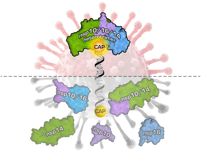
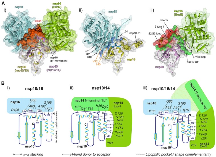
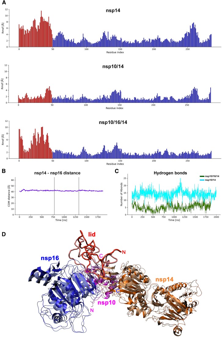
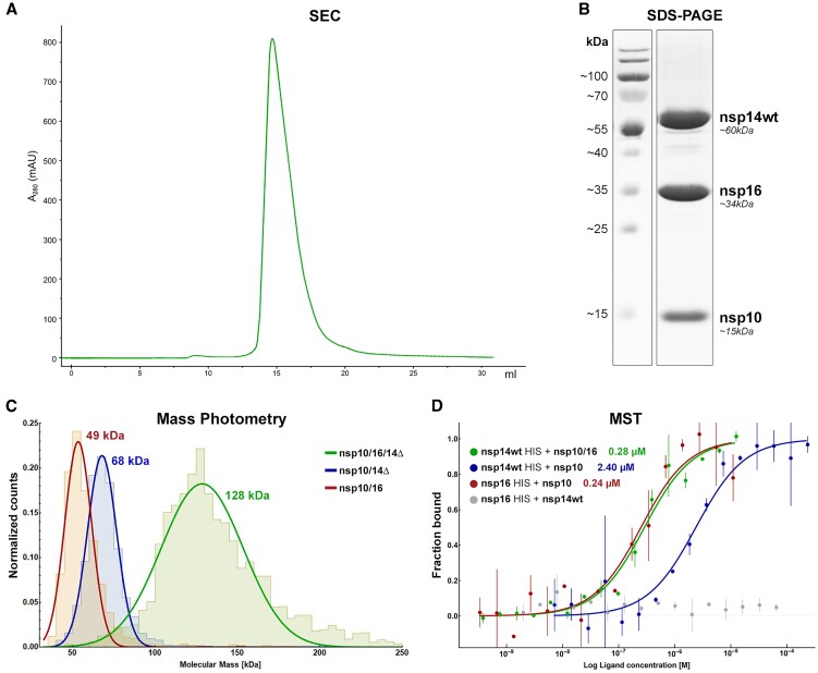
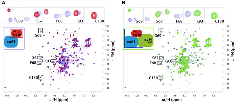
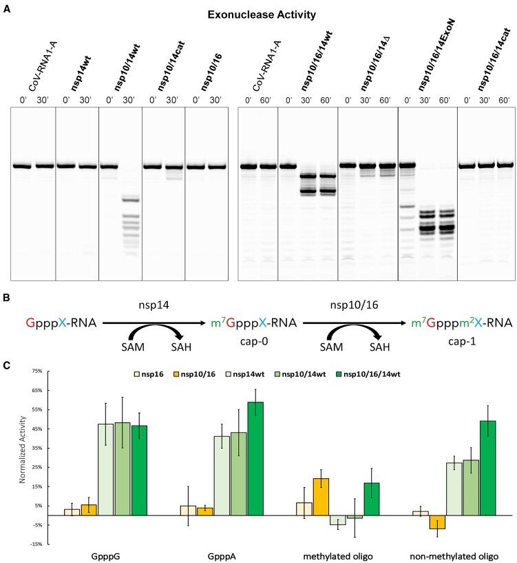
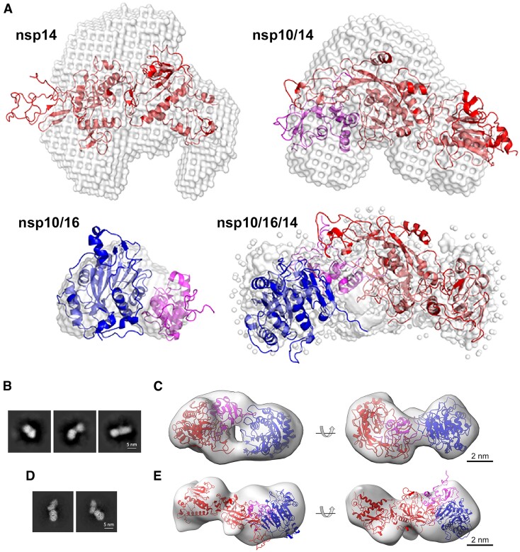
Coronaviruses modify their single-stranded RNA genome with a methylated cap during replication to mimic the eukaryotic mRNAs. The capping process is initiated by several nonstructural proteins (nsp) encoded in the viral genome. The methylation is performed by two methyltransferases, nsp14 and nsp16, while nsp10 acts as a co-factor to both. Additionally, nsp14 carries an exonuclease domain which operates in the proofreading system during RNA replication of the viral genome. Both nsp14 and nsp16 were reported to independently bind nsp10, but the available structural information suggests that the concomitant interaction between these three proteins would be impossible due to steric clashes. Here, we show that nsp14, nsp10, and nsp16 can form a heterotrimer complex upon significant allosteric change. This interaction is expected to encourage the formation of mature capped viral mRNA, modulating nsp14's exonuclease activity, and protecting the viral RNA. Our findings show that nsp14 is amenable to allosteric regulation and may serve as a novel target for therapeutic approaches.


Figures

Graphical Abstract
Graphical Abstract
Figure 1.
Figure 1.
Nsp10/16/14 heterotrimer complex formation and the lid hypothesis. (Ai) Overlay of the available high-resolution structures of SARS-CoV-2 nsp10/14 and nsp10/16 complexes with nsp10-centered alignment of nsp10/16 (PDB ID: 6WVN) and nsp10/14 (PDB ID: 7DIY). Nsp16 is shown in cyan and the associated nsp10 is shown in yellow. Nsp14 exonuclease domain is shown in green and the associated nsp10 is in magenta. The structural clash between nsp14 and nsp10/16 surfaces is shown in red. (Aii) Interface between nsp10 and nsp16. The strong hydrophobic interaction between nsp10 and nsp16 are shown in blue. The nsp10 residues V42 and L45 are indicated by the orange arrows. (Aiii) The interface between nsp10 and nsp14. The sidechains participating in hydrogen bonds between nsp10 and nsp14 are shown in bright green. The N-terminal region of nsp14, where the β turn 1 and β2/β3 loop are located is where the steric clash (red) is localized. All panels: Variable orientations of nsp10 α1′-helices seen in respective complexes with nsp16 and nsp14 are shown. Arrows indicate major structural features constituting the interface. (B) Schematic representation of interactions guiding the affinities of nsp10, nsp14 and nsp16. (Bi) Nsp16 binds nsp10 at the site overlapping that involved in binding of the lid of nsp14, but nsp10/16 interaction is characterized by a well-developed interface involving deep lipophilic pockets and solvent-shielded hydrogen bonds. The α1 helix of nsp10 is not defined in the crystal structures of the nsp10/16 complexes (PDB IDs: 6W4H, 6YZ1), indicating it is flexible and not involved in binding. (Bii) The α1 helix of nsp10 provides several deeply buried lipophilic and π-stacking interactions with the exonuclease domain of nsp14, and the interaction may be described in terms of shape complementarity. In turn, the interaction of the N-terminal lid region (amino acids 1–50) of nsp14 with nsp10 shows poor shape complementarity and is characterized only by a low number of solvent-exposed hydrogen bonds. (Biii) The formation of the nsp10/16/14 heterotrimer complex is accompanied by the lid displacement and stabilization of the α1 helix.
Figure 2.
Figure 2.
MD simulation. (A) RMSF against residue index for the nsp14 protein in each of the simulated cases with the lid part indicated as red bars. For each residue, the mean RMSF over all atoms is calculated. The lid part is highly flexible in free nsp14 simulation. Binding to nsp10 causes significant stabilization of this part. In the simulated heterotrimer, the lid is partially stabilized by interaction with nsp16 and additional interaction with the exonuclease domain but retains some degree of flexibility. (B) For the heterotrimer complex, the center of mass-distance between nsp14 and nsp16 stayed constant through the simulation time of total 1.7 μs and no dissociation happened. This indicates that nsp10 can accommodate both nsp14 (with lid readjustment) and nsp16 simultaneously. (C) The number of intermolecular hydrogen bonds between the lid part and the interacting proteins was calculated for nsp10/14 and the heterotrimer complex for each time frame. For nsp10/14, the lid was stabilized by on average 14 hydrogen bonds, and for the heterotrimer on average 5 hydrogen bonds appear between the proteins. (D) Heterotrimer structure simulated by MD. Nsp16 (blue), nsp10 (magenta), nsp14 (orange) and the lid region of nsp14 (red) are shown as ribbon plots. The first (dark hue, glossy) and last frame (light hue, matte) of the simulation are superimposed. The heterotrimer structure shows little structural deviations during the simulation except for the lid region that refolds visibly and a few loop regions. The overall shape of the complex remains stable.
Figure 3.
Figure 3.
In vitro formation of the nsp10/16/14 heterotrimer. (A) SEC chromatogram for the heterotrimer complex (nsp10/16/14wt) eluting at 14.7 ml on a Superdex 200 Increase 10/300 column. (B) SDS-PAGE analysis of the main peak from the SEC run of the heterotrimer. Three major proteins corresponding in size to nsp10, nsp16, and nsp14 (MW of 15, 34 and 60 kDa, respectively) are present in roughly equal quantities. (C) MP determination of molecular masses for nsp10/16 (red - MW 49 kDa), nsp10/14Δ (blue - MW 68 kDa), and for the heterotrimer (green - MW 128 kDa). (D) MST analysis of the intra-complex affinities to form the heterotrimer nsp10/16/14wt (green), heterodimers nsp10/14wt (blue), and nsp10/16 (red). Nsp16 does not interact with nsp14wt (gray). Data are represented as mean (dots) with error bars. The binding model fit is represented as solid line.
Figure 4.
Figure 4.
NMR titration of 2H15N-labeled nsp10 protein with unlabeled nsp16 and nsp14cat. Only resonances of the isotope-labeled nsp10 are observed in the NMR spectra. (A) The spectrum free nsp10 (100 μM; red peaks) is characteristic of a well-folded protein. The addition of approximately 200 μM of nsp16 (blue spectrum) causes the appearance of a second set of signals indicating the presence of free nsp10 and nsp10/16 in roughly equal amounts. Full saturation of nsp10 was not possible due to apo-nsp16 solubility limit. (B) To the nsp10/16 sample (blue), nsp14cat is added (green). The NMR signals of the free nsp10 population (the part of the blue spectrum that overlaps with the red one in panel (A) are not affected by the presence of nsp14cat (no change in free nsp10 amount and binding state is seen). The nsp10/16 complex population that appeared after nsp16 addition mostly disappeared in the presence of nsp14cat due to line broadening. This indicates that nsp14cat preferentially binds to the nsp10/16 heterodimer rather than the free nsp10. The inserts on top show magnified resonances marked on the whole spectrum for better readability.
Figure 5.
Figure 5.
The formation of nsp10/16/14wt heterotrimer modulates the ribonuclease, but not methyltransferase activities. (A) Exonuclease activity. CoV-RNA1-A degradation profiles of nsp14wt, nsp10/14 (wt, cat), nsp10/16, with 0- and 30-min incubation, and profiles of nsp10/16/14 (wt, Δ, ExoN, and cat) from 0-, 30- and 60-min incubation. Merged from Supplementary Figure S5A–C. (B) Schematic view of mRNA methylation. X represents a nascent nucleotide (adenine (A) or guanine (G)). m7 represents methylation of the first guanine at position N7 by nsp14. m2 indicates 2′-O methylation of the nascent mRNA nucleotide. (C) Methyltransferase activity of tested complexes. The results were normalized at SAH concentration. All experiments were performed in duplicate. Average values with error bars (SD) are shown. The Y-axis was normalized to the negative control in which the volume of the inhibitor was replaced with buffer supplemented with DMSO.
Figure 6.
Figure 6.
Heterotrimer structural characterization. (A) Molecular envelopes representing the experimental SAXS scattering profiles of nsp14wt (PDB ID: 7R2V), nsp10/14wt (PDB ID: 5C8U), nsp10/16 (PDB ID: 6W4H), nsp10/16/14wt (model generated using SREFLEX software). Overlaid are the best fits of crystallographic/theoretical models of relevant complexes. Color coding: nsp10 in magenta, nsp14 in red, nsp16 in blue, molecular envelopes in gray. (B) Transmission electron microscopy (TEM) characterization of the nsp10/16/14ExoN complex. Representative 2D classes obtained by template-free 2D classification of particles picked from NS-TEM micrographs. (C) Rigid body fit of SAXS-derived structure of heterotrimer complex into NS-TEM-derived 3D reconstitution map. Nsp14ExoN, nsp10, and nsp16 are represented as red, magenta, and blue ribbon models, respectively. The 3D reconstitution map is shown as transparent gray surface. (D) 2D classes obtained from particles from cryo-EM and the corresponding 3D volume (E). The size and shape of ab initio reconstituted density agrees with the expected heterotrimer structure. Significant dynamics of the complex prevented higher resolution analysis.

Similar articles

Cited by

References

    1. Pyrc K., Jebbink M.F., Berkhout B., van der Hoek L. Genome structure and transcriptional regulation of human coronavirus NL63. Virol. J. 2004; 1:7. - PMC - PubMed
    1. Gao Y., Yan L., Huang Y., Liu F., Zhao Y., Cao L., Wang T., Sun Q., Ming Z., Zhang L. et al. . Structure of the RNA-dependent RNA polymerase from COVID-19 virus. Science. 2020; 368:779–782. - PMC - PubMed
    1. Yan L., Ge J., Zheng L., Zhang Y., Gao Y., Wang T., Huang Y., Yang Y., Gao S., Li M. et al. . Cryo-EM structure of an extended SARS-CoV-2 replication and transcription complex reveals an intermediate State in cap synthesis. Cell. 2021; 184:184–193. - PMC - PubMed
    1. Wang Q., Wu J., Wang H., Gao Y., Liu Q., Mu A., Ji W., Yan L., Zhu Y., Zhu C. et al. . Structural basis for RNA replication by the SARS-CoV-2 polymerase. Cell. 2020; 182:417–428. - PMC - PubMed
    1. Kabinger F., Stiller C., Schmitzova J., Dienemann C., Kokic G., Hillen H.S., Hobartner C., Cramer P. Mechanism of molnupiravir-induced SARS-CoV-2 mutagenesis. Nat. Struct. Mol. Biol. 2021; 28:740–746. - PMC - PubMed

MeSH terms