. 2024 Jun 10;52(10):5572-5595.
doi: 10.1093/nar/gkae188.

Molecular insights into the fine-tuning of pH-dependent ArsR-mediated regulation of the SabA adhesin in Helicobacter pylori

Affiliations

Molecular insights into the fine-tuning of pH-dependent ArsR-mediated regulation of the SabA adhesin in Helicobacter pylori

Anna Åberg et al. Nucleic Acids Res. .

Abstract

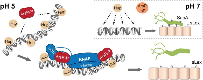
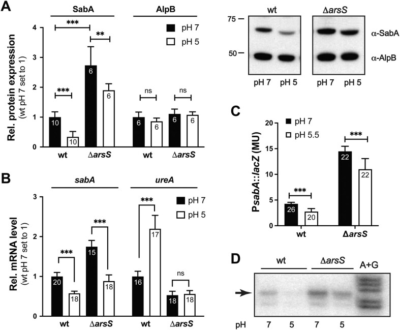
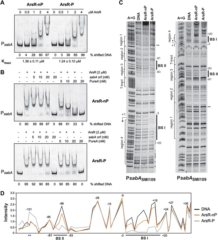
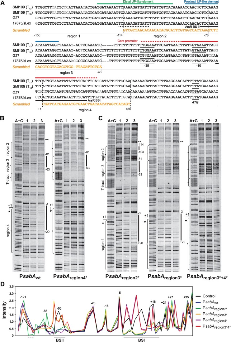
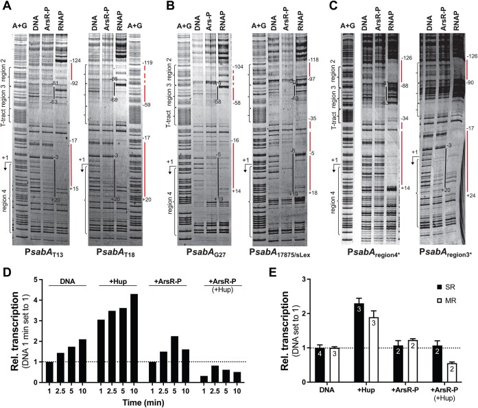
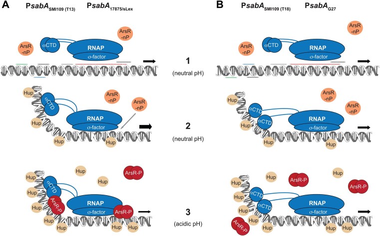
Adaptation to variations in pH is crucial for the ability of Helicobacter pylori to persist in the human stomach. The acid responsive two-component system ArsRS, constitutes the global regulon that responds to acidic conditions, but molecular details of how transcription is affected by the ArsR response regulator remains poorly understood. Using a combination of DNA-binding studies, in vitro transcription assays, and H. pylori mutants, we demonstrate that phosphorylated ArsR (ArsR-P) forms an active protein complex that binds DNA with high specificity in order to affect transcription. Our data showed that DNA topology is key for DNA binding. We found that AT-rich DNA sequences direct ArsR-P to specific sites and that DNA-bending proteins are important for the effect of ArsR-P on transcription regulation. The repression of sabA transcription is mediated by ArsR-P with the support of Hup and is affected by simple sequence repeats located upstream of the sabA promoter. Here stochastic events clearly contribute to the fine-tuning of pH-dependent gene regulation. Our results reveal important molecular aspects for how ArsR-P acts to repress transcription in response to acidic conditions. Such transcriptional control likely mediates shifts in bacterial positioning in the gastric mucus layer.


Figures

Graphical Abstract
Graphical Abstract
Figure 1.
Figure 1.
SabA expression is regulated by acidic pH at the transcriptional level. (A) Change in expression of SabA and AlpB protein in SMI109 wt and in a strain lacking ArsS sensor kinase (ΔarsS) after acid stress. Cultures were grown in Brucella broth to OD600 of 0.2–0.3 before the pH shift. Samples were collected 24 h after the shift to pH 5 (white bars) or as a control after growth at pH 7 (black bars). SabA and AlpB protein expression was analyzed by immunoblotting, quantified, and plotted relative to the expression observed in SMI109 wt at pH 7 (for each protein). The image to the right shows one representative immunoblot from the four independent experiments performed. The number of data points each average is based on are shown in the bars of the diagram. The statistical analysis was performed with the Mann-Whitney U-test (P< 0.001, ***; P< 0.01, **; P> 0.05, ns). (B) The effect of pH on sabA and ureA mRNA levels in SMI109 wt and ΔarsS strains as measured by RT-qPCR. Cultures were grown as described in (A). Data from three biological replicates, each with two technical replicates, were used for the quantification. The number of data points each average is based on are shown on the bars of the diagram. Statistical analysis was performed with the Mann–Whitney U-test (P< 0.001, ***; P< 0.01, **). (C) The effect on PsabA activity after acid stress as measured by β-galactosidase activity in the SMI109 PsabA::lacZ strain. Cultures of the wt and ΔarsS strains were initially grown as described in (A). The pH was kept at pH 7 as a control (black bars) or shifted to pH 5.5 (white bars) for 24 h. Quantifications were made from four independent experiments, and the number of data points each average is based on are written on the bars of the diagram. The statistical analysis was performed with the Mann–Whitney U-test (P< 0.001, ***). (D) Primer extension analysis of sabA using RNA isolated from SMI109 wt and ΔarsS after acid stress. Cultures were grown as described in (A). Total RNA was mixed with radiolabeled sabA-8 primer, and the samples were separated in a denaturing polyacrylamide gel. Maxam and Gilbert DNA sequencing reactions (A + G) were performed using the same primer and were loaded as size controls.
Figure 2.
Figure 2.
ArsR binds to PsabA DNA at two different positions. (A) Binding of His6-ArsR to PsabA DNA analyzed by EMSA. A total of 7 nM TET-labelled PsabA DNA (407 bp PCR; primers 486/485-TET; template pAAG264) was mixed with increasing concentrations (0–4 μM) of non-phosphorylated His6-ArsR (ArsR-nP) or phosphorylated His6-ArsR (ArsR-P). The amount of non-shifted DNA was quantified, and the percentage of shifted DNA was calculated. Average calculations of % shifted DNA from three independent experiments are shown below the picture with a standard deviation of ±3%. The Kd(app) (amount of protein needed to shift 50% of the DNA) was calculated separately for ArsR-nP and ArsR-P for each experiment. The mean and standard deviation are shown below the picture. Statistical analysis of the Kd(app) between ArsR-nP and ArsR-P, with the Mann–Whitney U-test showed P> 0.05 (ns). (B) Binding of His6-ArsR to PsabA DNA in the presence of non-specific or specific competitor DNA. A total of 5 nM TET-labelled PsabA DNA (407 bp, same as in A) was mixed with 2 μM ArsR-nP or ArsR-P and 0–20 nM non-specific (sabA ORF; 301 bp PCR; primers 486/485; template pAAG328) or specific competitor DNA (PureA; 424 bp PCR; primers 486/485, template pAAG261) was added to the reactions. The amount of non-shifted DNA was quantified, and the percentage of shifted DNA was calculated. The average calculations of the percentage of shifted DNA from two independent experiments are shown below each picture with standard deviations of ±2%. (C) DNase I footprint analysis of His6-ArsR binding to PsabA DNA from SMI109. The gel image to the left is with DNA of the coding strand (407 bp PCR; primers 486/485-TET; template pAAG264), and the gel image to the right is with DNA of the non-coding strand (407 bp PCR; primers 486-TET/485; template pAAG264). Transcriptional start site (+1) and T-tract are marked to the left of the gel images, as well as the regions described in Figure 3A. The Maxam and Gilbert DNA sequencing reaction (lane A + G) shows the sequence of the DNA used. A total of 25 nM DNA was mixed with protein storage buffer (lane DNA), 10 μM ArsR-nP (lane ArsR-nP), or 10 μM ArsR-P (lane ArsR-P). Binding sites are marked by solid lines, and the nucleotide positions of the binding sites are shown to the right of the gel images. The hypersensitive site is marked with **. Here is shown one representative gel of at least two independent experiments. (D) Line representation of the protection pattern of His6-ArsR on PsabA109 shown in C (black, free DNA; orange, ArsR-nP; grey, ArsR-P). The densities of the DNase I footprint bands (indicated by intensity) were quantified and plotted as migration (from the top to bottom of the DNase footprint). Nucleotide positions are written on top of the peaks, and the two ArsR binding sites are marked by solid lines below the diagram.
Figure 3.
Figure 3.
Sequences with long AT stretches are required for specific ArsR binding. (A) Sequence alignment of the sabA promoter region of the H. pylori strains used in this study; SMI109 (wt, T13), SMI109 (T18), G27 and 17875/sLex. The −35 and −10 regions are marked, as well as the transcriptional start site (+1). The solid black lines mark the two ArsR binding sites found by DNase I footprinting, namely BS II (−81 to -63) and BS I (−3 to + 20). The AT stretches in regions 2–4 were scrambled to reduce the length of AT in the DNase I footprint analysis (shown in B and C). The sequences of the scrambled regions are shown in orange text below each region. The additional binding site (−117 to −104) observed with region 2* DNA (shown in C) is marked by a dotted line. The distal and proximal UP-like elements and the core promoter where RNAP binds (Supplementary Figure S5A) are marked with green (distal), blue (proximal), and red (core) lines. The nucleotide positions relative to the +1 transcriptional start site are shown below indicating the sizes of the different regions. (B) DNase I footprint analysis of His6-ArsR binding to PsabA (407 bp PCR; primers 486/485-TET) wt DNA (template pAAG264) and scrambled region 4* (template pAAG267). Transcriptional start site (+1) and T-tract are marked along the left side, as well as the regions described in (A). Maxam and Gilbert DNA sequencing reaction (lanes A + G) showing the sequence of DNA used. DNA was mixed with protein storage buffer (lane 1), 10 μM ArsR-nP (lane 2), or 10 μM ArsR-P (lane 3). Binding sites are marked by solid lines along the right side. The image shows one representative gel of at least three independent experiments. (C) DNase I footprint analysis of His6-ArsR binding to scrambled region 2*, 3*, and 3*+4*. The same setup was used as described in (B), but different templates were used to make PsabA region 2* (template pAAG315), region 3* (template pAAG316), and region 3*+4* (template pAAG334). (D) Line representation of the protection pattern of ArsR-P on PsabA with scrambled region 2*, 3*, 4* and 3*+4* shown in (B) and (C). The control represents the DNase I footprint analysis of PsabAwt DNA without any other protein. The densities of the DNase I footprint bands were quantified and plotted as a migration (from the top to bottom of the DNase footprint). Nucleotide positions are written on top of the peaks, and the two ArsR binding sites are marked by solid lines below the diagram. The full line representation of each lane in the DNase I footprint gels shown in (B) and (C) can be found in Supplementary Figure S2B.
Figure 4.
Figure 4.
Small sequence variations and T-tract length affect ArsR binding to PsabA and the pH-dependent regulation of sabA expression. (A, B) Binding of His6-ArsR to PsabA DNA from different strains or with different T-tract length analyzed by EMSA. A total of 5 nM TET-labelled PsabA DNA (407–412 bp, 486/485-TET primers) with G27 (template pAAG265), 17875/sLex (template pAAG266), SMI109 T13 (template pAAG264), or SMI109 T18 (template pAAG286) was mixed with 0–2 μM ArsR-nP or ArsR-P. (A) The images show one representative gel from three to four independent experiments. The amount of non-shifted DNA was quantified, and the percentage of shifted DNA was calculated. Calculations of Kd(app) for each template DNA with ArsR-nP or ArsR-P were made for each gel image. The mean and standard deviation of four independent experiments is shown below each gel picture. (B) Kd(app) for ArsR-P to each PsabA DNA are plotted in the bar diagram and the number of data points each average is based on are shown on the bars of the diagram. Statistical analysis was performed with the Mann–Whitney U-test showing (P< 0.01,**, P> 0.05, ns). (C, D) Effect of pH on sabA and ureA mRNA levels in different H. pylori strains after acid stress and measured by RT-qPCR. Cultures of G27 or 17875/sLex (C) or isogenic variants of SMI109 with different T-tract lengths (D) were grown as described in Figure 1A. Data from two biological replicates, each including two technical replicates, were used for the quantification. The number of data points each average is based on are shown on the bars of the diagram. Statistical analysis was performed with the Mann–Whitney U-test (P< 0.01, **; P> 0.05, ns).
Figure 5.
Figure 5.
Transcription of sabA is enhanced by Hup and repressed by ArsR-P in vitro. (A) Set up of the in vitro transcription (IVT) assay with RNAP and PsabAwt DNA. A total of 2 nM of supercoiled template (pAAG264) and 0–10 nM of E. coli σ70-RNAP (New England Biolabs) was used in multiple-round IVT assays run at 37°C. (B) IVT assay with PsabAwt DNA and increasing concentrations of His6-ArsR. A total of 0.5 nM of supercoiled template (pAAG264) and 10 nM of RNAP was used in multiple-round IVT assays run at 37°C. A total of 0–1 μM ArsR-nP or 0–1 μM ArsR-P was added to each reaction. To one set of ArsR-P, 125 nM Hup-His6 was added to each reaction. The data were plotted relative to the transcript level with 0 nM ArsR set to 1. Black bars show the relative transcript level with ArsR-nP, grey bars show the relative transcript level with ArsR-P without Hup, and the white bars show the relative transcript level with ArsR-P and Hup. Images show one representative gel from at least four separate experiments, and the bar graph represents the combined results. The number of data points for each reaction are written in the bars of the diagram. Statistical analysis was performed on the raw data using the Mann–Whitney U-test (P< 0.01, **). (C) Binding of His6-ArsR to PsabA DNA in the presence of methyl green and netrospsin. A total of 5 nM TET-labelled PsabA DNA (407 bp, same as in Figure 2A) was mixed with 2 μM ArsR-nP (nP) or 2 μM ArsR-P (P). A total of 0–100 μM methyl green (left) or 0–100 μM netropsin (right) was added to each reaction. The amount of non-shifted DNA was quantified, and the percentage of shifted DNA was calculated. The mean values of the percentage of shifted DNA from three independent experiments are shown below each picture with a standard deviation of ±2%. (D) Binding of Hup-His6 to PsabA DNA as analyzed by EMSA. A total of 7 nM of TET-labelled PsabA DNA (407 bp, same as in Figure 2A) was mixed with increasing concentrations of Hup (0–1 μM) in the absence or presence of 0.5 μM ArsR-P. The amount of non-shifted DNA was quantified, and the percentage of shifted DNA was calculated. The mean values of the percentage of shifted DNA from four independent experiments are shown below each picture with a standard deviation of ±3%. (E) IVT assay with PsabAwt DNA and increasing concentrations of Hup-His6. A total of 0.5 nM template DNA (pAAG264) was mixed with 10 nM RNAP and 0–500 nM Hup. The experiment was performed as described in B. The amount of transcript was plotted relative to 0 nM Hup set to 1. The image shows one representative gel, and the bar graph shows the combined results from four independent experiments. The number of data points for each reaction is shown on the bars of the diagram. Statistical analysis was performed on the raw data using the Mann–Whitney U-test (P< 0.001, ***; P< 0.01, **; P< 0.05, *). (F) IVT assay using PsabA templates with scrambled DNA sequences in region 2–4 and Hup-His6. A total of 0.5 nM template DNA (PsabAwt; pAAG264, PsabAregion3*; pAAG316, PsabAregion4*; pAAG267, PsabAregion3*+4*; and pAAG334) was mixed with 10 nM RNAP and 0 or 500 nM Hup. Experimental procedures were as described in (B). The relative amount of transcript formed (500 nM/0 nM Hup) was calculated and plotted for each template DNA. The bar graph shows the combined results from at least two independent experiments. The number of data points for each reaction is shown on the bars of the diagram. Statistical analysis was performed on the raw data using the Mann–Whitney U-test (P< 0.001, ***; P< 0.01, **; P< 0.05, *). (G) IVT assay with PsabA templates from different strains (G27 and 17875/sLex) or with different T-tract lengths (SMI109/T13 and T18). A total of 0.5 nM template (PsabAG27; pAAG265, PsabA17875/sLex; pAAG266, or PsabASMI109; pAAG264 and PsabAT18; pAAG286) and 10 nM of RNAP were used in multiple-round IVT assays run at 37°C. The amount of transcript was quantified and plotted relative to the amount obtained with SMI109/T13 that was set to 1. The bar graph shows the combined results from three independent experiments. The number of data points for each reaction is shown on the bars of the diagram. Statistical analysis was performed on the raw data using the Mann–Whitney U-test (P< 0.01, **). (H) IVT assay with His6-ArsR and PsabA templates from different strains (G27 and 17875/sLex) or with different T-tract lengths (SMI109/T13 and T18). A total of 0.5 nM template (PsabAG27; pAAG265, PsabA17875/sLex; pAAG266, or PsabASMI109; pAAG264 and PsabAT18; pAAG286) was mixed with 10 nM of RNAP, 125 nM Hup, and 0 or 1 μM ArsR-P. Experimental procedures was as described in (B). The amount of transcript formed without ArsR-P (black bars, control) was set to 1 for each template DNA. The bar graph shows the combined results from at least two independent experiments. The number of data points for each reaction is shown on the bars of the diagram. Statistical analysis was performed on the raw data using the Mann–Whitney U-test (P< 0.01, **; P< 0.05, *; P> 0.05, ns).
Figure 6.
Figure 6.
pH-dependent regulation of sabA expression is abolished in a ΔarsSΔhup strain. (A) The effect of acid stress on sabA mRNA levels in the SMI109 wt, Δhup, and ΔarsS Δhup strains as measured by RT-qPCR. Cultures were grown as described in Figure 1A. Data are plotted relative to the mRNA levels in the SMI109 wt strain at pH 7, which was set to 1. Data from four biological replicates, each including two technical replicates, were used for the quantification. All data points included are shown as separate dots in the diagram. Statistical analysis was performed using the Mann–Whitney U-test (P< 0.0001, ****; P> 0.001, ***; P> 0.05, ns). (B) Effect of acid stress on SabA and AlpB protein in the SMI109 wt, Δhup, and ΔarsSΔhup strains, as monitored by immunoblot analysis. Normalized SabA expression levels (using expression levels of AlpB as reference) were calculated and plotted relative to the expression observed in SMI109 wt at pH 7, set to 1. The image above shows one representative immunoblot from the four independent experiments performed. All data points included are shown as separate dots in the diagram. Statistical analysis was performed using the Mann–Whitney U-test (P< 0.0001, ****; P> 0.05, ns). (C) The effect of acid stress on hup mRNA levels in SMI109 wt and ΔarsS strains as measured by RT-qPCR. Cultures were grown and analyzed as described in Figure 1A. The mRNA levels are plotted relative to the levels in SMI109 wt at pH 7, which was set to 1. Data from four biological replicates, each including two technical replicates, were used for the quantification. All data points included are shown as separate dots in the diagram. Statistical analysis was performed using the Mann–Whitney U-test (P< 0.0001, ****; P> 0.05, ns).
Figure 7.
Figure 7.
ArsR-mediated pH-dependent regulation of sabA occurs predominately via ArsR BS II. (A) The effect acid stress on sabA mRNA levels in SMI109 wt or isogeneic strains with scrambled region 3 (region 3*), region 4 (region 4*), or both (region 3* + 4*) as measured by RT-qPCR. Cultures were grown as described in Figure 1A. Data from four biological replicates, each including two technical replicates, were used for the quantification. All data points included are shown as separate dots in the diagram. Statistical analysis was performed using the Mann–Whitney U-test (P< 0.0001, ****; P> 0.05, ns). (B) Effect of acid stress on SabA and AlpB protein expression levels in SMI109 wt and isogeneic strains with scrambled DNA regions of the sabA promoter. From immunoblot analysis the normalized SabA expression levels (using AlpB expression levels as reference) were calculated and plotted relative to the expression observed in SMI109 wt at pH 7, which was set to 1. The image above shows one representative immunoblot from the four independent experiments. All data points included are shown as separate dots in the diagram. Statistical analysis was performed using the Mann–Whitney U-test (P< 0.0001, ****; P< 0.01, **; P> 0.05, ns). (C) IVT assay using PsabA templates with scrambled DNA sequences. A total of 0.5 nM template (PsabAwt; pAAG264, PsabAregion3*; pAAG316, or PsabAregion4*; pAAG267 and PsabAregion3*+4*; pAAG334) was mixed with 10 nM of RNAP. Experimental procedures were performed as described in Figure 5G. The amount of transcript was quantified and plotted relative to the amount obtained with PsabAwt, set to 1. The bar graph represents the combined results from three independent experiments. The number of data points for each sample is written in the bar. Statistical analysis was performed on the raw data using the Mann–Whitney U-test (P< 0.01, **; P< 0.05, *). (D) IVT assay using His6-ArsR and PsabA templates with scrambled DNA sequences. A total of 0.5 nM template (PsabAwt; pAAG264, PsabAregion3*; pAAG316, or PsabAregion4*; pAAG267 and PsabAregion3*+4*; pAAG334) was mixed with 10 nM of RNAP, 125 nM Hup, and 0 or 1 μM ArsR-P. Experimental procedures were performed as described in Figure 5B. The relative amount of transcript formed (ArsR-P/without ArsR-P) was calculated and plotted for each template DNA. The bar chart shows the combined results from at least two independent experiments. The number of data points for each reaction is written in the bars of the diagram. Statistical analysis was performed on the raw data using the Mann–Whitney U-test (P< 0.01, **; P> 0.05, ns). (E) Binding of His6-ArsR to PsabA with scrambled region 2–4 was analyzed by EMSA. A total of 7 nM TET-labelled PsabA DNA (407 bp PCR, 486/485-TET primers) with PsabAwt (template pAAG264), PsabAregion3* (template pAAG316), PsabAregion4* (template pAAG267) or PsabAregion3*+4* (template pAAG334) was mixed with 0–4 μM ArsR-P. The amount of non-shifted DNA was quantified, and the amount of shifted DNA was calculated for each DNA at each concentration of ArsR-P used. From the amount of shifted DNA, calculations of Kd(app) for ArsR-P with each template DNA were performed for each experiment. The bar graph represents the results from two independent experiments, and the number of data points for each sample is written in the bars of the diagram. Statistical analysis was performed on the raw data using the Mann–Whitney U-test (P< 0.05, *; P> 0.05, ns).
Figure 8.
Figure 8.
Binding of ArsR to BS II of PsabA DNA occurs near the binding site of the RNAP alpha subunit. (A–C) DNase I footprint analysis of His6-ArsR and RNAP binding to: (A) PsabA SMI109 DNA with different T-tract lengths (T13 and T18); (B) PsabA from different H. pylori strains (G27 and 17875sLex); (C) PsabA with scrambled DNA (region 3* or 4*). A total of 25 nM TET-labelled DNA (407–412 bp PCR 486/485-TET primers) was mixed with protein storage buffer (lane DNA), 10 μM ArsR-P (lane ArsR-P), or 300 nM E. coli σ70-RNAP (lane RNAP). The template plasmids used to generate the TET-labelled DNA were the same as the IVT plasmids used in Figures 5G and 7C. The Maxam and Gilbert DNA sequencing reaction (lane A + G) shows the sequences of DNA used. Binding sites are marked by solid lines (black for ArsR-P, red for RNAP), and the nucleotide positions of the binding sites are shown along the right side. Transcriptional start site (+1), T-tract, and the regions described in Figure 3A are indicated along the left side. The images show one representative gel of at least two independent experiments. The line representation of the protection pattern for ArsR-P and RNAP on PsabA is found in Supplementary Figure S6. (D) Time-course IVT assay with PsabA from SMI109. A total of 0.5 nM template (pAAG264) and 10 nM RNAP was used in multiple-round transcriptions run at 37°C together with 125 nM Hup or 1 μM ArsR-P. Time was recorded after the addition of nucleosides, and samples were removed for analysis after 1, 2.5, 5 and 10 min. The amount of transcript formed was quantified from the gel image. The transcription level without additional protein (DNA) at T = 1 min was set to 1, and the amount of transcript formed in the presence of only Hup or ArsR-P was plotted relative to that. The amount of transcript formed after the addition of both Hup and ArsR-P was plotted relative to the amount of transcript formed with only Hup at T = 1 min, which was set to 1. (E) Single and multiple-round IVT assays with PsabA from SMI109. A total of 0.5 nM template (pAAG264) and 10 nM of RNAP together with 125 nM Hup or 1 μM ArsR-P, was used in single (SR) or multiple round (MR) transcriptions run at 37°C. The amount of transcript formed after 10 min was quantified from the gel image. The transcription level without additional protein (DNA) was set to 1, and the amount of transcript formed in the presence of only Hup or ArsR-P was plotted relative to that for each assay. The amount of transcript formed after the addition of both Hup and ArsR-P was plotted relative to the amount of transcript formed with only Hup, which was set to 1. The bar graph shows the results from two independent experiments, and the number of data points for each reaction is written in the bars of the diagram.
Figure 9.
Figure 9.
Model for ArsR-mediated pH-dependent regulation of sabA expression in H. pylori. Schematic overview how regulation of sabA expression occurs via the repressing activity of phosphorylated ArsR (ArsR-P) and the DNA interaction by Hup in H. pylori. (A) A genetic variant with PsabA DNA that generates sabA high-expression (optimal T-tract length, SMI109 (T13) or 17875/sLex). (B) A genetic variant with PsabA DNA that generates sabA low-expression (suboptimal T-tract length, SMI109 (T18) or G27). The grey dotted line shows the position of the T-tract, the black dotted lines show the ArsR binding sites, the red dotted line shows the binding site of sigma-subunit (σ70) of the RNA polymerase (RNAP) and green or blue dotted lines show distal or proximal UP-like elements, binding site for the alpha-subunit C-terminal domains (αCTDs) of RNAP. Horizontal arrows ahead of the RNAP indicate relative expression levels. Regulatory interactions and expressions of the two genetic variants are illustrated in scenarios (1-3) of different pH conditions. 1) Transcription from a non-curved sabA promoter is very low as the RNAP αCTDs interaction with UP-like elements is not ideal due to poor DNA bending in the absence of Hup (A1) or a change in axial alignment due to T-tract length (B1). 2) At pH-neutral conditions, Hup together with an optimal T-tract length will mediate the correct curvature and spatial alignment needed for the optimal positioning of RNAP at the core promoter and thus increase transcription from the sabA promoter (A2). When a suboptimal T-tract length distorts the interaction of αCTDs to the UP-like elements, the positioning of RNAP at the core promoter will be changed, resulting in decreased transcription (B2). 3) Exposure to acidic pH, increase Hup expression, and ArsR becomes phosphorylated. Additional Hup will bind to PsabA DNA to change the DNA curvature by narrowing the size of the minor groove. This aid binding of ArsR-P to the minor groove of BS I and II (A3). The reduced change in DNA curvature with suboptimal T-tract length reduces the binding strength of ArsR-P to PsabA DNA (B3). Binding of ArsR-P to PsabA DNA will interact directly with the RNAP αCTDs at BS II or will change the interaction of αCTD with the UP-like element, and this will result in the repression of transcription from PsabA. Because binding of the RNAP σ70-subunit at the core promoter is very strong and partly overlaps with BS I, most of the ArsR-mediated repression occurs via BS II. However, depending on the positioning of RNAP, there might be a possibility for ArsR-P and/or ArsR-nP to bind to BS I, further repressing transcription by acting as a roadblock to RNAP (A3). For the sabA low-expressing strains (with suboptimal T-tract length), the binding of the two αCTDs at both the distal and proximal UP-like elements, in combination with the shifted σ70-subunit binding at the core promoter, overlaps with ArsR BS II and BS I. This results in displacement of ArsR-P at both binding sites, and no ArsR-mediated repression of transcription will occur (B3).

Similar articles

Cited by

References

    1. Salama N.R., Hartung M.L., Muller A. Life in the human stomach: persistence strategies of the bacterial pathogen Helicobacter pylori. Nat. Rev. Micro. 2013; 11:385–399. - PMC - PubMed
    1. Polk D.B., Peek R.M. Jr Helicobacter pylori: gastric cancer and beyond. Nat. Rev. Cancer. 2010; 10:403–414. - PMC - PubMed
    1. Keilberg D., Ottemann K.M. How Helicobacter pylori senses, targets and interacts with the gastric epithelium. Environ. Microbiol. 2016; 18:791–806. - PubMed
    1. Ansari S., Yamaoka Y. Survival of Helicobacter pylori in gastric acidic territory. Helicobacter. 2017; 22:e12386. - PMC - PubMed
    1. Scott D.R., Marcus E.A., Wen Y., Oh J., Sachs G. Gene expression in vivo shows that Helicobacter pylori colonizes an acidic niche on the gastric surface. Proc. Natl. Acad. Sci. U.S.A. 2007; 104:7235–7240. - PMC - PubMed

MeSH terms