Evolutionary origin of Hoxc13-dependent skin appendages in amphibians
- PMID: 38499530
- PMCID: PMC10948813
- DOI: 10.1038/s41467-024-46373-x
Evolutionary origin of Hoxc13-dependent skin appendages in amphibians
Abstract
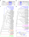
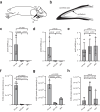
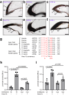
Cornified skin appendages, such as hair and nails, are major evolutionary innovations of terrestrial vertebrates. Human hair and nails consist largely of special intermediate filament proteins, known as hair keratins, which are expressed under the control of the transcription factor Hoxc13. Here, we show that the cornified claws of Xenopus frogs contain homologs of hair keratins and the genes encoding these keratins are flanked by promoters in which binding sites of Hoxc13 are conserved. Furthermore, these keratins and Hoxc13 are co-expressed in the claw-forming epithelium of frog toe tips. Upon deletion of hoxc13, the expression of hair keratin homologs is abolished and the development of cornified claws is abrogated in X. tropicalis. These results indicate that Hoxc13-dependent expression of hair keratin homologs evolved already in stem tetrapods, presumably as a mechanism for protecting toe tips, and that this ancestral genetic program was coopted to the growth of hair in mammals.
© 2024. The Author(s).
Conflict of interest statement
The authors declare no competing interests.
Figures


Similar articles
-
Skin Appendage Proteins of Tetrapods: Building Blocks of Claws, Feathers, Hair and Other Cornified Epithelial Structures.Animals (Basel). 2025 Feb 6;15(3):457. doi: 10.3390/ani15030457. Animals (Basel). 2025. PMID: 39943227 Free PMC article. Review.
-
Convergent Evolution of Cysteine-Rich Keratins in Hard Skin Appendages of Terrestrial Vertebrates.Mol Biol Evol. 2020 Apr 1;37(4):982-993. doi: 10.1093/molbev/msz279. Mol Biol Evol. 2020. PMID: 31822906 Free PMC article.
-
Recurrent functional divergence of early tetrapod keratins in amphibian toe pads and mammalian hair.Biol Lett. 2013 Mar 13;9(3):20130051. doi: 10.1098/rsbl.2013.0051. Print 2013 Jun 23. Biol Lett. 2013. PMID: 23485876 Free PMC article.
-
Ultrastructural localization of hair keratin homologs in the claw of the lizard Anolis carolinensis.J Morphol. 2011 Mar;272(3):363-70. doi: 10.1002/jmor.10920. Epub 2010 Dec 15. J Morphol. 2011. PMID: 21312232
-
Structure and functions of keratin proteins in simple, stratified, keratinized and cornified epithelia.J Anat. 2009 Apr;214(4):516-59. doi: 10.1111/j.1469-7580.2009.01066.x. J Anat. 2009. PMID: 19422428 Free PMC article. Review.
Cited by
-
Convergent Evolution of Cysteine-Rich Keratins in Horny Teeth of Jawless Vertebrates and in Cornified Skin Appendages of Amniotes.Mol Biol Evol. 2025 Feb 3;42(2):msaf028. doi: 10.1093/molbev/msaf028. Mol Biol Evol. 2025. PMID: 39891703 Free PMC article.
-
Skin Appendage Proteins of Tetrapods: Building Blocks of Claws, Feathers, Hair and Other Cornified Epithelial Structures.Animals (Basel). 2025 Feb 6;15(3):457. doi: 10.3390/ani15030457. Animals (Basel). 2025. PMID: 39943227 Free PMC article. Review.
-
The Evolution of Transglutaminases Underlies the Origin and Loss of Cornified Skin Appendages in Vertebrates.Mol Biol Evol. 2024 Jun 1;41(6):msae100. doi: 10.1093/molbev/msae100. Mol Biol Evol. 2024. PMID: 38781495 Free PMC article.
-
An immunotherapy guide constructed by cGAS-STING signature for breast cancer and the biofunction validation of the pivotal gene HOXC13 via in vitro experiments.Front Immunol. 2025 Aug 8;16:1586877. doi: 10.3389/fimmu.2025.1586877. eCollection 2025. Front Immunol. 2025. PMID: 40861477 Free PMC article.
-
Convergent Evolution Has Led to the Loss of Claw Proteins in Snakes and Worm Lizards.Genome Biol Evol. 2025 Jan 6;17(1):evae274. doi: 10.1093/gbe/evae274. Genome Biol Evol. 2025. PMID: 39696999 Free PMC article.
References
-
- Wagner, G. P. “Homology, Genes and Evolutionary Innovation” (Princeton University Press, Princeton, NJ, 2014).
MeSH terms
Substances
Grants and funding
LinkOut - more resources
Full Text Sources
Molecular Biology Databases

