. 2024 Mar 18;14(1):6476.
doi: 10.1038/s41598-024-56206-y.

Multiple-pathways light modulation in Pleurosigma strigosum bi-raphid diatom

Affiliations

Multiple-pathways light modulation in Pleurosigma strigosum bi-raphid diatom

Edoardo De Tommasi et al. Sci Rep. .

Abstract

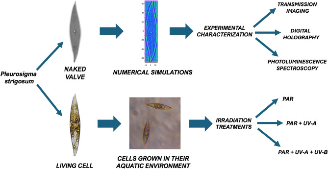
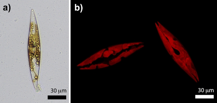
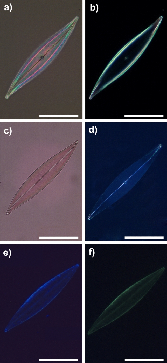
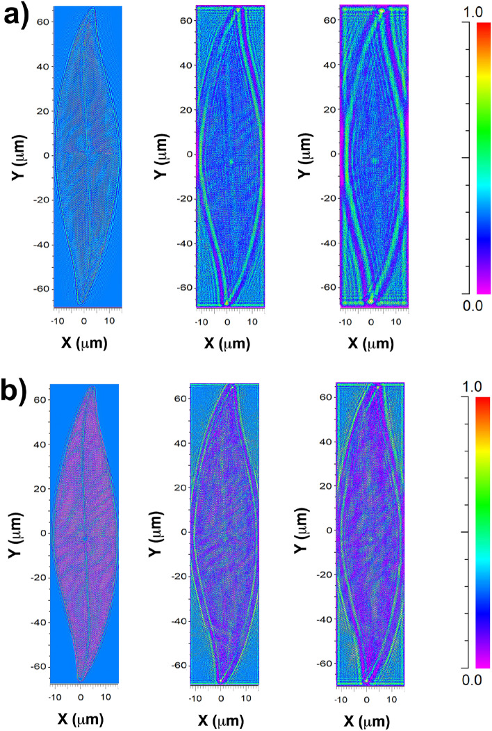
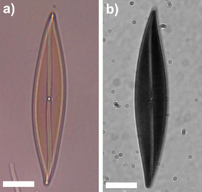
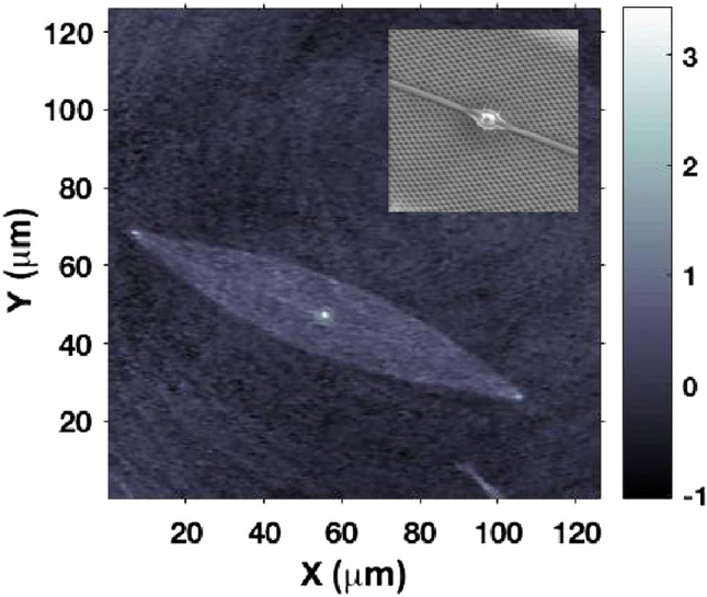
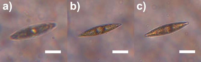
Ordered, quasi-ordered, and even disordered nanostructures can be identified as constituent components of several protists, plants and animals, making possible an efficient manipulation of light for intra- and inter- species communication, camouflage, or for the enhancement of primary production. Diatoms are ubiquitous unicellular microalgae inhabiting all the aquatic environments on Earth. They developed, through tens of millions of years of evolution, ultrastructured silica cell walls, the frustules, able to handle optical radiation through multiple diffractive, refractive, and wave-guiding processes, possibly at the basis of their high photosynthetic efficiency. In this study, we employed a range of imaging, spectroscopic and numerical techniques (including transmission imaging, digital holography, photoluminescence spectroscopy, and numerical simulations based on wide-angle beam propagation method) to identify and describe different mechanisms by which Pleurosigma strigosum frustules can modulate optical radiation of different spectral content. Finally, we correlated the optical response of the frustule to the interaction with light in living, individual cells within their aquatic environment following various irradiation treatments. The obtained results demonstrate the favorable transmission of photosynthetic active radiation inside the cell compared to potentially detrimental ultraviolet radiation.

Keywords: Biophotonics; Diatoms; Nanostructured biomaterials; Photonics in nature.


Conflict of interest statement

The authors declare no competing interests.

Figures

Figure 1
Figure 1
Conceptual scheme of the applied methodology used to study the photonic properties of Pleurosigma strigosum. Following the removal of organic content from the cell, individual valves are extracted and morphologically characterized using scanning electron microscopy (SEM). SEM characterization facilitates the retrieval of accurate CAD models for simulating light propagation through single valves in different spectral ranges. Numerical results are then compared with transmission imaging (for both PAR and UVR). The optical characterization of the valve is further complemented by digital holography and photoluminescence spectroscopy. Simultaneously, living cells are cultivated in their aquatic medium and subjected to various irradiation treatments, including UVR. The obtained results, encompassing growth rates and photosynthetic parameters, are analyzed, taking into consideration the optical properties of the valve. This analysis aims to highlight the potential role of the valve in photosynthesis and photoprotection within the living organism.
Figure 2
Figure 2
Optical micrograph of a single P. strigosum cell (a) and examples of 3D plastids reconstruction obtained by confocal laser scanning microscopy (b). Plastids autofluorescence is peaked at about λ=680 nm following excitation by a diode laser emitting at λ=637 nm.
Figure 3
Figure 3
SEM images of a P. strigosum single valve at different magnifications. Outer (ac, left column) and inner (df, right column) layer. Inset in (e): central oval nodule.
Figure 4
Figure 4
Bright field (a), dark field (b), transmission (c), cross-polarization (d), and fluorescence (e, f) images of a single P. strigosum valve. Scale bar: 50 µm. Excitation wavelengths employed in fluorescence imaging: 365 nm in (e) and 470 nm in (f).
Figure 5
Figure 5
Color-encoded intensity transmitted by a single P. strigosum valve evaluated by WA-BPM for an incident wavelength of λ=633 nm (a) and λ=280 nm (b) at different distances along the optical axis z^: z=0.4 µm immediately after the valve (first column); z=5 µm (second column); z=10 µm (third column). Incident intensity: 0.3 (a.u.). Fields propagating in air.
Figure 6
Figure 6
Transmission micrographs of a single P. strigosum valve when irradiated by PAR (a, λ=400–700 nm) and UV-B radiation (b, λ=280–315 nm). Scale bar: 25 µm.
Figure 7
Figure 7
Phase map reconstructed at a distance z=5 µm from a valve irradiated by a laser beam at λ=660 nm. The bright spots at the center and the apices of the valve correspond to higher values of the optical path length respect to the surrounding area. Phase is expressed in radiants. Inset: a detail of the central area of the valve (inner layer) as detected by SEM.
Figure 8
Figure 8
Photoluminescence emission spectra of a sample of dense frustules deposited onto a silicon wafer after excitation at λ=325 nm (a, P0= 3.2 mW, t=0.1 s) and at λ=442 nm (b, P0= 3.4 mW, t=1 s). The spectra have been acquired from the same sample. P0: incident laser power; t: integration time.
Figure 9
Figure 9
Absorbance spectra of a P-treated sample (black line) and a PAB-treated sample (red line) of P. strigosum revealing low contribution around λ=334 nm (ascribable to MAAs) if compared to absorption by other pigments.
Figure 10
Figure 10
Transmission micrographs of single living P. strigosum cells immersed in their growth medium when illuminated by PAR (left column, λ=400–700 nm) and UV-B radiation (right column, λ=280-315 nm). Scale bar: 50 µm.
Figure 11
Figure 11
Transmission micrographs of a single living P. strigosum cell immersed in its growth medium when illuminated by PAR (λ=400–700 nm) at three different positions along the propagation axis z^: far-field in backward direction (z0 µm, a); focal plane (z=0 µm, b); far-field in forward direction (z0 µm, c). Scale bar: 50 µm.
Figure 12
Figure 12
Intensity (left column) and phase (right column) maps relative to a single living P. strigosum cell immersed in its growth medium numerically reconstructed at different distances along the propagation axis z^ after acquisition of a hologram: z=-30 µm (a), z=0 µm (b), and z=20 µm (c). Irradiation wavelength: λ=660 nm. Intensity is expressed in arbitrary units while phase in radiants.

References

    1. De Tommasi E, et al. Frontiers of light manipulation in natural, metallic, and dielectric nanostructures. La Rivista del Nuovo Cimento. 2021;44:1–68. doi: 10.1007/s40766-021-00015-w. - DOI
    1. Wu J, et al. Two-dimensional materials for integrated photonics: Recent advances and future challenges. Small Sci. 2021;1:2000053. doi: 10.1002/smsc.202000053. - DOI
    1. Chen S, Li G, Cheah KW, Zentgraf T, Zhang S. Controlling the phase of optical nonlinearity with plasmonic metasurfaces. Nanophotonics. 2018;7:1013–1024. doi: 10.1515/nanoph-2018-0011. - DOI
    1. Liu M, et al. MuMultifunctional metasurfaces enabled by simultaneous and independent control of phase and amplitude for orthogonal polarization states. Light Sci. Appl. 2021;10:107. doi: 10.1038/s41377-021-00552-3. - DOI - PMC - PubMed
    1. Johansen VE, Onelli OD, Steiner LM, Vignolini S. Photonics in nature: From order to disorder. In: Gorb S, Gorb E, editors. Functional Surfaces in Biology III. Diversity of the Physical Phenomena. Springer; 2017. pp. 53–89.

Substances