. 2024 Apr;25(4):1859-1885.
doi: 10.1038/s44319-024-00103-y. Epub 2024 Mar 18.

New plastids, old proteins: repeated endosymbiotic acquisitions in kareniacean dinoflagellates

Affiliations

New plastids, old proteins: repeated endosymbiotic acquisitions in kareniacean dinoflagellates

Anna Mg Novák Vanclová et al. EMBO Rep. 2024 Apr.

Abstract

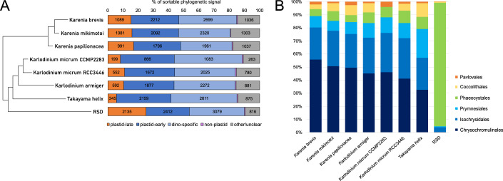
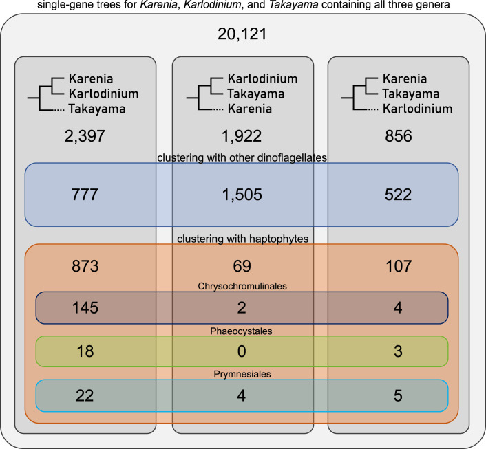
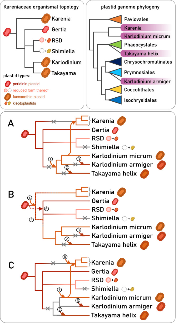
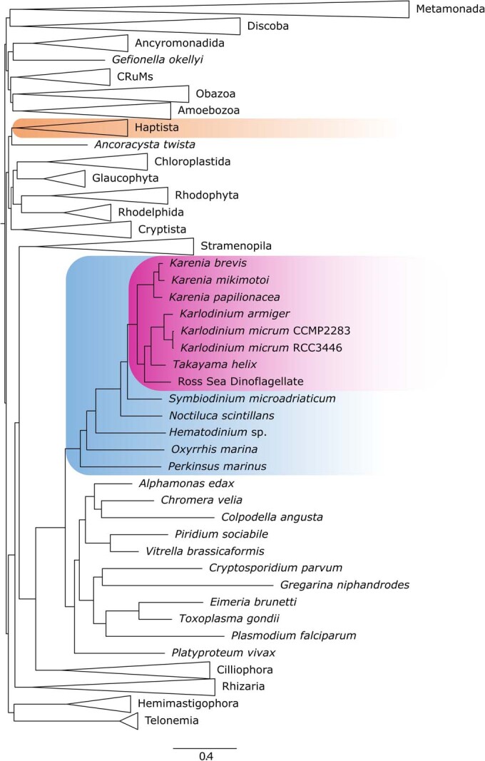
Dinoflagellates are a diverse group of ecologically significant micro-eukaryotes that can serve as a model system for plastid symbiogenesis due to their susceptibility to plastid loss and replacement via serial endosymbiosis. Kareniaceae harbor fucoxanthin-pigmented plastids instead of the ancestral peridinin-pigmented ones and support them with a diverse range of nucleus-encoded plastid-targeted proteins originating from the haptophyte endosymbiont, dinoflagellate host, and/or lateral gene transfers (LGT). Here, we present predicted plastid proteomes from seven distantly related kareniaceans in three genera (Karenia, Karlodinium, and Takayama) and analyze their evolutionary patterns using automated tree building and sorting. We project a relatively limited ( ~ 10%) haptophyte signal pointing towards a shared origin in the family Chrysochromulinaceae. Our data establish significant variations in the functional distributions of these signals, emphasizing the importance of micro-evolutionary processes in shaping the chimeric proteomes. Analysis of plastid genome sequences recontextualizes these results by a striking finding the extant kareniacean plastids are in fact not all of the same origin, as two of the studied species (Karlodinium armiger, Takayama helix) possess plastids from different haptophyte orders than the rest.

Keywords: Automated Tree Sorting; Myzozoa; Post-Endosymbiotic Organelle Evolution; Protists; Shopping Bag Model.


Conflict of interest statement

The authors declare no competing interests.

Figures

Figure 1
Figure 1. Ratios of phylogenetic signal of plastid proteins with detectable homologs in Kareniacean transcriptomes.
This plot shows:plastid-late (haptophyte-like; orange), plastid-early (dinoflagellate-like or dinoflagellate-specific; blue), ancestrally alveolate-like (purple), and other or unclear (gray) (A), and further breakdown of the plastid-late signal traceable to a concrete haptophyte family (Chrysochromulinaceae (dark blue), Isochrysidales (blue), Prymnesiales (light blue), Phaeocystales (green), Coccolithales (yellow), and Pavlovales (orange)) (B).
Figure 2
Figure 2. Proportions of single-gene trees supporting three different internal topologies of the three studied Kareniacean genera, grouped by broader phylogenetic origin.
The plastid-early genes clustering with other dinoflagellates primarily support the topology obtained from 18S trees and PhyloFisher trees (i.e., with Karenia sister to Takayama/Karlodinium). In contrast, the plastid-late genes clustering with haptophytes predominantly support Takayama as an outgroup of Karenia and Karlodinium. Karlodinium is less frequently recovered as an outgroup to a monophyletic clade of Takayama/Karenia.
Figure 3
Figure 3. Reconstruction of major metabolic pathways of plastids of the seven kareniaceans, adapted from KEGG (Kanehisa and Goto, 2000).
Plastid proteins are arranged by major metabolic pathway or biological process, with each protein shown as rosettes. Each rosette (described in the legend) relates to the homologs of a specific nucleus-encoded and plastid-targeted protein, with each circle within each rosette corresponding to a different species, and different colors corresponding to different evolutionary affiliations. Proteins of plastid-late (haptophyte) origin, such as are concentrated in photosystem and ribosomal processes, are colored red; and proteins of plastid-early (dinoflagellate) origin, such as are concentrated in carbon and amino acid metabolism are colored blue. Additional phylogenetic origins are denoted by further examples in the legend, e.g., LGT shown in orange, green, and brown. Proteins encoded in plastid genomes (red with black border) are provided for the incomplete genome assembly for Karlodinium micrum, and partial plastid transcriptomes are identified for Karenia mikimotoi and Karenia brevis. In certain cases (shown as rosettes with multiple colors), homologs from different species have different evolutionary origins, e.g., Karenia possessing plastid-late and Karlodinium/Takayama plastid-early. A detailed breakdown of the individual sequences with their predicted origins used for this reconstruction are available in Dataset EV3, and an alternative graphic, including the RSD, is available as Appendix Fig. S13.
Figure 4
Figure 4. Sequence logos for aligned signal peptides associated with kareniacean plastid proteomes.
These plots show comparable signal peptides for four datasets of different evolutionary identity: haptophyte, peridinin dinoflagellate, plastid-late and plastid-early kareniacean signal peptides (AD), occurrences of three-letter sequence motifs in signal peptides of each dataset, represented as a percentage of signal peptides in which at least one such motif occurs (E), and the sequence logo of the signal peptide and partial transit peptide for kareniacean plastidial proteins regardless of their phylogenetic origin with a red arrow denoting the signal peptide cleavage site (F).
Figure 5
Figure 5. Polyphyletic origins of kareniacean plastid genomes.
Phylogenetic tree reconstructed by IQ-TREE based on five plastid-encoded proteins (PsaA, PsbA, PsbC, PsbD, and RbcL) of six kareniaceans and 20 haptophytes for which partial or complete plastid genomic datasets are available, alongside 13 further algae with primary and complex red plastids; bootstrap support is expressed by the branch color (black for ≥90%, dark gray for ≥75%, and light gray for <75%). Phylogeny based on plastid genome sequences does not support the monophyly of kareniacean plastids and their close relationship to Chrysochromulinaceae retrieved based on nucleus-encoded plastid proteins. Instead, Takayama helix and Karlodinium armiger split from the rest, placing within Phaeocystales and Prymnesiales, respectively, with high support.
Figure 6
Figure 6. Relative abundances of the three studied kareniacean genera in Tara Oceans stations based on ribosomal 18S SSU V9 metabarcoding—Karenia exhibits higher abundance compared to both Karlodinium and Takayama, the latter displaying the lowest.
Karlodinium exhibits more cosmopolitan distributions in comparison to the others. A Correlation heatmap of each species against one another, showing a significant positive correlation between the relative abundances of Karlodinium and Takayama is provided in Appendix Fig. S17, while correlation circles against individual environmental variables are provided in Appendix Fig. S16.
Figure 7
Figure 7. Non-coincidence of kareniaceae and haptophytes in global meta-genome datasets.
Correlation circle based on the partial least square analysis of the Tara Oceans relative abundances (surface depth only) of the three studied kareniacean genera against those of different haptophyte families (color-coding is the same as in Fig. 1B) as observable variables; all three kareniaceans are negatively correlated to all families and most strongly to Chrysochromulinaceae.
Figure 8
Figure 8. Proposed scenarios of plastid evolutionary history in Kareniaceae integrating previously published organismal topology with our data based on plastid protein and plastid genome phylogeny.
Scenario (A) proposes that the first fucoxanthin plastid was acquired by the common ancestor of Karlodinium and Takayma (1) and later transferred to Karenia and independently lost and replaced in Takayama and Karlodinium armiger (2, 3). This scenario best explains the shared Chrysochromulinaceae-like signal and tree topologies recovered by our phylogenetic analyses (Figs. 2 and EV2) but requires a higher number of plastid losses. Scenario (B) places the fucoxanthin plastid origin at the base of all Kareniaceae (4) and requires the peridinin plastid to either have coexisted with the fucoxanthin one for a period of time (5) or have been lost and re-acquired (6) to explain its presence in Gertia and RSD. It proposes less plastid losses and no additional endosymbiotic transfer between fucoxanthin species, but it explains the shared Chrysochromulinaceae-like signal only partially as it is congruent with its monophyly but not the inner relationships. Scenario (C) proposes an independent plastid origin for all four lineages (2,3,7,8) or for three lineages with subsequent transfer between Karlodinium and Karenia (2,3,7) or vice versa (only one direction is shown in the schematic). This scenario considers the Chrysochromulinaceae-like signal a phylogenetic artifact caused by the under-sampling of the plastid donors or sources of non-specific LGT.
Figure EV1
Figure EV1. The eight kareniaceans in the pan-eukaryotic phylogenetic context as reconstructed by IQ-TREE based on a matrix of 241 genes prepared using PhyloFisher toolkit.
The inner relationships between the studied kareniaceans are resolved with maximum support and confirm the results of previous phylogenetic analyses based on ribosomal subunits (Takahashi et al, ; Ok et al, 2021).
Figure EV2
Figure EV2. Example single-gene trees for proteins of plastid-late origin, with specifically plastidial function and present in all studied organisms.
The non-RSD kareniacean sequences typically resolve adjacent to Chrysochromulinales or a mixed Chrysochromulinales/Prymnesiales clade. Their inner topology varies and typically differs from the organismal one (Figure EV1), with Takayama often branching as sister to the rest (A, B) and sometimes even separately (C, D), albeit with low support, or inside the Karenia/Karlodinium clade but sister to Karenia rather than Karlodinium (E, F). All species of Karenia and Karlodinium are, however, consistently retrieved as monophyletic, suggesting a common chrysochromulinalean-like origin of their nucleus-encoded plastid protein inventory, which is for the most part, also shared with Takayama.
Figure EV3
Figure EV3. Automatically generated phylogenetic tree showing the green origin of one form of heme oxygenase in Karenia, Karlodinium, and the RSD (no Takayama homolog was retrieved); the second form is of plastid-late origin in all genera.
Bootstrap support is expressed by the branch color (black for ≥90%, dark gray for ≥75%, and light gray for <75%). Kareniacean sequences are colored pink with proteins with predicted plastid-targeting signal in darker shades and annotated “CP” suffix; dinoflagellates are coded blue; haptophytes are coded orange; green algae (Chloroplastida) are coded green.
Figure EV4
Figure EV4. Automatically generated phylogenetic tree showing the green origin of plastidial histidyl-tRNA synthetases of Karenia and Karlodinium and brown origin of the plastidial homolog in Takayama.
A green-like homolog present in Takayama is not predicted as plastidial. The Karenia brevis SP3 homolog clustering with the dinoflagellates was likely falsely predicted as plastid-targeted due to an artificial N-terminal extension of the transcript. Bootstrap support is expressed by the branch color (black for ≥90%, dark gray for ≥75%, light gray for <75%). Kareniacean sequences are colored pink with proteins with predicted plastid-targeting signal in darker shades and annotated “CP” suffix; dinoflagellates are coded blue; haptophytes are coded orange; green algae (Chloroplastida) are coded green; and brown algae (Ochrophyta) are coded brown.
Figure EV5
Figure EV5. Automatically generated phylogenetic tree showing the brown origin of plastidial methionyl-tRNA synthetase in all three genera.
Bootstrap support is expressed by the branch color (black for ≥90%, dark gray for ≥75%, and light gray for <75%). Kareniacean sequences are colored pink with proteins with predicted plastid-targeting signal in darker shades and annotated “CP” suffix; dinoflagellates are coded blue; haptophytes are coded orange; and brown algae (Ochrophyta) are coded brown.

Similar articles

Cited by

References

    1. Almagro Armenteros JJ, Tsirigos KD, Sønderby CK, Petersen TN, Winther O, Brunak S, von Heijne G, Nielsen H. SignalP 5.0 improves signal peptide predictions using deep neural networks. Nat Biotechnol. 2019;37:420–423. doi: 10.1038/s41587-019-0036-z. - DOI - PubMed
    1. Ardyna, M, D’Ovidio, F, Speich, S, Leconte, J, Chaffron, S, Audic, S, Garczarek, L, Pesant, S, Tara Oceans Consortium, C., & Tara Oceans Expedition, P. (2017). Environmental context of all samples from the Tara Oceans Expedition (2009-2013), about mesoscale features at the sampling location. In: Tara Oceans Consortium, Coordinators; Tara Oceans Expedition, Participants (2017): Registry of all samples from the Tara Oceans Expedition (2009-2013).
    1. Bannerman BP, Kramer S, Dorrell RG, Carrington M. Multispecies reconstructions uncover widespread conservation, and lineage-specific elaborations in eukaryotic mRNA metabolism. PLoS ONE. 2018;13:e0192633. doi: 10.1371/journal.pone.0192633. - DOI - PMC - PubMed
    1. Beauchemin M, Morse D. A proteomic portrait of dinoflagellate chromatin reveals abundant RNA-binding proteins. Chromosoma. 2018;127:29–43. doi: 10.1007/s00412-017-0643-8. - DOI - PubMed
    1. Bentlage B, Rogers TS, Bachvaroff TR, Delwiche CF. Complex ancestries of isoprenoid synthesis in dinoflagellates. J Eukaryot Microbiol. 2016;63:123–137. doi: 10.1111/jeu.12261. - DOI - PMC - PubMed