Review
. 2024 Jun;49(6):2074-2082.
doi: 10.1007/s00261-024-04189-9. Epub 2024 Mar 19.

Imaging features of intra-abdominal and intra-pelvic causes of hirsutism

Affiliations
Review

Imaging features of intra-abdominal and intra-pelvic causes of hirsutism

Arleen Li et al. Abdom Radiol (NY). 2024 Jun.

Abstract

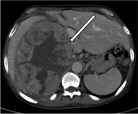
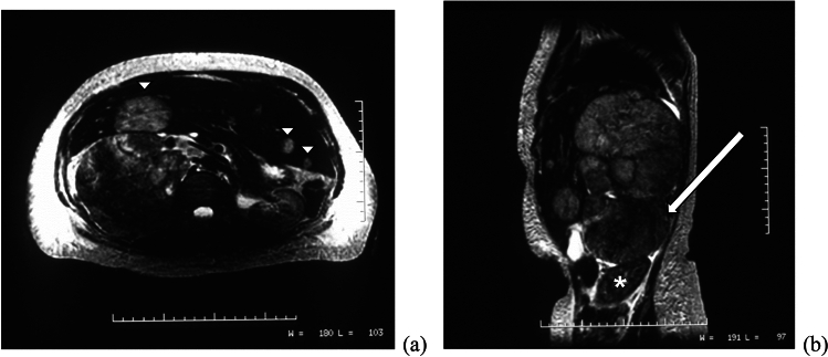
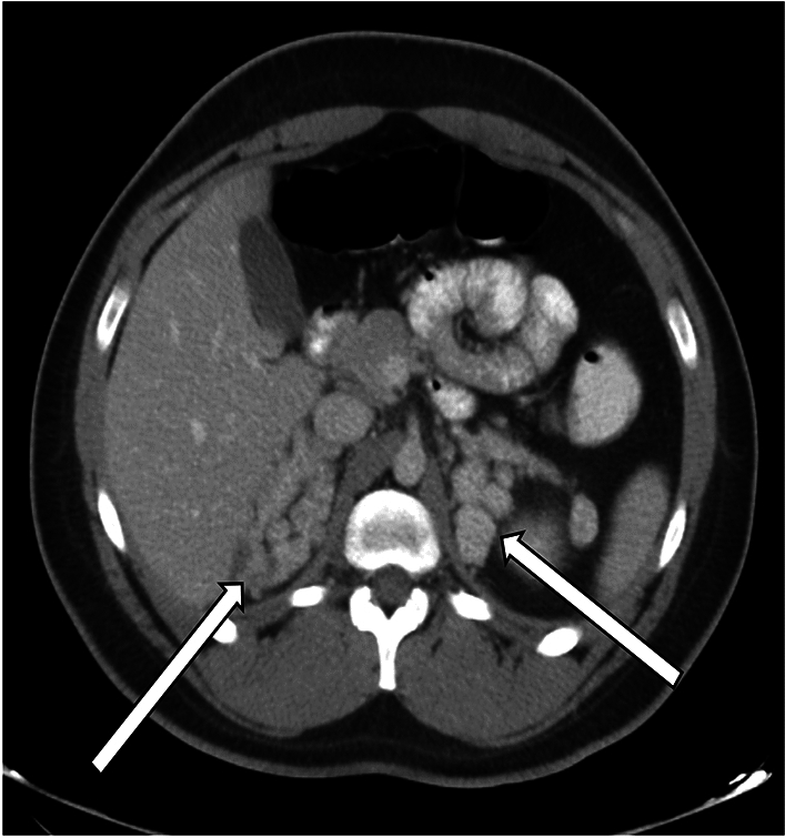
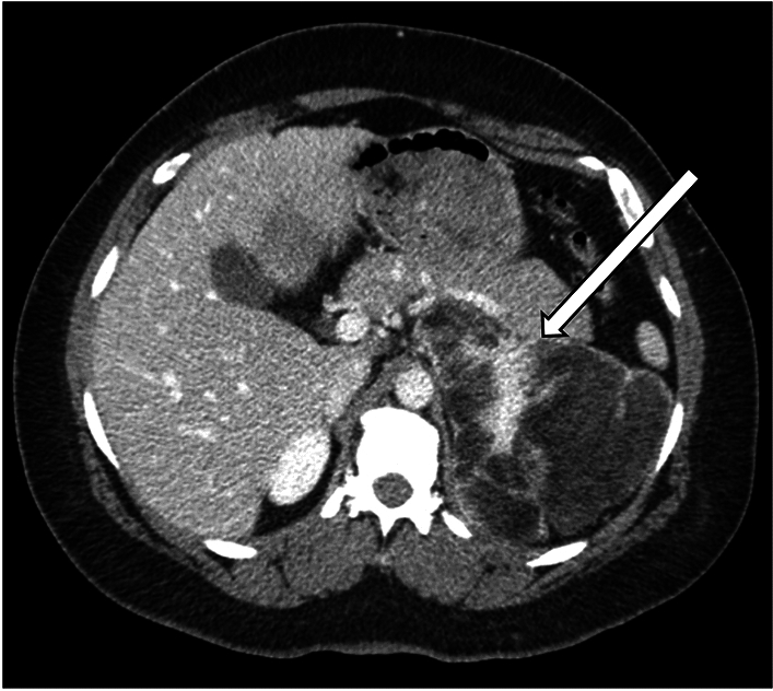
Hirsutism is a relatively common disorder which affects approximately 5% to 15% of women. It is defined by excessive growth of terminal hair in women, which primarily affects areas dependent on androgens, such as the face, abdomen, buttocks, and thighs. Hirsutism can be caused by a variety of etiologies, which are most often not lifethreatening. However, in some cases, hirsutism can be an indicator of more serious underlying pathology, such as a neoplasm, which may require further elucidation with imaging. Within the abdomen and pelvis, adrenal and ovarian pathologies are the primary consideration. The goal of this manuscript is to review the etiologies and imaging features of various intra-abdominal and intra-pelvic causes of hirsutism.

Keywords: Adrenal; Hirsutism; Hyperandrogenism; Imaging features; Ovarian.


Figures

Fig. 1
Fig. 1
A representative case of ACC in a 64-year-old man with abdominal pain. The axial CT image of the right adrenal gland demonstrated a large necrotic mass (arrow) with associated mass effect on the adjacent viscera. Retroperitoneal lymphadenopathy was present as well (not shown). Pathology was consistent with primary adrenocortical carcinoma, with invasion into the periadrenal/renal adipose tissue
Fig. 2
Fig. 2
A 60-year-old woman presented with marked hirsutism and elevated androgen levels. Axial (a) and sagittal (b) T2-weighted MRI images demonstrate a large right adrenal mass (arrow) which displaces the kidney (*) inferiorly. Multiple liver metastases are also present (arrowheads)
Fig. 3
Fig. 3
A representative case of CAH in a 21-year-old man, with axial CT showing nodular thickening of both adrenal glands (arrows)
Fig. 4
Fig. 4
A representative case of adrenal myelolipoma in the setting of CAH in a 42-year-old man with history of ambiguous genitalia who underwent CT for a suspected adrenal mass. CT showed a large left adrenal mass (arrow) with areas of fat attenuation and enhancement, consistent with adrenal myelolipoma. The right adrenal gland had previously been resected due to hemorrhage, with surgical pathology of the right adrenal gland consistent with myelolipoma and diffuse cortical hyperplasia of the adrenal gland
Fig. 5
Fig. 5
A 28-year-old woman with no significant past medical history presented with irregular menstrual periods. She underwent pelvic ultrasound which demonstrated mildly enlarged ovaries with numerous peripherally oriented small follicles (arrowheads). The right ovary measured 22 mL in volume, while the left ovary measured 16 mL (not shown)
Fig. 6
Fig. 6
T2-weighted coronal MRI of the pelvis of a 29-year-old woman who presented with hirsutism and right ovarian cyst on ultrasound. Enlargement of both ovaries, which contain numerous peripheral small follicles in a “string of pearls” distribution, is seen (arrows). This appearance is compatible with PCOS
Fig. 7
Fig. 7
A 69-year-old woman with a history of hypertension, diabetes mellitus, and hepatitis C, who presented with 2 years of hirsutism, with laboratory results demonstrating elevated total testosterone levels and low DHEAS level. MRI abdomen demonstrated stable mild thickening of the left adrenal gland and normal right adrenal gland. Subsequent pelvic MRI was significant for enlargement of both ovaries (arrows) with low to intermediate signal on T1-weighted sequences (image a). On T2-weighted images, the ovaries demonstrated relatively decreased signal intensity (image b). Contrast-enhanced images (image c) demonstrated mild enhancement of the ovaries. The decision was made for the patient to undergo bilateral salpingo-oophorectomy. Final pathology was consistent with ovarian stromal hyperplasia and stromal hyperthecosis
Fig. 8
Fig. 8
A 21-year-old woman with no significant past medical history, who presented with menstrual irregularity and pelvic pain. Pelvic ultrasound demonstrated a solid left ovarian mass (image a) with internal vascular flow (image b), most consistent with a solid ovarian lesion such as fibrothecoma
Fig. 9
Fig. 9
Pelvic MRI in the same patient shows a solid left ovarian lesion with low to intermediate T2 signal intensity (arrow), as well as mild uniform enhancement, consistent with fibrous tumor such as fibrothecoma. The patient underwent left salpingo-oophorectomy, with final pathology consistent with ovarian fibrothecoma
Fig. 10
Fig. 10
A 57-year-old woman underwent pelvic ultrasound exam showing a solid left ovarian mass with internal vascularity on Doppler ultrasound exam
Fig. 11
Fig. 11
The patient subsequently underwent contrast-enhanced CT examination, which demonstrated a corresponding heterogeneously enhancing left ovarian lesion (arrow). Final pathology confirmed Leydig cell tumor
Fig. 12
Fig. 12
A 68-year-old woman with a past medical history of hypertension and uterine fibroids presented with a 3-year history of hirsutism, with increased hair on shoulders. Laboratory results showed elevated testosterone, suppressed luteinizing hormone (LH) and follicle-stimulating hormone (FSH), and low DHEAS levels. Pelvic MRI showed a right ovarian lesion (arrow) with low to intermediate T1 signal intensity (image a), low T2 signal intensity (image b), and heterogeneous enhancement (image c). On surgical resection, a 2.5 cm right ovarian mass was noted, with final pathology consistent with benign Sertoli–Leydig cell tumor. On follow up visits, the patient reported improving hirsutism and had decreasing testosterone levels
Fig. 13
Fig. 13
A 15-year-old female with a past medical history of epilepsy presented with hirsutism and elevated testosterone levels. Pelvic ultrasound demonstrated a large cystic structure in the right adnexum containing internal low-level echoes
Fig. 14
Fig. 14
Contrast-enhanced CT of the pelvis in the same patient as Fig. 13, which demonstrates a large fluid-filled cystic structure (arrow) with adjacent solid nodule (arrowhead) containing punctate calcification. Macroscopic fat (*) is present anteriorly in the mass. Final pathology was consistent with mature teratoma. The patient’s testosterone levels normalized after surgery

Similar articles

References

    1. Matheson E, Bain J. Hirsutism in Women. Am Fam Physician. 2019; 100(3): 168–175. https://www.aafp.org/pubs/afp/issues/2019/0801/p168.html - PubMed
    1. Hafsi W, Badri T. Hirsutism. StatPearls. Treasure Island, FL: StatPearls Publishing; 2023. https://www.ncbi.nlm.nih.gov/books/NBK470417/
    1. Sachdeva S. Hirsutism: Evaluation and Treatment. Indian J Dermatol. 2010;55(1):3–7. doi: 10.4103/0019-5154.60342. - DOI - PMC - PubMed
    1. Franks S. The investigation and management of hirsutism. Journal of Family Planning and Reproductive Health Care. 2012; 38: 182–186. https://srh.bmj.com/content/38/3/182 - PubMed
    1. Winterson E, Eyster K, Hansen KA. An Uncommon Etiology For A Common Problem: Hirsutism. S D Med. 2011; 64(5): 161–3, 165. - PubMed

LinkOut - more resources