Accelerated IVIM-corrected DTI in acute hamstring injury: towards a clinically feasible acquisition time
- PMID: 38499843
- PMCID: PMC10948680
- DOI: 10.1186/s41747-024-00437-1
Accelerated IVIM-corrected DTI in acute hamstring injury: towards a clinically feasible acquisition time
Abstract
Background: Intravoxel incoherent motion (IVIM)-corrected diffusion tensor imaging (DTI) potentially enhances return-to-play (RTP) prediction after hamstring injuries. However, the long scan times hamper clinical implementation. We assessed accelerated IVIM-corrected DTI approaches in acute hamstring injuries and explore the sensitivity of the perfusion fraction (f) to acute muscle damage.
Methods: Athletes with acute hamstring injury received DTI scans of both thighs < 7 days after injury and at RTP. For a subset, DTI scans were repeated with multiband (MB) acceleration. Data from standard and MB-accelerated scans were fitted with standard and accelerated IVIM-corrected DTI approach using high b-values only. Segmentations of the injury and contralateral healthy muscles were contoured. The fitting methods as well as the standard and MB-accelerated scan were compared using linear regression analysis. For sensitivity to injury, Δ(injured minus healthy) DTI parameters between the methods and the differences between injured and healthy muscles were compared (Wilcoxon signed-rank test).
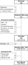
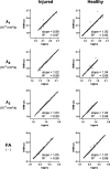
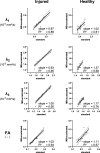
Results: The baseline dataset consisted of 109 athletes (16 with MB acceleration); 64 of them received an RTP scan (8 with MB acceleration). Linear regression of the standard and high-b DTI fitting showed excellent agreement. With both fitting methods, standard and MB-accelerated scans were comparable. Δ(injured minus healthy) was similar between standard and accelerated methods. For all methods, all IVIM-DTI parameters except f were significantly different between injured and healthy muscles.
Conclusions: High-b DTI fitting with MB acceleration reduced the scan time from 11:08 to 3:40 min:s while maintaining sensitivity to hamstring injuries; f was not different between healthy and injured muscles.
Relevance statement: The accelerated IVIM-corrected DTI protocol, using fewer b-values and MB acceleration, reduced the scan time to under 4 min without affecting the sensitivity of the quantitative outcome parameters to hamstring injuries. This allows for routine clinical monitoring of hamstring injuries, which could directly benefit injury treatment and monitoring.
Key points: • Combining high-b DTI-fitting and multiband-acceleration dramatically reduced by two thirds the scan time. • The accelerated IVIM-corrected DTI approaches maintained the sensitivity to hamstring injuries. • The IVIM-derived perfusion fraction was not sensitive to hamstring injuries.
Keywords: Athletic injuries; Diffusion magnetic resonance imaging; Diffusion tensor imaging; Muscle (skeletal); Return to sport.
© 2024. The Author(s).
Conflict of interest statement
The authors declare that they have no competing interests.
Figures


References
-
- Ekstrand J, Bengtsson H, Waldén M, Davison M, Khan KM, Hägglund M (2022) Hamstring injury rates have increased during recent seasons and now constitute 24% of all injuries in men’s professional football: the UEFA Elite Club Injury Study from 2001/02 to 2021/22. Br J Sports Med 292–298. 10.1136/bjsports-2021-105407 - PMC - PubMed
-
- Paton BM, Read P, van Dyk N, et al (2023) London International Consensus and Delphi study on hamstring injuries part 3: rehabilitation, running and return to sport. Br J Sports Med. 10.1136/bjsports-2021-105384 - PubMed
-
- Paton BM, Court N, Giakoumis M, et al (2023) London International Consensus and Delphi study on hamstring injuries part 1: classification. Br J Sports Med. 10.1136/bjsports-2021-105371 - PubMed
MeSH terms
Grants and funding
LinkOut - more resources
Full Text Sources
Miscellaneous
