

Fractal analysis of left ventricular trabeculae in post-STEMI: from acute to chronic phase
- PMID: 38499900
- PMCID: PMC10948656
- DOI: 10.1186/s13244-024-01641-8
Fractal analysis of left ventricular trabeculae in post-STEMI: from acute to chronic phase
Abstract
Purpose: The temporal evolution of ventricular trabecular complexity and its correlation with major adverse cardiovascular events (MACE) remain indeterminate in patients presenting with acute ST elevation myocardial infarction (STEMI).
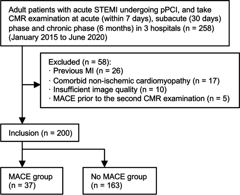
Methods: This retrospective analysis enrolled patients undergoing primary percutaneous coronary intervention (pPCI) for acute STEMI, possessing cardiac magnetic resonance (CMR) data in the acute (within 7 days), subacute (1 month after pPCI), and chronic phases (6 months after pPCI) from January 2015 to January 2020 at the three participating sites. Fractal dimensions (FD) were measured for the global, infarct, and remote regions of left ventricular trabeculae during each phase. The potential association of FD with MACE was analyzed using multivariate Cox regression.
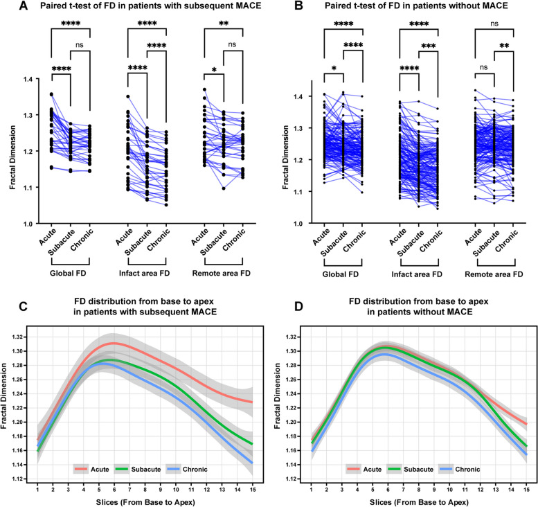
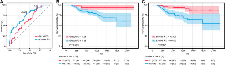
Results: Among the 200 analyzed patients (182 men; median age, 61 years; age range, 50-66 years), 37 (18.5%) encountered MACE during a median follow-up of 31.2 months. FD exhibited a gradual decrement (global FD at acute, subacute, and chronic phases: 1.253 ± 0.049, 1.239 ± 0.046, 1.230 ± 0.045, p < 0.0001), with a more pronounced decrease observed in patients subsequently experiencing MACE (p < 0.001). The global FD at the subacute phase correlated with MACE (hazard ratio 0.89 (0.82, 0.97), p = 0.01), and a global FD value below 1.26 was associated with a heightened risk.
Conclusion: In patients post-STEMI, the global FD, serving as an indicator of left ventricular trabeculae complexity, independently demonstrated an association with subsequent major adverse cardiovascular events, beyond factors encompassing left ventricular ejection fraction, indexed left ventricular end-diastolic volume, infarct size, heart rate, NYHA class, and post-pPCI TIMI flow.
Critical relevance statement: In patients who have had an ST-segment elevation myocardial infarction, global fractal dimension, as a measure of left ventricular trabeculae complexity, provided independent association with subsequent major adverse cardiovascular event.
Key points: • Global and regional FD decreased after STEMI, and more so in patients with subsequent MACE. • Lower global FD at the subacute phase and Δglobal FD from acute to subacute phase were associated with subsequent MACE besides clinical and CMR factors. • Global FD at the subacute phase independently correlated with MACE and global FD value below 1.26 was associated with higher risk.
Keywords: Cine; Fractals; Magnetic resonance imaging; Myocardium; ST elevation myocardial infarction.
© 2024. The Author(s).
Conflict of interest statement
L.D. works for Shanghai Robotics Institute. The remaining authors declare that they have no competing interests.
Figures


Similar articles
-
Left ventricular thrombus after acute ST-segment elevation myocardial infarction: multi-parametric cardiac magnetic resonance imaging with long-term outcomes.Int J Cardiovasc Imaging. 2022 Nov;38(11):2373-2384. doi: 10.1007/s10554-022-02598-9. Epub 2022 Mar 27. Int J Cardiovasc Imaging. 2022. PMID: 36434326
-
Prognostic impact of remote myocardium changes using T1 mapping in patients with ST-segment elevation myocardial infarction.Eur Radiol. 2025 Jun 4. doi: 10.1007/s00330-025-11711-0. Online ahead of print. Eur Radiol. 2025. PMID: 40464914
-
Prognostic value of left ventricular global function index in patients after ST-segment elevation myocardial infarction.Eur Heart J Cardiovasc Imaging. 2016 Feb;17(2):169-76. doi: 10.1093/ehjci/jev129. Epub 2015 Jun 7. Eur Heart J Cardiovasc Imaging. 2016. PMID: 26056134 Free PMC article.
-
Acute Response in the Noninfarcted Myocardium Predicts Long-Term Major Adverse Cardiac Events After STEMI.JACC Cardiovasc Imaging. 2023 Jan;16(1):46-59. doi: 10.1016/j.jcmg.2022.09.015. Epub 2022 Dec 14. JACC Cardiovasc Imaging. 2023. PMID: 36599569 Free PMC article.
-
The prognostic value of myocardial salvage index by cardiac magnetic resonance in ST-segment elevation myocardial infarction patients: a systematic review and meta-analysis.Eur Radiol. 2023 Nov;33(11):8214-8225. doi: 10.1007/s00330-023-09739-1. Epub 2023 Jun 16. Eur Radiol. 2023. PMID: 37328640
References
-
- Paun B, Bijnens B, Butakoff C (2018) Relationship between the left ventricular size and the amount of trabeculations. Int J Numer Method Biomed Eng 34(3) - PubMed
Grants and funding
- YG2022QN020/Shanghai Jiao Tong University Medical Engineering cross fund
- 22YF1438600/Shanghai Sailing Program
- 82171884/National Natural Science Foundation of China
- 82302174/National Natural Science Foundation of China Youth Project
- 19DZ2203800/Shanghai science and technology innovation action plan, technology standard project
LinkOut - more resources
Full Text Sources
