

Evaluation of canine adipose-derived mesenchymal stem cells for neurological functional recovery in a rat model of traumatic brain injury
- PMID: 38500105
- PMCID: PMC10946090
- DOI: 10.1186/s12917-024-03912-4
Evaluation of canine adipose-derived mesenchymal stem cells for neurological functional recovery in a rat model of traumatic brain injury
Abstract
Background: Traumatic brain injury (TBI) is a common condition in veterinary medicine that is difficult to manage.Veterinary regenerative therapy based on adipose mesenchymal stem cells seem to be an effective strategy for the treatment of traumatic brain injury. In this study, we evaluated therapeutic efficacy of canine Adipose-derived mesenchymal stem cells (AD-MSCs)in a rat TBI model, in terms of improved nerve function and anti-neuroinflammation.
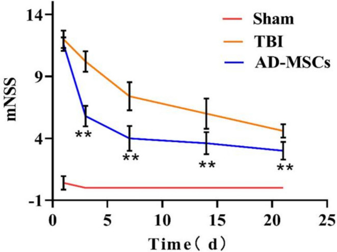
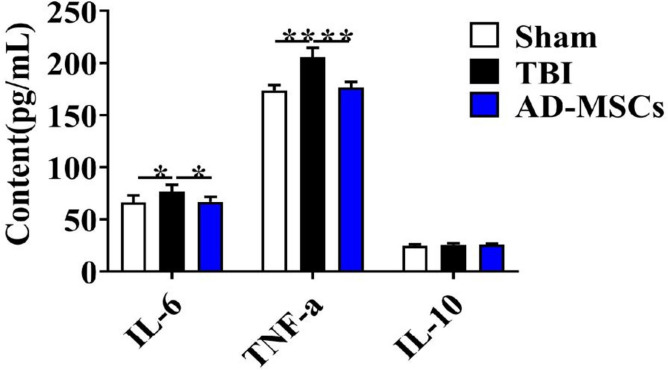
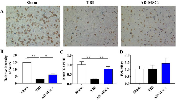
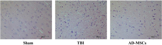
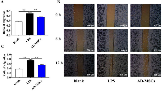
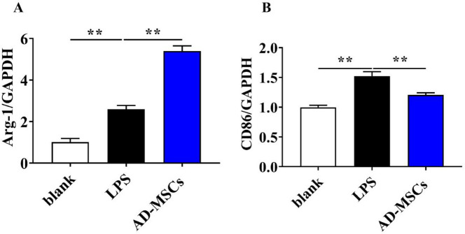
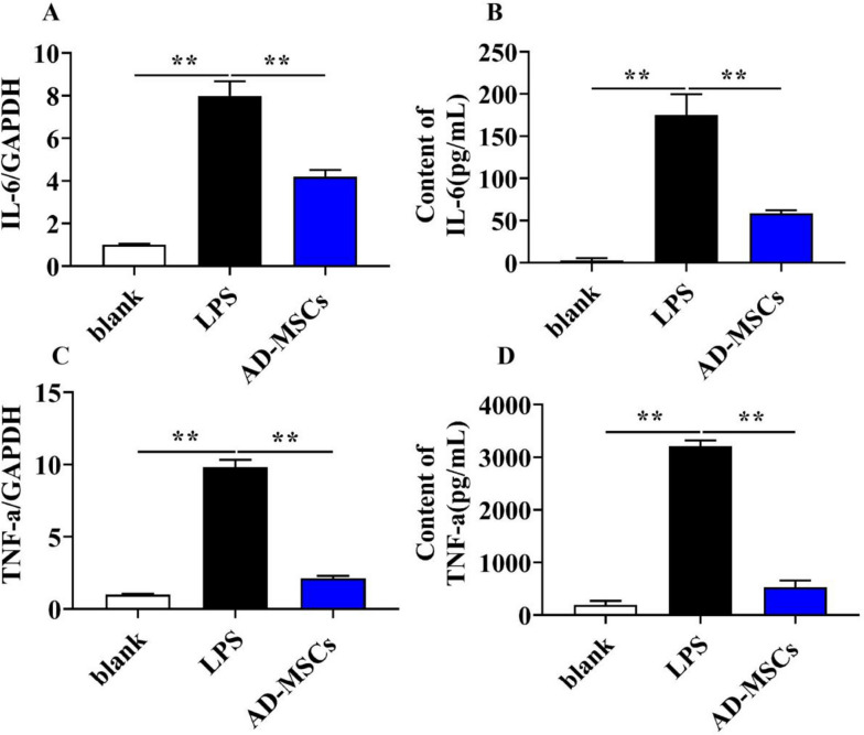
Results: Canine AD-MSCs promoted neural functional recovery, reduced neuronal apoptosis, and inhibited the activation of microglia and astrocytes in TBI rats. According to the results in vivo, we further investigated the regulatory mechanism of AD-MSCs on activated microglia by co-culture in vitro. Finally, we found that canine AD-MSCs promoted their polarization to the M2 phenotype, and inhibited their polarization to the M1 phenotype. What's more, AD-MSCs could reduce the migration, proliferation and Inflammatory cytokines of activated microglia, which is able to inhibit inflammation in the central system.
Conclusions: Collectively, the present study demonstrates that transplantation of canine AD-MSCs can promote functional recovery in TBI rats via inhibition of neuronal apoptosis, glial cell activation and central system inflammation, thus providing a theoretical basis for canine AD-MSCs therapy for TBI in veterinary clinic.
Keywords: Adipose-derived mesenchymal stem cells; Attenuating inflammation; Microglia; Therapy; Traumatic brain injury.
© 2024. The Author(s).
Conflict of interest statement
The authors declare no competing interests.
Figures


Similar articles
-
Intravenous administration of human chorionic membrane mesenchymal stem cells promotes functional recovery in a rat traumatic brain injury model.Neuroreport. 2024 Feb 7;35(2):81-89. doi: 10.1097/WNR.0000000000001981. Epub 2023 Dec 12. Neuroreport. 2024. PMID: 38109419
-
Transplantation of RADA16-BDNF peptide scaffold with human umbilical cord mesenchymal stem cells forced with CXCR4 and activated astrocytes for repair of traumatic brain injury.Acta Biomater. 2016 Nov;45:247-261. doi: 10.1016/j.actbio.2016.09.001. Epub 2016 Sep 2. Acta Biomater. 2016. PMID: 27592818
-
Exosomal miRNA-21 derived from umbilical cord mesenchymal stem cells inhibits microglial overactivation to counteract nerve damage.Mol Biol Rep. 2024 Aug 28;51(1):941. doi: 10.1007/s11033-024-09878-8. Mol Biol Rep. 2024. PMID: 39196412
-
Mesenchymal stem cells maintain the microenvironment of central nervous system by regulating the polarization of macrophages/microglia after traumatic brain injury.Int J Neurosci. 2017 Dec;127(12):1124-1135. doi: 10.1080/00207454.2017.1325884. Epub 2017 May 19. Int J Neurosci. 2017. PMID: 28464695 Review.
-
Mesenchymal stem cell therapy for the treatment of traumatic brain injury: progress and prospects.Rev Neurosci. 2019 Nov 26;30(8):839-855. doi: 10.1515/revneuro-2019-0002. Rev Neurosci. 2019. PMID: 31203262 Review.
References
MeSH terms
LinkOut - more resources
Full Text Sources
Medical
