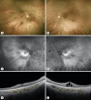
GNB1-Related Rod-Cone Dystrophy: A Case Report
- PMID: 38500542
- PMCID: PMC10948171
- DOI: 10.1159/000537997
GNB1-Related Rod-Cone Dystrophy: A Case Report
Abstract
Introduction: The GNB1 (guanine nucleotide-binding protein, β1) gene encodes for the ubiquitous β1 subunit of heterotrimeric G proteins, which are associated with G-protein-coupled receptors (GPCRs). GNB1 mutations cause a neurodevelopmental disorder characterized by a broad clinical spectrum. A novel variant has recently been confirmed in a case of rod-cone dystrophy.
Case presentation: We describe the second confirmed case of a classical rod-cone dystrophy associated with a mutation located in exon 6 of GNB1 [NM_002074.5:c.217G>C, p.(Ala73Pro)] in a 56-year-old patient also presenting mild intellectual disability, attention deficit/hyperactivity disorder, and truncal obesity.
Conclusion: This paper confirms the role of GNB1 in the pathogenesis of a classic rod-cone dystrophy and highlights the importance of including this gene in the genetic analysis panel for inherited retinal diseases.
Keywords: Case report; GNB1; Inherited retinal disease; Retinitis pigmentosa; Rod-cone dystrophy.
© 2024 The Author(s). Published by S. Karger AG, Basel.
Conflict of interest statement
The authors have no conflicts of interest to declare.
Figures


References
-
- Ford CE, Skiba NP, Daaka Y, Reuveny E, Shekter LR, Rosal R, et al. . Molecular basis for interactions of G protein betagamma subunits with effectors. Science. 1998;280(5367):1271–4. - PubMed
-
- Uhlén M, Fagerberg L, Hallström BM, Lindskog C, Oksvold P, Mardinoglu A, et al. . Tissue-based map of the human proteome. Science. 2015;347(6220):1260419. - PubMed
-
- Peng YW, Robishaw JD, Levine MA, Yau KW. Peng YW, Robishaw JD, Levine MA, Yau KW. Retinal Rods and Cones Have Distinct G Protein β and γ Subunits [Internet]. Vol. 89, Source. 1992. Available from: http://www.jstor.orgURL:http://www.jstor.org/stable/2362015. - PMC - PubMed
Publication types
LinkOut - more resources
Full Text Sources

