. 2024:2:1-13.
doi: 10.1016/j.mitoco.2024.01.001. Epub 2024 Jan 10.

Structural determinants of mitochondrial STAT3 targeting and function

Affiliations

Structural determinants of mitochondrial STAT3 targeting and function

Isabelle J Marié et al. Mitochondrial Commun. 2024.

Abstract

Signal transducer and activator of transcription (STAT) 3 has been found within mitochondria in addition to its canonical role of shuttling between cytoplasm and nucleus during cytokine signaling. Mitochondrial STAT3 has been implicated in modulation of cellular metabolism, largely through effects on the respiratory electron transport chain. However, the structural requirements underlying mitochondrial targeting and function have remained unclear. Here, we show that mitochondrial STAT3 partitions between mitochondrial compartments defined by differential detergent solubility, suggesting that mitochondrial STAT3 is membrane associated. The majority of STAT3 was found in an SDS soluble fraction copurifying with respiratory chain proteins, including numerous components of the complex I NADH dehydrogenase, while a minor component was found with proteins of the mitochondrial translation machinery. Mitochondrial targeting of STAT3 required the amino-terminal domain, and an internal linker domain motif also directed mitochondrial translocation. However, neither the phosphorylation of serine 727 nor the presence of mitochondrial DNA was required for the mitochondrial localization of STAT3. Two cysteine residues in the STAT3 SH2 domain, which have been previously suggested to be targets for protein palmitoylation, were also not required for mitochondrial translocation, but were required for its function as an enhancer of complex I activity. These structural determinants of STAT3 mitochondrial targeting and function provide potential therapeutic targets for disrupting the activity of mitochondrial STAT3 in diseases such as cancer.

Keywords: Electron transport chain; Mitochondrial import; Stat3.


Conflict of interest statement

Declaration of competing interest The authors declare no conflicts of interest.

Figures

Fig. 1.
Fig. 1.. Mitochondrial STAT3 partitions between an NP40-soluble and an SDS-soluble compartment.
Purified mitochondria from mouse liver (A), heart (B) or 143b cells (C) were sequentially extracted in the presence of NP-40 (NP-40) or SDS (SDS) as indicated. Protein extracts were separated on SDS-PAGE and analyzed by immunoblotting using the indicated antibodies. (D) Mitochondrial extracts from MDA-MB-231 cells (left panel) or MDA-MB-231 cells lacking mitochondrial DNA (MDA-MB-231 rho0 (ρ0)) (right panel) were performed as in A-C before being resolved on SDS-PAGE and probed with the indicated antibodies. (E) Quantification of mitochondrial DNA (mtDNA) in MDA-MB-231 rho0 (ρ0) and parental MDA-MB-231 cells by qPCR, p-value <0.05.
Fig. 2.
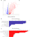
Fig. 2.. Mass spectrometry analysis of NP-40 and SDS mitochondrial fractions.
Volcano plot of protein abundance found respectively in NP-40 extracted fraction (NP-40) and SDS-extracted fraction (SDS). Arrow indicates STAT3. Reactome 2022 pathway analysis of proteins enriched in the NP-40 fractions (B) or SDS fractions (C).
Fig. 3.
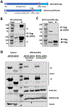
Fig. 3.. STAT3 exists in a multimeric form in the mitochondria and its N-terminal domain plays a role in its mitochondrial localization.
(A) Graphic representation of recombinant STAT3 constructs. Mitochondrial extracts from MDA-MB-231 cells expressing MTS.STAT3.Bio (B) or MTS.TruncSTAT3.Bio (C) were immunoprecipitated with anti-Flag antibodies and immunoblotted with either STAT3 or Flag antibodies as indicated. Asterix (*) marks endogenous STAT3. (D) Cytosolic (Cytosol) or mitochondrial (Mitochondria) extracts generated by 2 distinct purification protocols from HEK293T cells expressing either full length STAT3 (WTSTAT3) or STAT3 missing the first 132 amino acids (ΔNST3) were analyzed by SDS-PAGE followed by immunoblotting using antibodies against STAT3, SDHA, UBQCR2 and Tubulin. SE: short exposure. LE: long exposure.
Fig. 4.
Fig. 4.. A region of the linker domain of STAT3 contains a functional mitochondrial targeting motif.
In vitro translated 35S-labeled GFP, STAT3, Slc and Slc-GFP were incubated with freshly prepared mitochondria. Input fractions (I) were resolved on SDS-page alongside the mitochondria imported fraction (M) to probe mitochondrial import efficiency. (B) Graphic map of the different functional domains of STAT3 (upper panel). Description of STAT3 linker domain mutants M1 to M5. Residues highlighted in red were mutated to alanine (lower panel). (C) In vitro mitochondrial import of STAT3 domains F1 to F6 fused to GFP (as described in A). (D) In vitro mitochondrial import of mutant M1 to M5 (as described in B) of linker domain F4 fused to GFP (as described in A). Lanes 1–2 showing fragment F4 (delineated by a dotted line) is the same image as lanes 11–12 of panel C, reshown to allow direct comparison. (E) As in (D), in vitro mitochondrial export of F4-GFP compared to its N-terminal fragment (F4-GFP N-term) and C-terminal fragment (F4-GFP C-term).
Fig. 5.
Fig. 5.. C687/712 are not essential for mitochondrial import of STAT3.
(A) Cytoplasmic (CYTO) and mitochondrial extracts (MITO) as well as remaining residual pellet (PEL) of MDA-MB-231 cells deficient for endogenous STAT3 but ectopically expressing WT STAT3 (WT) or STAT3 mutated on C687 (C687S) or STAT3 doubly mutated on C687 and C712 (C687/C712S) were analyzed by SDS-PAGE and western blotting and probed with antibodies against STAT3, Cox4l1 and calreticulin (CalR). (B) Quantification of the STAT3 target gene SOCS3 mRNA levels by RT-qPCR normalized to B2M mRNA levels in STAT3 WT cells (WT), STAT3 KO cells (KO), STAT3 KO cells ectopically expressing either WT STAT3 (KO + WT) or STAT3 carrying the double mutation C6787S/C712S (KO+2CS). (C) Complex I activity of the set of cell lines described in B.
Fig. 6.
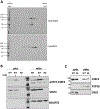
Fig. 6.. S727 is not essential for mitochondrial localization.
(A) Cytoplasmic (CytoSTAT3) and mitochondrial (MitoSTAT3) protein extracts from MDA-MB-231 cells were separated by isoelectric focusing followed by SDS-PAGE and then analyzed by immunoblotting for STAT3. (B) MDA-MB-231 cells either carrying WT STAT3 (WT) or deleted for STAT3 (KO) were treated with Calyculin A (CalA) or left untreated. Cytoplasmic (cyto) and mitochondrial (mito) extracts were separated on SDS-PAGE and probed for pS727-STAT3, total STAT3 (STAT3) or NDUFV2 by western blot. (C) Cytoplasmic and mitochondrial extracts from livers of WT mice (WT) and mice homozygous for a knock-in mutant of STAT3 where serine727 is replaced by alanine (SA) were analyzed by SDS-PAGE and immunoblotting using antibodies against STAT3, F1 ATPase and ERK1.

References

    1. Akira S, Nishio Y, Inoue M, et al. Molecular cloning of APRF, a novel IFN-stimulated gene factor 3 p91-related transcription factor involved in the gp130-mediated signaling pathway. Cell. 1994;77:63–71. - PubMed
    1. Zhong Z, Wen Z, Darnell JE. Stat3: a STAT family member activated by tyrosine phosphorylation in response to epidermal growth factor and interleukin-6. Science. 1994;264:95–98. - PubMed
    1. Raz R, Durbin JE, Levy DE. Acute phase response factor and additional members of the interferon-stimulated gene factor 3 family integrate diverse signals from cytokines, interferons, and growth factors. J Biol Chem. 1994;269:24391–24395. - PubMed
    1. Schaefer TS, Sanders LK, Park OK, Nathans D. Functional differences between Stat3alpha and Stat3beta. Mol Cell Biol. 1997;17:5307–5316. - PMC - PubMed
    1. Levy DE, Darnell JE Jr. STATs: transcriptional control and biological impact. Nat Rev Mol Cell Biol. 2002;3(9):651–662. - PubMed

LinkOut - more resources