. 2024 Mar 1;17(3):dmm050412.
doi: 10.1242/dmm.050412. Epub 2024 Apr 2.

Cross-species modeling of muscular dystrophy in Caenorhabditis elegans using patient-derived extracellular vesicles

Affiliations

Cross-species modeling of muscular dystrophy in Caenorhabditis elegans using patient-derived extracellular vesicles

Rewayd Shalash et al. Dis Model Mech. .

Abstract

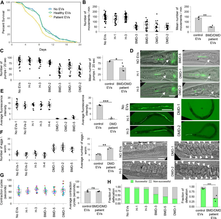
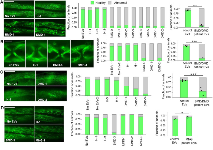
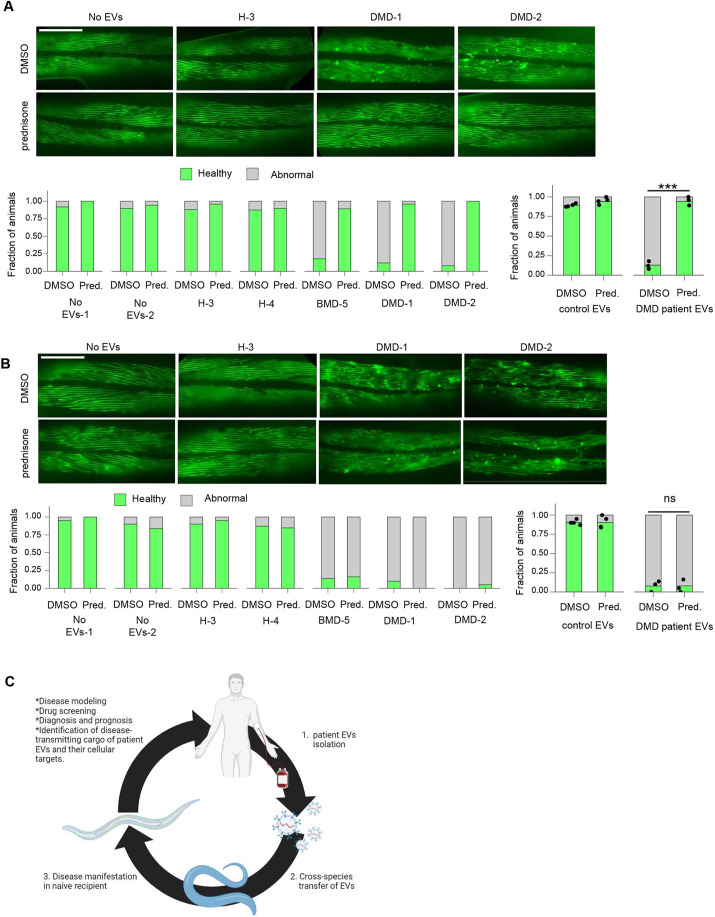
Reliable disease models are critical for medicine advancement. Here, we established a versatile human disease model system using patient-derived extracellular vesicles (EVs), which transfer a pathology-inducing cargo from a patient to a recipient naïve model organism. As a proof of principle, we applied EVs from the serum of patients with muscular dystrophy to Caenorhabditis elegans and demonstrated their capability to induce a spectrum of muscle pathologies, including lifespan shortening and robust impairment of muscle organization and function. This demonstrates that patient-derived EVs can deliver disease-relevant pathologies between species and can be exploited for establishing novel and personalized models of human disease. Such models can potentially be used for disease diagnosis, prognosis, analyzing treatment responses, drug screening and identification of the disease-transmitting cargo of patient-derived EVs and their cellular targets. This system complements traditional genetic disease models and enables modeling of multifactorial diseases and of those not yet associated with specific genetic mutations.

Keywords: Caenorhabditis elegans; Disease modeling; Duchenne muscular dystrophy; Extracellular vesicles.


Conflict of interest statement

Competing interests The authors declare no competing or financial interests.

Figures

Fig. 1.
Fig. 1.
EVs from patients with BMD/DMD impair C. elegans muscle functionality and decrease survival. (A) Lifespan assay for C. elegans treated with no EVs (N=1, n=105), EVs from unaffected individuals (‘healthy EVs’) (N=2, n=195; P=0.902 compared to the ‘No EVs’ condition), EVs from patients with Duchenne muscular dystrophy (DMD) or Becker muscular dystrophy (BMD) (‘patient EVs’) (N=7, n=735; P=0.0000036). (B) Thrashing assay for C. elegans treated with control EVs (N=3, n=127) and patient EVs (N=4, n=103); P=0.001171. (C-E) Pharynx muscle functionality was assessed by pharynx contraction (pumps) (C) and by monitoring fluorescent food uptake past the grinder of the pharynx (D, arrows mark pharynx grinder) and into the intestine (E). (C) Control EVs (N=3, n=93), patient EVs (N=4, n=121); P=0.03898. (E) Control EVs (N=5, n=115), patient EVs (N=3, n=70); P=0.0004444. (F) Egg-laying defects. In-uterus embryos are marked by arrowheads. Control EVs (N=4, n=100), patient EVs (N=3, n=75); P=0.001265. (G) Defecation assays. The time between posterior intestinal contractions was scored for a total of 105 contractions. Control EVs (N=3, n=15), patient EVs (N=3, n=15); P=0.8386. (H) Defecation assays scoring the fraction of successful defecation events. Bars (left) represent proportions of successful defecation events in animals upon each EV treatment. Bars (Right) represent predicted proportions obtained from mixed-effects binary logistic regression of the control EV- or patient EV-treated animals. Control EVs (N=3, n=15), patient EVs (N=3, n=15). P<0.0001. Boxplots in B,C,E-G present the score distribution of individual animals upon each EV treatment. Each boxplot represents an independent biological replicate. Boxes show the interquartile range, whiskers are 1.5 times the interquartile range, and the median is marked with a line. The bar graphs for ‘control EVs’ average the mean scores of treatments with healthy EVs and control untreated animals and those for ‘BMD/DMD patient EVs’ average the mean scores of treatments with patient EVs, as indicated in the neighboring boxplots. N represents the number of treatment conditions, n represents the number of animals analyzed. Scale bars: 20 µm (D,F); 200 µm (E). ns, not significant; *P<0.05; **P<0.01; ***P<0.0001. Statistical tests: log-rank test (A); unpaired two-tailed Welch's two-sample t-test (B,C,E-G); binary logistic regression (H).
Fig. 2.
Fig. 2.
EVs derived from patients with BMD/DMD impair C. elegans muscle structure. (A-D) Body wall muscle myofilaments (A,D) and vulva muscle myofilaments (B) visualized by a MYO-3::GFP transgene, and body wall muscle F-actin filaments visualized by phalloidin staining (C). Scale bars: 100 µm (A,C,D); 20 µm (B). Bar graphs (left) represent proportions of healthy or abnormal muscle filaments upon each EV treatment, each bar representing an independent biological replicate. Bar graphs (right) represent predicted proportions obtained from mixed-effects binary logistic regression of the control EV (treatments with healthy EVs and control untreated animals, or patient EV-treated animals, as indicated in the neighboring bar graphs). (A) Control EVs (N=4, n=224), patient EVs (N=8, n=480); ***P<0.0001. (B) Control EVs (N=4, n=97), patient EVs (N=3, n=79); ***P<0.0001. (C) Control EVs (N=4, n=116), patient EVs (N=3, n=97); ***P<0.0001. (D) Control EVs (N=3, n=120), EVs from patients with meningioma (MNG) (N=3, n=115); ns, not significant, P<0.0546. N represents the number of treatment conditions, n represents the number of animals analyzed.
Fig. 3.
Fig. 3.
Prednisone inhibits the effects of EVs derived from patients with BMD/DMD on the impairment of muscle structure in C. elegans, but does not resolve muscle atrophy. (A,B) Body wall muscle myofilaments visualized by a MYO-3::GFP transgene in animals treated with DMSO or prednisone after incubation with control or patient EVs. Prednisone treatment was initiated either at L4 stage, prior to the first signs of muscle disorganization (A), or at day 2, after muscle disorganization had commenced (B). Scale bars: 100 µm. Bar graphs (left) represent proportions of healthy or abnormal muscle filaments upon each EV treatment, each bar representing an independent biological replicate. Bar graphs (right) represent predicted proportions obtained from mixed-effects binary logistic regression of the control EV- or patient EV-treated animals (as indicated in the neighboring bar graphs). (A) Control EVs DMSO (N=4, n=121), patient EVs DMSO (N=3, n=78), control EVs prednisone (N=4, n=125; P=0.1954 compared to the ‘control EVs DMSO’ condition), patient EVs prednisone (N=3, n=87; ***P=0.0001 compared to the ‘patient EVs DMSO’ condition). (B) Control EVs DMSO (N=4, n=109), patient EVs DMSO (N=3, n=69), control EVs prednisone (N=4, n=104; P=0.9772 compared to the ‘control EVs DMSO’ condition), patient EVs prednisone (N=3, n=77; ns, not significant, P=0.9772 compared to the ‘patient EVs DMSO’ condition). N represents the number of treatment conditions, n represents the number of animals analyzed. (C) Interspecies modeling of human diseases using patient-derived EVs. EVs isolated from patient serum can induce pathological processes associated with the disease in simple model organisms such as C. elegans, generating an interspecies disease modeling system that can be used for analyzing the roles of EVs as mediators of disease pathological processes and for therapeutic screening. Image created with BioRender.com.

Similar articles

Cited by

References

    1. Aartsma-Rus, A., Straub, V., Hemmings, R., Haas, M., Schlosser-Weber, G., Stoyanova-Beninska, V., Mercuri, E., Muntoni, F., Sepodes, B., Vroom, E.et al. (2017). Development of exon skipping therapies for duchenne muscular dystrophy: a critical review and a perspective on the outstanding issues. Nucleic Acid Ther. 27, 251-259. 10.1089/nat.2017.0682 - DOI - PMC - PubMed
    1. Annexstad, E. J., Lund-Petersen, I. and Rasmussen, M. (2014). Duchenne muscular dystrophy. Tidsskr. Nor. Laegeforen. 134, 1361-1364. 10.4045/tidsskr.13.0836 - DOI - PubMed
    1. Apfeld, J. and Alper, S. (2018). What can we learn about human disease from the nematode C. elegans? Methods Mol. Biol. 1706, 53-75. 10.1007/978-1-4939-7471-9_4 - DOI - PMC - PubMed
    1. Azkona, G. and Sanchez-Pernaute, R. (2022). Mice in translational neuroscience: what R we doing? Prog. Neurobiol. 217, 102330. - PubMed
    1. Baldridge, D., Wangler, M. F., Bowman, A. N., Yamamoto, S., Schedl, T., Pak, S. C., Postlethwait, J. H., Shin, J., Solnica-Krezel, L., Bellen, H. J.et al. (2021). Model organisms contribute to diagnosis and discovery in the undiagnosed diseases network: current state and a future vision. Orphanet J. Rare Dis. 16, 206. 10.1186/s13023-021-01839-9 - DOI - PMC - PubMed

Substances