

[PCID2 is highly expressed in gastric cancer and affects the prognosis by regulating cancer cell cycle and proliferation]
- PMID: 38501418
- PMCID: PMC10954517
- DOI: 10.12122/j.issn.1673-4254.2024.02.15
[PCID2 is highly expressed in gastric cancer and affects the prognosis by regulating cancer cell cycle and proliferation]
Abstract
Objective: To investigate the expression of PCI Domain Containing 2 (PCID2) in gastric cancer, its effect on gastric cancer cell cycle and proliferation and the possible molecular mechanisms.
Methods: We examined PCID2 expression levels in gastric cancer and adjacent tissues from 100 patients undergoing radical gastrectomy in our hospital between January, 2012 and December, 2016, and analyzed the correlation of PCID2 expression level with cancer progression and postoperative 5-year survival rate of the patients. GO enrichment analysis was performed to identify the possible pathways that mediated the effect of PCID2 in gastric cancer progression. The effects of lentivirus-mediated PCID2 knockdown and overexpression on cell proliferation and cell cycle were analyzed in gastric cancer MGC-803 cells and in nude mice.
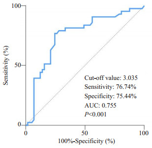
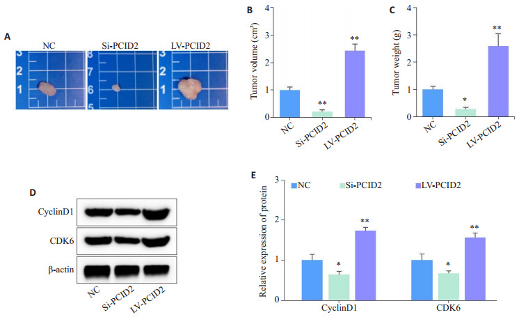
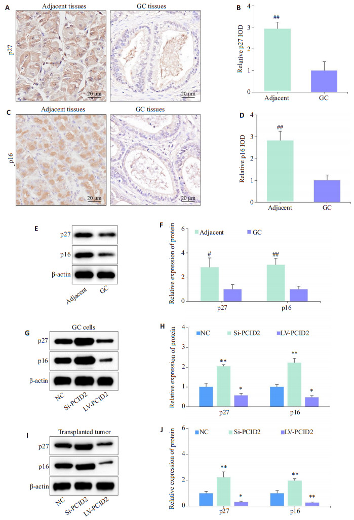
Results: PCID2 was highly expressed in gastric cancer tissues and positively correlated with peripheral blood levels of CA19-9 and CEA (P < 0.01). In gastric cancer patients, a high PCID2 expression was associated with a significantly lowered postoperative 5-year survival rate (P < 0.001) as an independent risk factor for postoperative survival (HR: 2.987, 95% CI: 1.616-5.519). The sensitivity, specificity, and area under the curve of PCID2 for predicting postoperative 5-year survival were 76.74%, 75.44%, and 0.755 (P < 0.001), respectively. GO enrichment analysis suggested that PCID2 was associated with gastric cancer cell cycle progression. PCID2 overexpression in MGC-803 cells significantly promoted cell proliferation, G1/S phase transition, expressions of cyclin D1 and CDK6, and the growth of transplanted xenograft in nude mice (P < 0.05). The expressions of p27 and p16 were significantly lowered in gastric cancer tissues, and their expression levels were negatively regulated by PCID2 expression in MGC-803 cells (P < 0.05).
Conclusion: PCID2 is highly expressed in gastric cancer tissues in close correlation with poor prognosis of the patients. High PCID2 expression promotes gastric cancer proliferation and cell cycle progression by inhibiting the expression of p27 and p16.
目的: 探讨扩增基因PCI结构域2(PCID2)在胃癌中的表达情况及其对细胞周期进程和增殖的影响,并分析其可能的分子机制。
方法: 纳入2012年1月~2016年12月在我院接受胃癌根治术的患者100例,检测胃癌和癌旁组织中PCID2的表达差异;统计学分析PCID2表达水平对胃癌进展及术后5年生存率的影响;采用GO富集分析预测PCID2参与胃癌恶性进展的可能作用途径;体外采用慢病毒转染特异性敲低和过表达胃癌细胞系(MGC-803)中PCID2表达,并分析其对细胞增殖及周期的影响;进一步通过裸鼠移植瘤模型进行在体验证;最后通过组织样本分析结合体内外研究,分析PCID2影响胃癌细胞的分子机制。
结果: 相较于癌旁组织,PCID2在胃癌组织中高表达(P < 0.01);胃癌组织中PCID2表达水平与外周血中CA19-9及CEA水平呈正相关(P < 0.001);以胃癌组织中PCID2相对表达水平(IOD值)的中位数(3.085)为界,将纳入研究的患者分为PCID2高表达组(n=50)和PCID2低表达组(n=50),PCID2高表达组患者术后5年生存率显著低于PCID2低表达组(P < 0.001);单因素结合Cox多因素分析表明胃癌组织中PCID2高表达是影响患者术后5年生存率的独立危险因素(HR:2.987,95% CI:1.616-5.519);ROC曲线分析显示,以PCID2相对表达水平(3.035)为截点值,评估胃癌患者术后5年生存率的敏感性为76.74%,特异性为75.44%,曲线下面积为0.755(P < 0.001)。GO功能富集分析提示PCID2可能与胃癌细胞周期进程有关(P < 0.01)。体外实验证实,PCID2过表达可促进胃癌细胞增殖、G1/S期转变和细胞周期相关蛋白(CyclinD1、CDK6)的表达,而敲低则反之(P < 0.05)。裸鼠成瘤实验证明,PCID2过表达可促进裸鼠移植瘤的生长,而敲低则抑制(P < 0.05)。组织样本分析联合体内外研究表明,胃癌组织中p27、p16蛋白表达水平较癌旁组织均明显降低(P < 0.05)。此外,PCID2过表达抑制细胞周期调控蛋白p27、p16的表达,敲低则相反(P < 0.05)。
结论: PCID2在胃癌组织中高表达且与患者预后不良密切相关,其可能通过抑制p27、p16表达促进胃癌细胞增殖和细胞周期进程。
Keywords: PCID2; cell cycle; gastric cancer; prognosis; proliferation.
Figures


References
-
- Song ZY, Wu YY, Yang JB, et al. Progress in the treatment of advanced gastric cancer. Tumour Biol. 2017;39(7):1010428317714626. - PubMed
Publication types
MeSH terms
Substances
LinkOut - more resources
Full Text Sources
Medical
Molecular Biology Databases
Research Materials
Miscellaneous