. 2024 Apr;33(4):e4937.
doi: 10.1002/pro.4937.

A cellulosomal double-dockerin module from Clostridium thermocellum shows distinct structural and cohesin-binding features

Affiliations

A cellulosomal double-dockerin module from Clostridium thermocellum shows distinct structural and cohesin-binding features

Chao Chen et al. Protein Sci. 2024 Apr.

Abstract

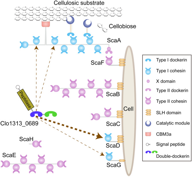
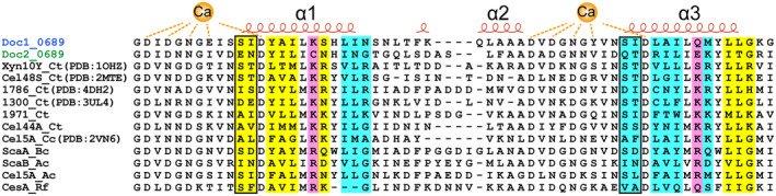
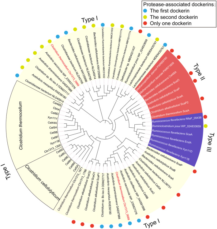
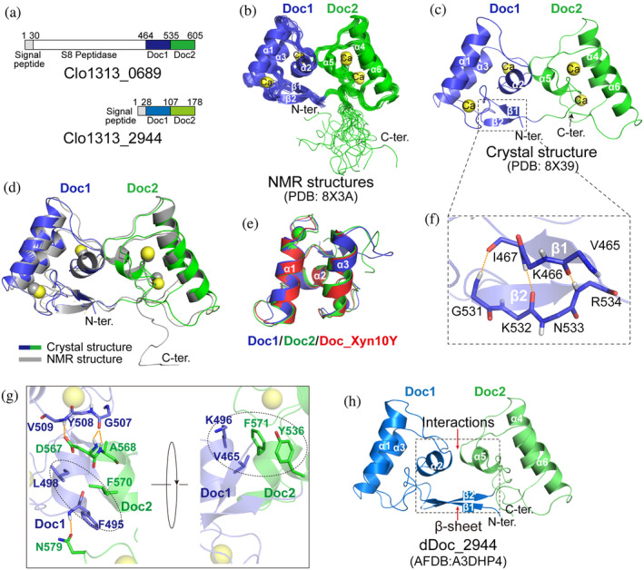
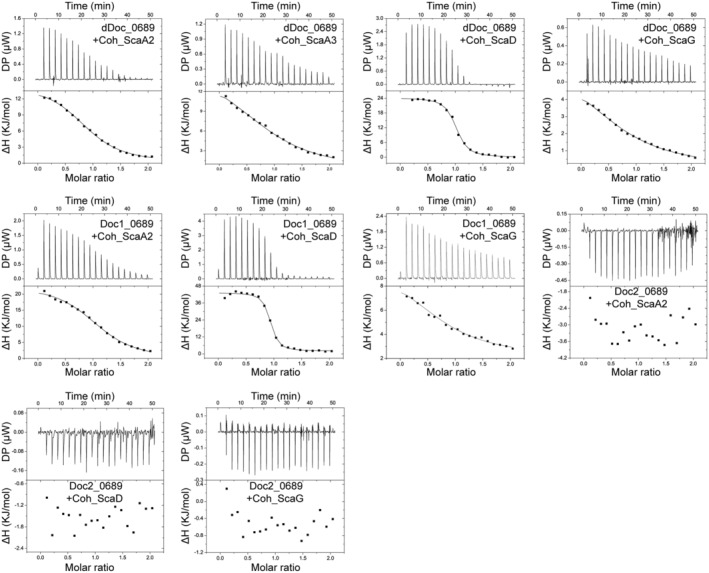
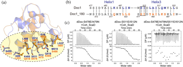
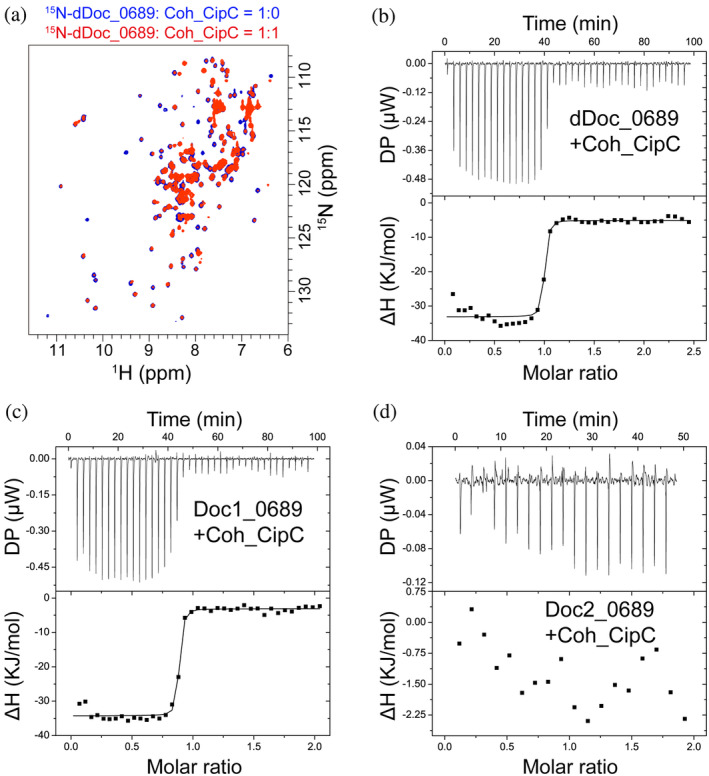
Cellulosomes are intricate cellulose-degrading multi-enzymatic complexes produced by anaerobic bacteria, which are valuable for bioenergy development and biotechnology. Cellulosome assembly relies on the selective interaction between cohesin modules in structural scaffolding proteins (scaffoldins) and dockerin modules in enzymes. Although the number of tandem cohesins in the scaffoldins is believed to determine the complexity of the cellulosomes, tandem dockerins also exist, albeit very rare, in some cellulosomal components whose assembly and functional roles are currently unclear. In this study, we characterized the structure and mode of assembly of a tandem bimodular double-dockerin, which is connected to a putative S8 protease in the cellulosome-producing bacterium, Clostridium thermocellum. Crystal and NMR structures of the double-dockerin revealed two typical type I dockerin folds with significant interactions between them. Interaction analysis by isothermal titration calorimetry and NMR titration experiments revealed that the double-dockerin displays a preference for binding to the cell-wall anchoring scaffoldin ScaD through the first dockerin with a canonical dual-binding mode, while the second dockerin module was unable to bind to any of the tested cohesins. Surprisingly, the double-dockerin showed a much higher affinity to a cohesin from the CipC scaffoldin of Clostridium cellulolyticum than to the resident cohesins from C. thermocellum. These results contribute valuable insights into the structure and assembly of the double-dockerin module, and provide the basis for further functional studies on multiple-dockerin modules and cellulosomal proteases, thus highlighting the complexity and diversity of cellulosomal components.

Keywords: NMR; X-ray crystallography; binding affinity; protein complex; protein-protein interaction; scaffolding protein; tandem modules.


Conflict of interest statement

There are no conflicts of interest to declare.

Figures

FIGURE 1
FIGURE 1
Schematic representation of the cellulosome from C. thermocellum DSM1313. The proposed binding position of Clo1313_0689 on scaffoldins are indicated by dashed arrows and the thickness of arrow lines indicate the experimentally determined differences in binding affinities.
FIGURE 2
FIGURE 2
Sequence alignments of type I dockerins. Secondary structure elements of Doc_Xyn10Y (PDB: 1OHZ) are shown at the top of the alignment. The putative residues involved in Coh‐binding are highlighted with yellow background (for the N‐terminal binding site), cyan background (for the C‐terminal binding site), or magenta background (for both the N‐terminal and C‐terminal binding sites) according to the interaction of Doc_Xyn10Y and Coh_ScaA2 (Carvalho et al., 2003). The characteristic key residues are indicated by black rectangles.
FIGURE 3
FIGURE 3
Molecular evolutionary tree of dockerin modules. All protease‐associated dockerins from several species are labeled by colored dots. The light blue and yellow green dots represent the first and second dockerin modules of the double‐dockerin modules connected to the protease domains, respectively. The red dots denote the single dockerin module of the protease‐containing proteins. Doc1_0689 and Doc2_0689 are highlighted with species names in red. The type I, II, and III dockerin modules are shaded in light yellow, red, and purple, respectively.
FIGURE 4
FIGURE 4
Structure of dDoc_0689. (a) Schematic representation of the composition of the double‐dockerin‐containing proteins of C. thermocellum. (b, c) Ribbon representation of the ensemble of 20 NMR structures (b) and the crystal structure (c) of dDoc_0689. Doc1_0689 and Doc2_0689 are colored in blue and green, respectively. (d) Superposition of the NMR (gray) and crystal (blue for Doc1_0689 and green for Doc2_0689) structures of dDoc_0689. (e) Superposition of the crystal structures of separated Doc1_0689 (blue), Doc2_0689 (green), and DocI_Xyn10Y (red, PDB: 1OHZ‐B). (f) Representation of the intramolecular clasp formed by the anti‐parallel β‐sheet of Doc1_0689, which is enlarged from panel (c) into the dashed box. The residues involved in the formation of the intramolecular clasp are shown as sticks. (g) Interactions between Doc1_0689 and Doc2_0689 in dDoc_0689. The residues involved in the interactions are shown as blue (Doc1_0689) and green (Doc2_0689) sticks. (h) AlphaFold2 structural model of dDoc_2944.
FIGURE 5
FIGURE 5
Representative ITC data for titrations of various cohesins into dockerins. ITC traces are shown on top, and integrated binding isotherms are shown on the bottom. The curve represents the best fit to a single‐site‐binding model. The dockerin and cohesin proteins used for the titrations are shown in each ITC trace. All ITC experiments were carried out at 298 K.
FIGURE 6
FIGURE 6
Identification of the “dual binding mode” of Doc1_0689. (a) Structural superposition of Doc1_0689 (blue) and a 180°‐rotated Doc1_0689 (orange). The putative residues involved in C‐ and N‐terminal cohesin binding sites are shown as blue and orange sticks, respectively. (b) Sequence alignment of α‐helix 1 and 3 of Doc1_0689 and its rotated state. The key sites for species specificity are indicated by a black rectangle. (c) ITC titration of the mutants of dDoc_0689 with Coh_ScaD.
FIGURE 7
FIGURE 7
The interaction of dDoc_0689 and the cohesin of C. cellulolyticum Coh_CipC. (a) NMR titration of dDoc_0689 with Coh_CipC. Overlaid 1H–15N HSQC spectra of 15N‐labeled dDoc_0689 without and with unlabeled Coh_CipC are shown in blue and red, respectively. (b–d) ITC titrations of dDoc_0689 and its two individual dockerins with Coh_CipC. ITC traces are shown at the top, and integrated binding isotherms are shown at the bottom. The curve represents the best fit to a single‐site‐binding model. All NMR titration and ITC experiments were carried out at 298 K.

Similar articles

Cited by

References

    1. Adams JJ, Pal G, Jia Z, Smith SP. Mechanism of bacterial cell‐surface attachment revealed by the structure of cellulosomal type II cohesin‐dockerin complex. Proc Natl Acad Sci U S A. 2006;103:305–310. - PMC - PubMed
    1. Adams PD, Afonine PV, Bunkóczi G, Chen VB, Davis IW, Echols N, et al. PHENIX: a comprehensive Python‐based system for macromolecular structure solution. Acta Crystallogr D Biol Crystallogr. 2010;66:213–221. - PMC - PubMed
    1. Alves VD, Fontes C, Bule P. Cellulosomes: highly efficient cellulolytic complexes. Subcell Biochem. 2021;96:323–354. - PubMed
    1. Artzi L, Bayer EA, Moraïs S. Cellulosomes: bacterial nanomachines for dismantling plant polysaccharides. Nat Rev Microbiol. 2017;15:83–95. - PubMed
    1. Artzi L, Dassa B, Borovok I, Shamshoum M, Lamed R, Bayer EA. Cellulosomics of the cellulolytic thermophile Clostridium clariflavum . Biotechnol Biofuels. 2014;7:100. - PMC - PubMed

MeSH terms

LinkOut - more resources