

Pyroptosis and polarization of macrophages in septic acute lung injury induced by lipopolysaccharide in mice
- PMID: 38501547
- PMCID: PMC10949386
- DOI: 10.1002/iid3.1197
Pyroptosis and polarization of macrophages in septic acute lung injury induced by lipopolysaccharide in mice
Abstract
Background: Pyroptosis and polarization are significant contributors to the onset and development of many diseases. At present, the relationship between pyroptosis and polarization in acute lung injury (ALI) caused by sepsis remains unclear.
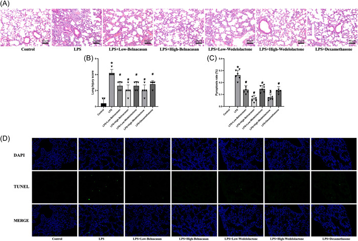
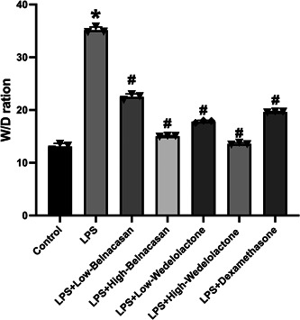
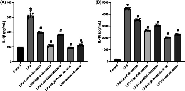
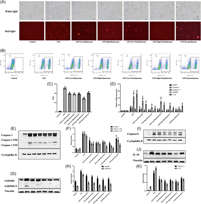
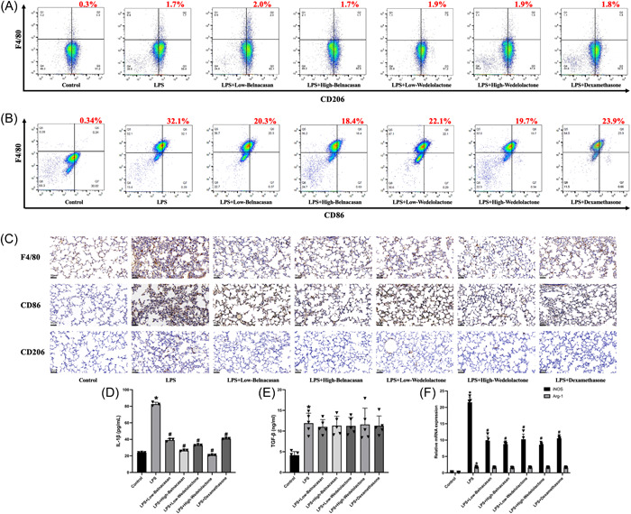
Methods: The ALI model for sepsis was created in mice and categorized into the blank control, lipopolysaccharide (LPS) group, LPS + low-dose Belnacasan group, LPS + high-dose Belnacasan group, LPS + low-dose Wedelolactone group, LPS + high-dose Wedelolactone group, and positive control group. The wet-dry specific gravity was evaluated to compare pulmonary edema. Hematoxylin-eosin, Masson, and terminal deoxynucleotidyl transferase dUTP nick end labeling staining techniques were conducted to observe and contrast the pathological changes in lung tissue. ELISA was utilized to identify M1 and M2 macrophages and correlated inflammatory factors. Immunohistochemical staining and flow cytometry were employed to identify markers of M1 and M2 macrophages in lung tissue. Propidium iodide staining, together with flow cytometry, was utilized to observe the degree and positive rate of pyroptosis of alveolar macrophages. Western blot analysis was conducted to detect the expression levels of Caspase 1, Caspase 11, GSDMD, and IL-18 in the lung tissues of each group. The real-time quantitative polymerase chain reaction method was used to ascertain relative expression levels of NLRP3, Caspase 1, Caspase 11, GSDMD, IL-18, iNOS, and Arg-1 in lung tissues of all groups.
Results: In mice with sepsis-induced ALI, both classical and nonclassical pathways of pyroptosis are observed. Inhibiting pyroptosis has been found to ameliorate lung injury, pulmonary edema, and inflammation induced by LPS. Notably, the expression of NLRP3, Caspase 1, Caspase 11, GSDMD, IL-1β, IL-18, TGF-β, CD86, CD206, iNOS, and Arg-1 were all altered in this process. Additionally, alveolar macrophages were polarized along with pyroptosis in mice with ALI caused by sepsis.
Conclusion: Pyroptosis of alveolar macrophages in the context of ALI in mice infected with sepsis has been linked to the polarization of alveolar macrophages toward type M1.
Keywords: acute lung injury; alveolar macrophage; polarization; pyroptosis; sepsis.
© 2024 The Authors. Immunity, Inflammation and Disease published by John Wiley & Sons Ltd.
Conflict of interest statement
The authors declare no conflict of interest.
Figures
Similar articles
-
Knockdown of angiopoietin-like 4 suppresses sepsis-induced acute lung injury by blocking the NF-κB pathway activation and hindering macrophage M1 polarization and pyroptosis.Toxicol In Vitro. 2024 Feb;94:105709. doi: 10.1016/j.tiv.2023.105709. Epub 2023 Oct 10. Toxicol In Vitro. 2024. PMID: 37820748
-
Mdivi-1 Attenuates Sepsis-Associated Acute Lung Injury by Inhibiting M1 Alveolar Macrophage Polarization and Pyroptosis.Mediators Inflamm. 2025 Mar 30;2025:3675276. doi: 10.1155/mi/3675276. eCollection 2025. Mediators Inflamm. 2025. PMID: 40196168 Free PMC article.
-
Alpha-linolenic acid pretreatment alleviates NETs-induced alveolar macrophage pyroptosis by inhibiting pyrin inflammasome activation in a mouse model of sepsis-induced ALI/ARDS.Front Immunol. 2023 Mar 27;14:1146612. doi: 10.3389/fimmu.2023.1146612. eCollection 2023. Front Immunol. 2023. PMID: 37051243 Free PMC article.
-
Regulated Cell Death of Alveolar Macrophages in Acute Lung Inflammation: Current Knowledge and Perspectives.J Inflamm Res. 2024 Dec 21;17:11419-11436. doi: 10.2147/JIR.S497775. eCollection 2024. J Inflamm Res. 2024. PMID: 39722732 Free PMC article. Review.
-
Immunologic role of macrophages in sepsis-induced acute liver injury.Int Immunopharmacol. 2024 Dec 25;143(Pt 2):113492. doi: 10.1016/j.intimp.2024.113492. Epub 2024 Oct 29. Int Immunopharmacol. 2024. PMID: 39471696 Review.
Cited by
-
Differences in macrophage pyroptosis and polarization induced by nano-/micro-calcium oxalate crystals.J Nanobiotechnology. 2025 Jul 10;23(1):499. doi: 10.1186/s12951-025-03549-x. J Nanobiotechnology. 2025. PMID: 40640899 Free PMC article.
-
Kirenol ameliorates endotoxin-induced acute lung injury by inhibiting the ERK and JNK phosphorylation-mediated NFκB pathway in mice.Inflammopharmacology. 2025 Apr;33(4):2069-2081. doi: 10.1007/s10787-025-01693-2. Epub 2025 Mar 4. Inflammopharmacology. 2025. PMID: 40035943
-
Macrophages in sepsis-induced acute lung injury: exosomal modulation and therapeutic potential.Front Immunol. 2025 Jan 7;15:1518008. doi: 10.3389/fimmu.2024.1518008. eCollection 2024. Front Immunol. 2025. PMID: 39840035 Free PMC article. Review.
-
[Amentoflavone alleviates acute lung injury in mice by inhibiting cell pyroptosis].Nan Fang Yi Ke Da Xue Xue Bao. 2025 Apr 20;45(4):692-701. doi: 10.12122/j.issn.1673-4254.2025.04.03. Nan Fang Yi Ke Da Xue Xue Bao. 2025. PMID: 40294918 Free PMC article. Chinese.
References
-
- Hao LI, Wang Zihao, Lu Yiming. Roles of pyroptosis‐related genes in immunotherapy and prognosis in hepatocellular carcinoma. Mil Med. 2023;47(03):196‐204.
Publication types
MeSH terms
Substances
Grants and funding
LinkOut - more resources
Full Text Sources
Medical
Research Materials
Miscellaneous
