. 2024 Oct 3;147(10):3583-3595.
doi: 10.1093/brain/awae088.

A direct spinal cord-computer interface enables the control of the paralysed hand in spinal cord injury

Affiliations

A direct spinal cord-computer interface enables the control of the paralysed hand in spinal cord injury

Daniela Souza Oliveira et al. Brain. .

Abstract

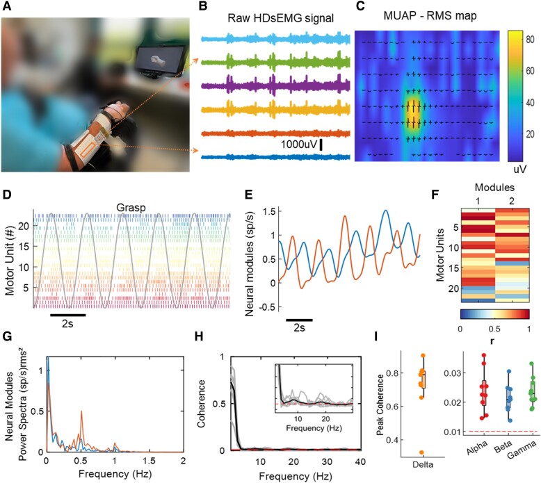
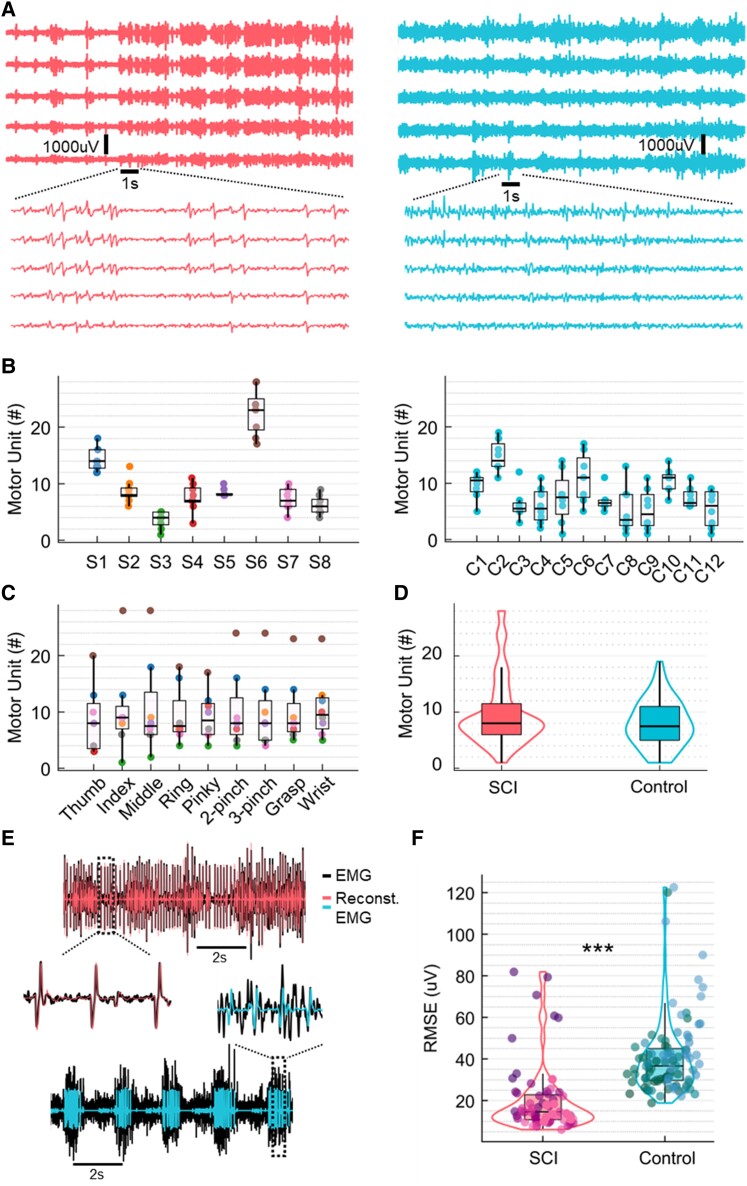
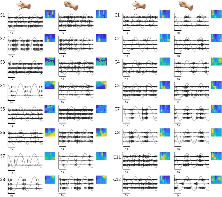
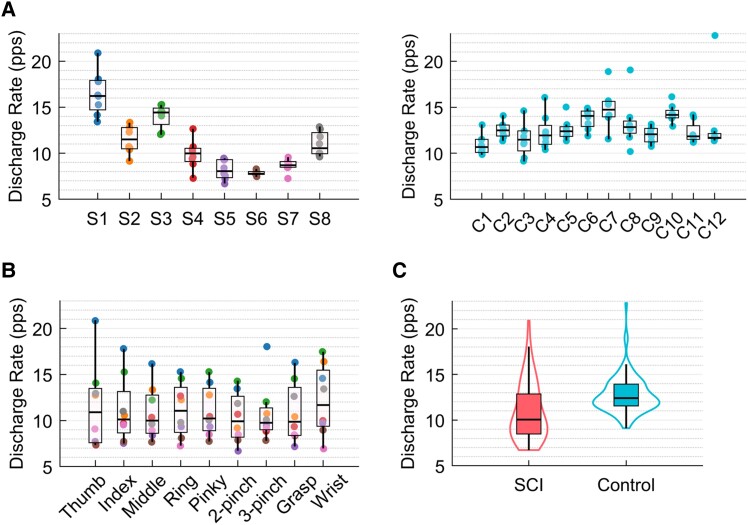
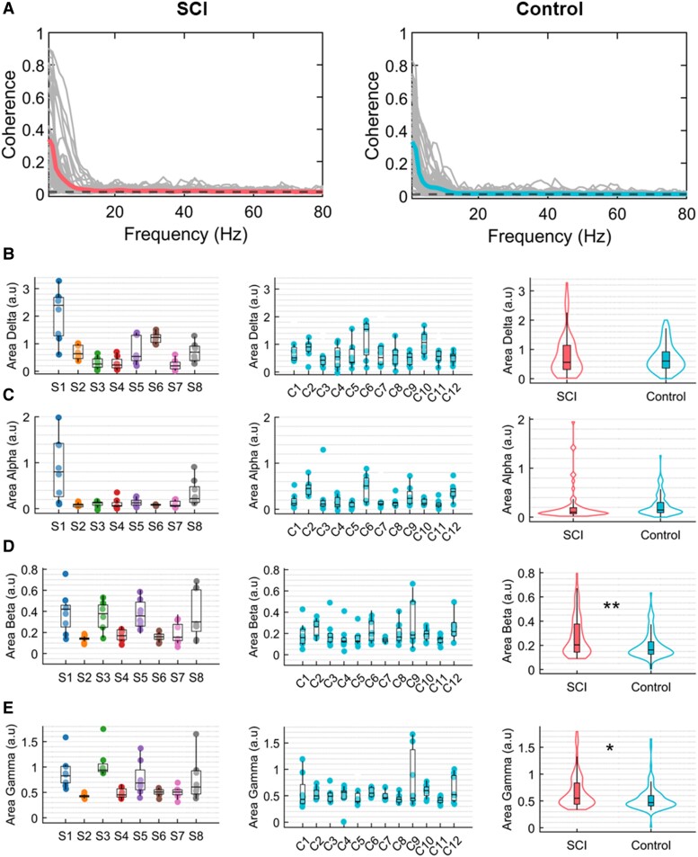
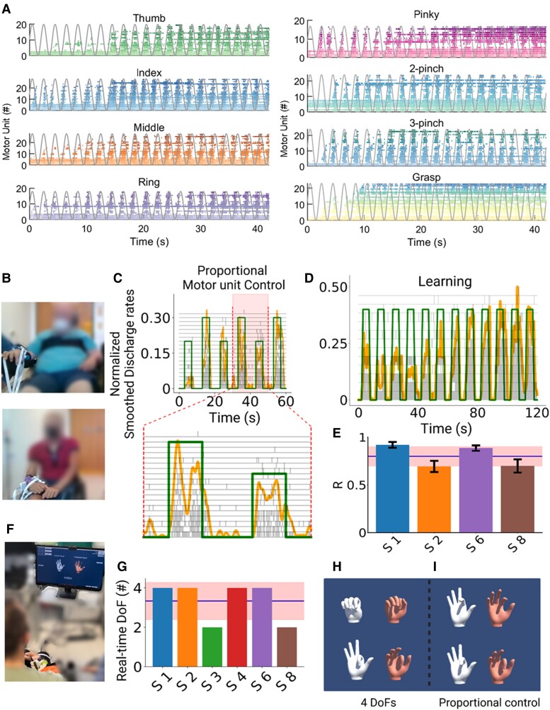
Paralysis of the muscles controlling the hand dramatically limits the quality of life for individuals living with spinal cord injury (SCI). Here, with a non-invasive neural interface, we demonstrate that eight motor complete SCI individuals (C5-C6) are still able to task-modulate in real-time the activity of populations of spinal motor neurons with residual neural pathways. In all SCI participants tested, we identified groups of motor units under voluntary control that encoded various hand movements. The motor unit discharges were mapped into more than 10 degrees of freedom, ranging from grasping to individual hand-digit flexion and extension. We then mapped the neural dynamics into a real-time controlled virtual hand. The SCI participants were able to match the cue hand posture by proportionally controlling four degrees of freedom (opening and closing the hand and index flexion/extension). These results demonstrate that wearable muscle sensors provide access to spared motor neurons that are fully under voluntary control in complete cervical SCI individuals. This non-invasive neural interface allows the investigation of motor neuron changes after the injury and has the potential to promote movement restoration when integrated with assistive devices.

Keywords: high density surface electromyography; motor neuron; motor unit; neural interface; spinal cord injury.


Conflict of interest statement

The authors report no competing interests.

Figures

Figure 1
Figure 1
Overview of experimental setup and motor unit data analysis. (A) Experimental setup consisting of 320 surface electromyogram (EMG) electrodes placed in the forearm muscles. The movement instructions were guided by a virtual hand video displayed on a monitor in front of the subject. (B) A few example electrodes show raw high-density surface EMG (HDsEMG) signals while the subject attempts a grasp task (flexion and extension of the fingers, 0.5 Hz). (C) Example of spatial mapping based on the root mean square (RMS) values of the motor unit action potential (MUAP). (D) Raster plot of motor unit firings (colour-coded) identified during 10 s of a grasp task. (E) Neural modules extracted for the same task, using factorization analysis. (F) Pearson correlation values (r) of the individual motor units with the two neural modules. (G) Neural modules’ power spectra, showing a peak at the movement frequency (0.5 Hz). (H) Coherence between cumulative spike trains of motor units across all tasks of Subject 6 (S6), highlighting alpha and beta bands. (I) Coherence peak across all tasks of Subject S6 for delta (1–5 Hz), alpha (6–12 Hz), beta (15–30 Hz) and gamma (31–80 Hz) bandwidths. The dashed line in red in H and I indicates the coherence threshold (average coherence between 100–250 Hz).
Figure 2
Figure 2
Number of detected motor units and residual high-density surface electromyogram signals. (A) Example of raw high-density surface electromyogram (HDsEMG) signals for both groups, spinal cord injury (SCI; left) and control (right). The signals are shown in time windows of 20 and 1 s. (B) Number of detected motor units across subjects for both groups, SCI and control (the dots are colour-coded for the subjects of the SCI group). (C) Number of detected motor units across all tasks (the dots represent the tasks). (D) Distribution of the total number of motor units across groups, SCI (left) and control (right). (E) Example of EMG channels from both SCI and control groups overlayed with the reconstructed EMG. (F) Root mean square error (RMSE) between EMG and reconstructed EMG, representing the residual EMG activity for both groups. ***P < 0.001.
Figure 6
Figure 6
Examples of raw high-density surface electromyograph (HDsEMG) signals and spatial amplitude maps. We report examples of EMG signals for all subjects of the spinal cord injury (SCI) group (S1–S8) during index and grasp tasks. The normalized signals from the three EMG channels with higher root mean square (RMS) values (in black) are presented over 5 s, together with the virtual hand kinematics (in grey). For each subject, we show a spatial map based on the RMS values of each EMG channel. For brevity, we only present data from eight control group participants for comparison.
Figure 3
Figure 3
Discharge rate. (A) Average discharge rate across subjects for both groups [the dots are colour-coded for the subjects of the spinal cord injury (SCI) group]. (B) Average discharge rate across all tasks (the dots represent the tasks). (C) Distribution of the total number of motor units across groups, SCI in pink and control in blue.
Figure 4
Figure 4
Coherence. (A) Average coherence across all participants and all tasks for both groups, spinal cord injury (SCI) in pink and control in blue. The black dashed line represents the coherence threshold (average coherence between 100–250 Hz). Each curve in grey represents the coherence for one subject. (BE) Area under coherence curve across all subjects and groups for delta (1–5 Hz), alpha (6–12 Hz), beta (15–30 Hz) and gamma (31–80 Hz) bands, respectively (the dots represent the tasks and are colour-coded for the subjects of the SCI group). For each frequency band, we also show the group distribution of the coherence area values across all tasks and subjects. *0.01 < P < 0.05, **0.001 < P < 0.01.
Figure 5
Figure 5
Real-time control of motor units and virtual hand. (A) Raster plot for all motor units identified for Subject S6 (S6) during the respective task (colour-coded) and the virtual hand movement trajectories (grey line). Note the task-modulated activity of the motor unit firing patterns that encoded flexion and extension movements. (B) Real-time tasks for two participants (Subjects S1 and S6). (C) The participants were asked to follow a trajectory on a screen (green line) by attempting a grasp movement. The motor units were decomposed online and the cumulative smoothed discharge rate (yellow line) was used as biofeedback. After a few seconds of training (D), the subjects could track the trajectories with high accuracy and at different target levels (C). (E) Cross-correlation coefficient (R) between the smoothed discharge rate and the requested tasks for four subjects. (F) After the online motor unit decomposition, we used a supervised machine learning method to proportionally control the movement of a virtual hand. Four of six subjects were able to proportionally open and close the hand (GI) and proportionally control in both movement directions (flexion and extension) the index finger (H and I). These subjects were able to control four degrees of freedom (DoFs) that corresponded to hand opening, closing, index flexion and extension. These subjects were able to control four DoFs that corresponded to hand opening, closing, index flexion and extension.

Similar articles

Cited by

References

    1. Snoek GJ, Ijzerman MJ, Hermens HJ, Maxwell D, Biering-Sorensen F. Survey of the needs of patients with spinal cord injury: Impact and priority for improvement in hand function in tetraplegics. Spinal Cord. 2004;42:526–532. - PubMed
    1. Fridén J, House J, Keith M, Schibli S, van Zyl N. Improving hand function after spinal cord injury. J Hand Surg Eur. 2022;47:105–116. - PubMed
    1. Collinger JL, Boninger ML, Bruns TM, Curley K, Wang W, Weber DJ. Functional priorities, assistive technology and brain-computer interfaces after spinal cord injury. J Rehabil Res Dev. 2013;50:145–160. - PMC - PubMed
    1. Bouton CE, Shaikhouni A, Annetta NV, et al. . Restoring cortical control of functional movement in a human with quadriplegia. Nature. 2016;533:247–250. - PubMed
    1. Benabid AL, Costecalde T, Eliseyev A, et al. . An exoskeleton controlled by an epidural wireless brain–machine interface in a tetraplegic patient: A proof-of-concept demonstration. Lancet Neurol. 2019;18:1112–1122. - PubMed