. 2024 Apr 16;98(4):e0013924.
doi: 10.1128/jvi.00139-24. Epub 2024 Mar 19.

Functional dissection of the spike glycoprotein S1 subunit and identification of cellular cofactors for regulation of swine acute diarrhea syndrome coronavirus entry

Affiliations

Functional dissection of the spike glycoprotein S1 subunit and identification of cellular cofactors for regulation of swine acute diarrhea syndrome coronavirus entry

Yong-Le Yang et al. J Virol. .

Abstract

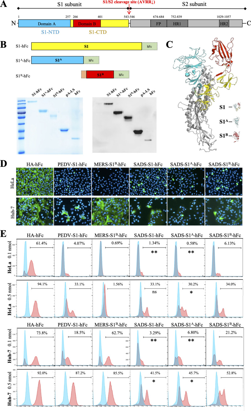
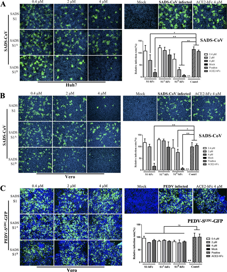
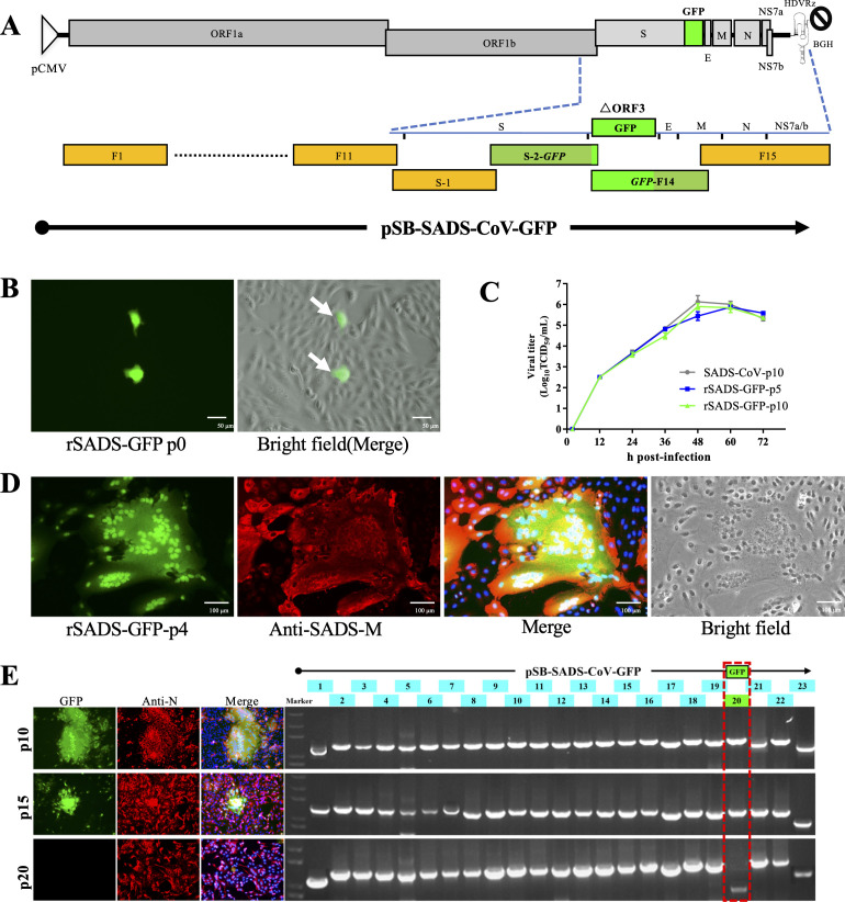
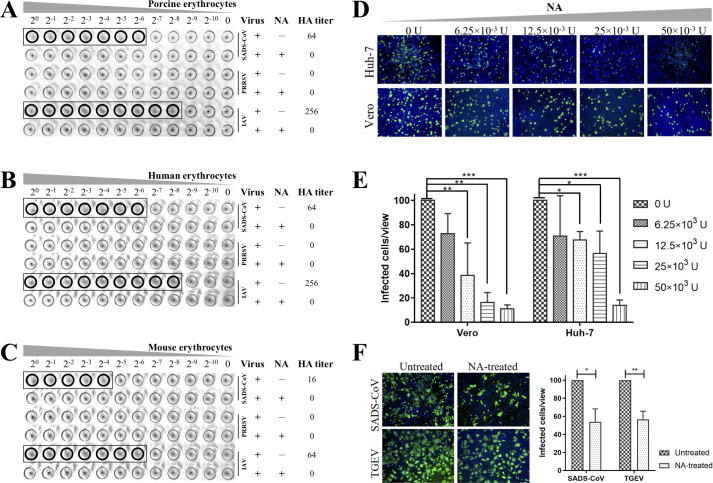
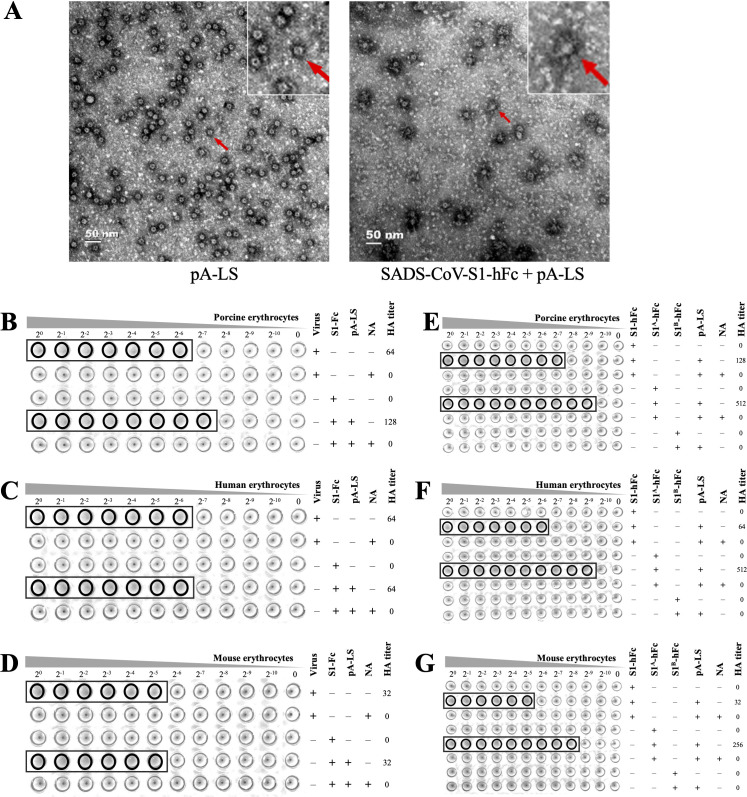
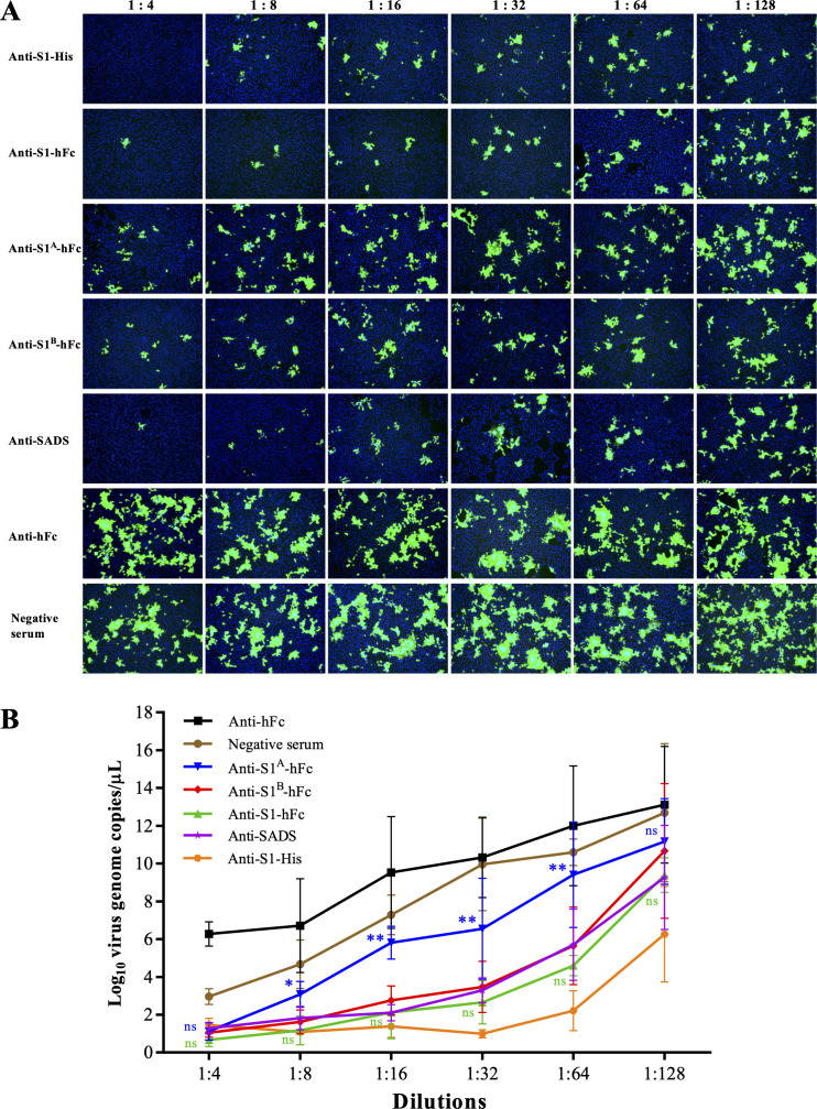
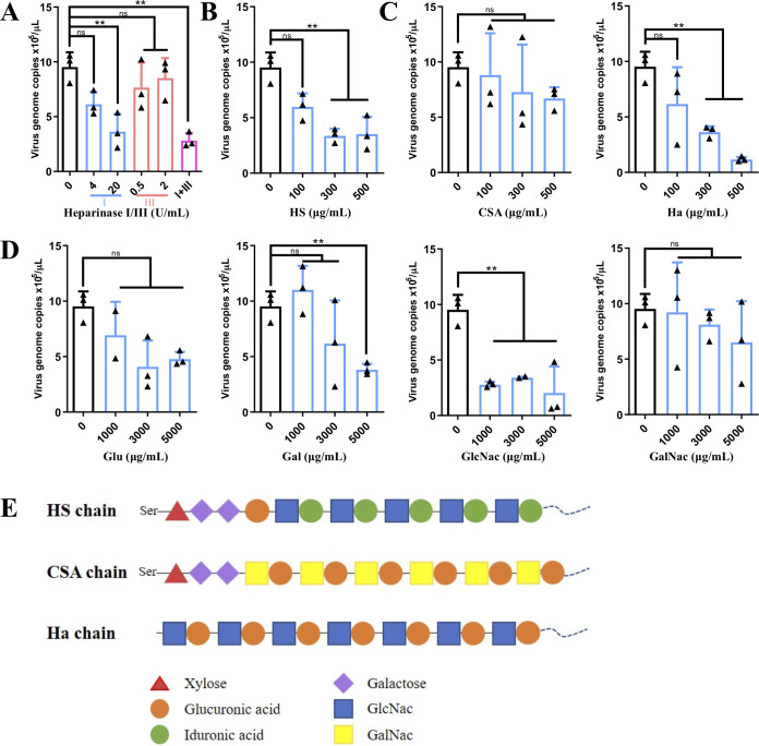
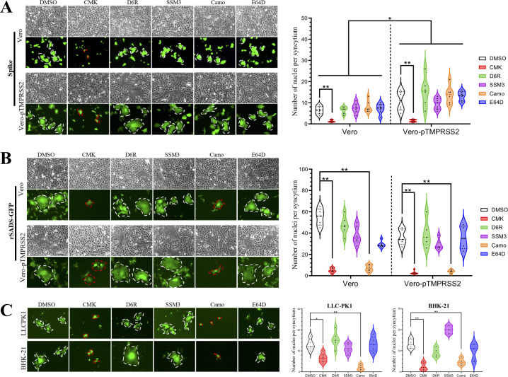
Swine acute diarrhea syndrome coronavirus (SADS-CoV) is a novel porcine enteric coronavirus, and the broad interspecies infection of SADS-CoV poses a potential threat to human health. This study provides experimental evidence to dissect the roles of distinct domains within the SADS-CoV spike S1 subunit in cellular entry. Specifically, we expressed the S1 and its subdomains, S1A and S1B. Cell binding and invasion inhibition assays revealed a preference for the S1B subdomain in binding to the receptors on the cell surface, and this unknown receptor is not utilized by the porcine epidemic diarrhea virus. Nanoparticle display demonstrated hemagglutination of erythrocytes from pigs, humans, and mice, linking the S1A subdomain to the binding of sialic acid (Sia) involved in virus attachment. We successfully rescued GFP-labeled SADS-CoV (rSADS-GFP) from a recombinant cDNA clone to track viral infection. Antisera raised against S1, S1A, or S1B contained highly potent neutralizing antibodies, with anti-S1B showing better efficiency in neutralizing rSADS-GFP infection compared to anti-S1A. Furthermore, depletion of heparan sulfate (HS) by heparinase treatment or pre-incubation of rSADS-GFP with HS or constituent monosaccharides could inhibit SADS-CoV entry. Finally, we demonstrated that active furin cleavage of S glycoprotein and the presence of type II transmembrane serine protease (TMPRSS2) are essential for SADS-CoV infection. These combined observations suggest that the wide cell tropism of SADS-CoV may be related to the distribution of Sia or HS on the cell surface, whereas the S1B contains the main protein receptor binding site. Specific host proteases also play important roles in facilitating SADS-CoV entry.IMPORTANCESwine acute diarrhea syndrome coronavirus (SADS-CoV) is a novel pathogen infecting piglet, and its unique genetic evolution characteristics and broad species tropism suggest the potential for cross-species transmission. The virus enters cells through its spike (S) glycoprotein. In this study, we identify the receptor binding domain on the C-terminal part of the S1 subunit (S1B) of SADS-CoV, whereas the sugar-binding domain located at the S1 N-terminal part of S1 (S1A). Sialic acid, heparan sulfate, and specific host proteases play essential roles in viral attachment and entry. The dissection of SADS-CoV S1 subunit's functional domains and identification of cellular entry cofactors will help to explore the receptors used by SADS-CoV, which may contribute to exploring the mechanisms behind cross-species transmission and host tropism.

Keywords: TMPRSS2; heparan sulfate; receptor-binding domain (RBD); sialic acid; spike; swine acute diarrhea syndrome coronavirus (SADS-CoV).


Conflict of interest statement

The authors declare no conflict of interest.

Figures

Fig 1
Fig 1
Binding of the SADS-CoV S protein to the host cell surface. (A) Schematic representation of the SADS-CoV S protein (drawn to scale). S1, receptor-binding subunit; S2, membrane-fusion subunit; S1-NTD, N-terminal domain of S1 (S1A); S1-CTD, C-terminal domain of S1 (S1B); FP, fusion peptide; HR1 and HR2, heptad repeats. Furin cleavage site at the boundary between the S1 and S2 subunits is 543–546 (AVRR↓). (B) Schematic of different expression peptides used in the study (upper). SADS-CoV full-length S1 subunit (residues 1–544), S1A subdomain (residues 1–257), and S1B subdomain (residues 266–401) were each expressed with a C-terminal fusion to human Fc-tag; S1B alone included an N-terminal CD5 signal peptide (pink box). Besides, lumazine synthase (LS) was constructed with N-terminal fusion to domain B of protein A (pA-LS). (lower) Protein expression was determined by SDS-PAGE (left) or western blot using an anti-Fc-tag antibody (right). (C) Structural view of the SADS-CoV S protein with S1 subunit (yellow plus blue and red), S1A domain (blue), S1B domain (red), and S2 subunit (gray). (D and E) Binding of SADS-CoV S1, S1A, or S1B subunit to the cell surface. Purified peptides were incubated with HeLa or Huh-7 cells without permeablization treatment, followed by IFA ((D); magnification = 200× ) or flow cytometry assay (E) using anti-Fc-tag antibody. Hemagglutinin (HA) from influenza virus (binds to both HeLa and Huh-7) and S1B of MERS-CoV (binds Huh-7 only) were used as controls, and S1 protein from PEDV was used as a comparison. Two doses (0.1 or 0.5 nmol) of each bind protein were used in flow cytometry binding assay. Statistical analysis between SADS-S1 and SADS-S1B, or SADS- S1A and SADS-S1B was conducted. *: P ≤ 0.05; **: P ≤ 0.01; ns: not significant.
Fig 2
Fig 2
Domain S1B of the SADS-CoV S protein may be primarily responsible for receptor binding. (A and B) Receptors on the cell surface were blocked by incubation with different concentrations of purified SADS-CoV S1-hFc, S1A-hFc, or S1B-hFc peptides (nonspecific ACE2-hFc as negative control) on Vero (A) or Huh-7 cells (B). After discarding the supernatant, cells were infected with SADS-CoV at MOI = 0.5 and stained at 16 h post-infection with anti-SADS-CoV N protein antibody. Relative infection rate was compared based on fluorescent signal in SADS-CoV-infected Vero or Huh-7 cells. (C) Vero cells were incubated with the same combination of SADS-CoV S peptides as in (A and B) and subsequently infected with PEDV-SGDU-GFP at MOI = 0.5, and GFP signal was monitored by fluorescence microscopy. The relative infection rate in Vero cells based on GFP signal. Magnification = 100× ; *: P ≤ 0.05; **: P ≤ 0.01; ***: P ≤ 0.001; ns: not significant.
Fig 3
Fig 3
Rescue of recombinant SADS-GFP from a full-length cDNA clone. (A) The SADS-CoV genome was divided into 15 overlapping amplicons, and ORF3 was replaced by GFP during assembly into a full-length cDNA clone. Since the 3′-end of ORF3 (1–690 nt) has a 29-nt overlap with the TRS-containing subgenome of the E gene, the last 51 bases of ORF3 were retained, replacing nt 1–639 with the GFP gene. A cytomegalovirus promoter (pCMV) was engineered at the 5′-end of the genomic cDNA, and a hepatitis delta virus ribozyme (HDVRz), followed by bovine growth hormone polyadenylation and termination sequences (BGH) engineered at the 3′-end. (B) Rescue of infectious virus by cotransfection of rSADS-GFP cDNA clone plasmid plus a helper plasmid expressing SADS-CoV N protein in Vero cells; GFP signal was monitored by fluorescence microscopy at 48 h post-transfection. (C) Comparison of growth kinetics between rSADS-GFP-p5 (passage 5), rSADS-GFP-p10 (passage 10), and SADS-CoV-p10 (passage 10) at MOI = 0.1. (D) Rescued rSADS-GFP virus after four continuous passages on Vero cells produced obvious syncytia. Expression of SADS-CoV M protein was confirmed using a specific antibody. (E) Determination of the stability of rSADS-GFP at p10, p15, or p20. The viral infection was examined by GFP signal and staining of anti-N antibody. The complete genome of recombinant virus was identified by RT-PCR amplification and sequencing of a total of 23 overlapping genome fragments (including the fragment #20 corresponding to the inserted GFP gene).
Fig 4
Fig 4
Hemagglutination and viral infection of SADS-CoV could be reduced with neuraminidase treatment. (A–C) SADS-CoV, PRRSV, or IAV were incubated with 0.5% washed erythrocytes from porcine (A), human (B), or mice (C) with or without neuraminidase (NA). Hemagglutination (HA) titer was scored according to the presence of agglutination in 96-well V-bottom plates. (D and E) Vero and Huh-7 cells were treated with NA in different concentrations to deplete the cell-surface sialic acids, followed by infection with rSADS-GFP at MOI = 0.1. At 24 h post-infection, GFP signal was detected (D), and infected cells were quantified (E). (F) NA treatment reduced infection by SADS-CoV or TGEV on Vero-pAPN cells. GFP expression of rSADS-GFP or IFA staining of TGEV N protein was quantified by counting a series of fields for each; magnification = 100× ; *: P ≤ 0.05; **: P ≤ 0.01; ***: P ≤ 0.001.
Fig 5
Fig 5
SADS-CoV S1-mediated Sia-binding activity resides in the S1A domain. (A) Electron microscopy image of pA-LS nanoparticles alone (left) or after incubation with SADS-CoV S1-hFc (right), which is visible as “bumps” on the surface; scale bar = 50 nm. SADS-CoV S1-hFc peptide or SADS-CoV virions (control) (B–D) or soluble S1-hFc, S1A-hFc, and S1B-hFc peptides (E–G) were incubated with 0.5% washed erythrocytes from porcine (B and E), human (C and F), or mice (D and G) with or without pA-LS and/or neuraminidase (NA). Hemagglutination (HA) titer was scored according to the presence of agglutination in 96-well V-bottom plates.
Fig 6
Fig 6
Domain S1B of the SADS-CoV S protein elicits neutralizing antibodies. Antisera against SADS-CoV S1-hFc, S1A-hFc, and S1B-hFc peptides were prepared by immunizing BALB/c mice, for comparison against anti-hFc, anti-S1-His antibodies as well as negative serum. (A) For neutralization assays, 100 TCID50 of rSADS-GFP was incubated with twofold serial dilutions of each antibody for 2 h at 37°C prior to infection of Vero cells. SADS-CoV infection was measured by GFP signal observed on a fluorescent microscope at 16 h post-infection (hpi); magnification = 100× . (B) The viral genome copy number at 16 hpi was determined by qRT-PCR in the culture supernatants from the infections in (A). Statistical analyses of neutralizing titers at each dilution between the anti-S1B and anti-S1A (blue), and between the anti-S1B and anti-S1 (green) were conducted. *: P ≤ 0.05; **: P ≤ 0.01; ns: not significant.
Fig 7
Fig 7
Heparan sulfate serves as an essential factor during SADS-CoV attachment. (A) Vero cells were pretreated with heparinase I or III at 37°C for 2 h to remove the heparan sulfate (HS) on the cell surface, and then infected with rSADS-GFP at MOI = 0.1. Alternatively, HS (B), glycosaminoglycans (C) such as chondroitin sulfate A (CSA) or hyaluronic acid (Ha), or monosaccharides (D) including galactose (Gal), glucose (Glu), N-acetyl-D-glucosamine (GlcNac), or N-acetyl-D-galactosamine (GalNac) were incubated with rSADS-GFP prior to infection of Vero cells. Supernatants were harvested at 24 h post-infection (hpi) to determine virus titer by qRT-PCR analysis targeting the N gene. **: P ≤ 0.01; ns: not significant. (E) Schematic representation of glycosaminoglycan chains of HS, CSA, and Ha consisting of disaccharide units with various monosaccharides. HS and CSA are connected to proteoglycan via a serine residue, while Ha was found as a free sugar chain in the extracellular matrix.
Fig 8
Fig 8
Furin cleavage of the SADS-CoV S protein is essential for cell-cell fusion. (A) Vero cells were co-transfected pRK5-eGFP with expression plasmids encoding wild-type (543-AVRR-546) or furin cleavage site mutants (ΔAVRR or 543-AVAA-546) of the SADS-CoV S protein or the empty pRK5 (vector), with rSADS-GFP infection (MOI = 0.5) as positive control. IFA was used to detect S proteins (red), using polyclonal antibodies against SADS-CoV. (B) (upper) The furin cleavage site of the SADS-CoV S1 subunit (543-AVRR-546) was deleted (ΔAVRR) or mutated (AVAA) and coexpressed with eGFP in Vero or Vero-pTMPRSS2 cells. Only the wild-type S protein was able to induce syncytium formation and the area range was larger in Vero-pTMPRSS2 cells. rSADS-GFP virus and pRK5 vector served as positive and negative controls, respectively. Green fluorescence allows clear observation of syncytia formation (dashed outlines). (lower) Violin plots showing number of nuclei per syncytium induced by S protein expression; each dot represents a single GFP+ syncytium. Magnification = 200× ; *: P ≤ 0.05; **: P ≤ 0.01; ns: not significant.
Fig 9
Fig 9
pTMPRSS2 is essential for SADS-CoV infection. (A) (left) Vero or Vero-pTMPRSS2 cells co-transfected with wild-type S protein (AVRR) and eGFP were pretreated with furin inhibitor CMK (50 mM), proprotein convertase inhibitor D6R (50 mM), furin inhibitor SSM3 (25 mM), TMPRSS2 inhibitor Camo (500 mM), or cathepsin inhibitor E64D (20 mM) for 12 h, and DMSO was used as the vehicle control. (right) Violin plots show the number of nuclei per syncytium during inhibitor treatment in cells expressing S protein. (B) (left) Vero or Vero-pTMPRSS2 cells infected by rSADS-GFP were pretreated with the same inhibitors as in (A). (right) Violin plots show the number of nuclei per syncytium during inhibitor treatment in cells infected with rSADS-GFP. (C) (left) LLC-PK1 or BHK-21 cells infected by rSADS-GFP were pretreated with the same inhibitors as in (A). (right) Violin plots show the number of nuclei per syncytium during inhibitor treatment in cells infected with rSADS-GFP. Magnification = 200× ; *: P ≤ 0.05; **: P ≤ 0.01; ns: not significant.

Similar articles

Cited by

References

    1. Damas J, Hughes GM, Keough KC, Painter CA, Persky NS, Corbo M, Hiller M, Koepfli KP, Pfenning AR, Zhao H, Genereux DP, Swofford R, Pollard KS, Ryder OA, Nweeia MT, Lindblad-Toh K, Teeling EC, Karlsson EK, Lewin HA. 2020. Broad host range of SARS-CoV-2 predicted by comparative and structural analysis of ACE2 in vertebrates. Proc Natl Acad Sci U S A 117:22311–22322. doi:10.1073/pnas.2010146117 - DOI - PMC - PubMed
    1. Starr TN, Greaney AJ, Hilton SK, Ellis D, Crawford KHD, Dingens AS, Navarro MJ, Bowen JE, Tortorici MA, Walls AC, King NP, Veesler D, Bloom JD. 2020. Deep mutational scanning of SARS-CoV-2 receptor binding domain reveals constraints on folding and ACE2 binding. Cell 182:1295–1310. doi:10.1016/j.cell.2020.08.012 - DOI - PMC - PubMed
    1. Li F. 2016. Structure, function, and evolution of coronavirus spike proteins. Annu Rev Virol 3:237–261. doi:10.1146/annurev-virology-110615-042301 - DOI - PMC - PubMed
    1. Forster P, Forster L, Renfrew C, Forster M. 2020. Phylogenetic network analysis of SARS-CoV-2 genomes. Proc Natl Acad Sci U S A 117:9241–9243. doi:10.1073/pnas.2004999117 - DOI - PMC - PubMed
    1. Hodcroft EB, Zuber M, Nadeau S, Vaughan TG, Crawford KHD, Althaus CL, Reichmuth ML, Bowen JE, Walls AC, Corti D, Bloom JD, Veesler D, Mateo D, Hernando A, Comas I, González-Candelas F, Stadler T, Neher RA, SeqCOVID-SPAIN consortium . 2021. Spread of a SARS-CoV-2 variant through Europe in the summer of 2020. Nature 595:707–712. doi:10.1038/s41586-021-03677-y - DOI - PubMed

Supplementary concepts

LinkOut - more resources