. 2024 Apr 10;15(4):e0030324.
doi: 10.1128/mbio.00303-24. Epub 2024 Mar 19.

Irgm proteins attenuate inflammatory disease in mouse models of genital Chlamydia infection

Affiliations

Irgm proteins attenuate inflammatory disease in mouse models of genital Chlamydia infection

Jacob Dockterman et al. mBio. .

Abstract

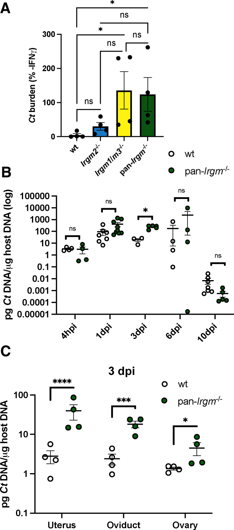
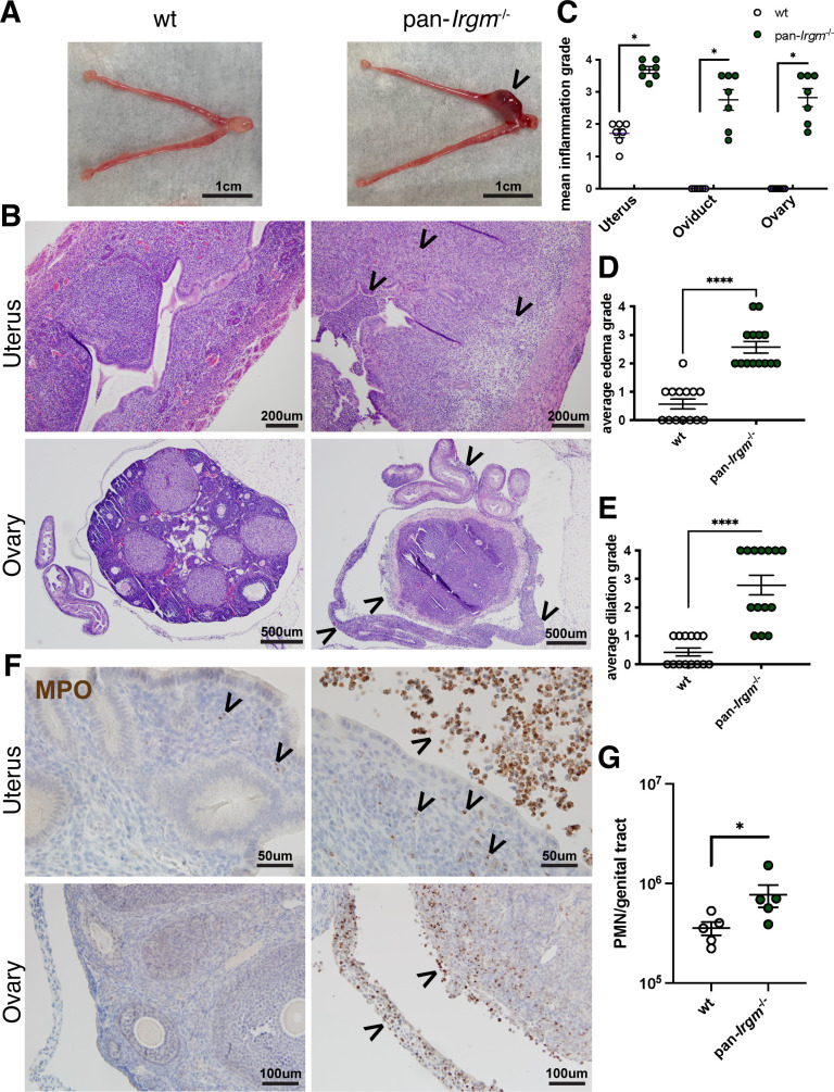
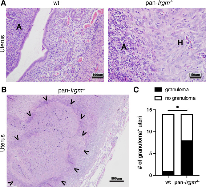
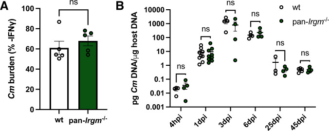
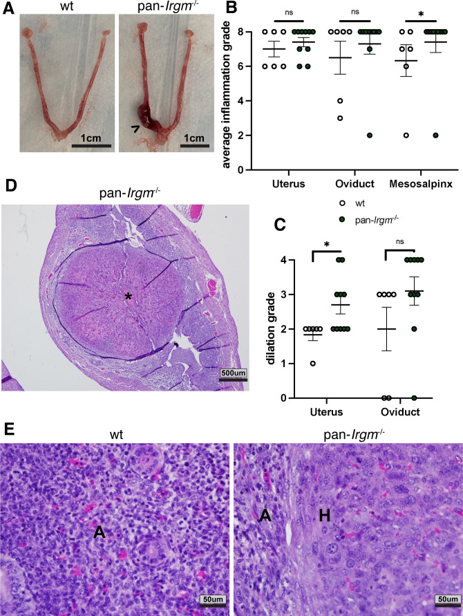
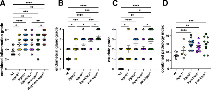
Chlamydiae are obligate intracellular bacterial pathogens that may cause genital pathology via induction of destructive host immune responses. Human-adapted Chlamydia trachomatis causes inflammatory disease in human hosts but is easily cleared in mice, and mouse-adapted Chlamydia muridarum establishes a productive and pathogenic infection in murine hosts. While numerous anti-chlamydial host resistance factors have been discovered in mice and humans alike, little is known about host factors promoting host fitness independent of host resistance. Here, we show that interferon-inducible immunity-related GTPase M (Irgm) proteins function as such host factors ameliorating infection-associated sequalae in the murine female genital tract, thus characterizing Irgm proteins as mediators of disease tolerance. Specifically, we demonstrate that mice deficient for all three murine Irgm paralogs (pan-Irgm-/-) are defective for cell-autonomous immunity to C. trachomatis, which correlates with an early and transient increase in bacterial burden and sustained hyperinflammation in vivo. In contrast, upon infection of pan-Irgm-/- mice with C. muridarum, bacterial burden is unaffected, yet genital inflammation and scarring pathology are nonetheless increased, demonstrating that Irgm proteins can promote host fitness without altering bacterial burden. Additionally, pan-Irgm-/- mice display increased granulomatous inflammation in genital Chlamydia infection, implicating Irgm proteins in the regulation of granuloma formation and maintenance. These findings demonstrate that Irgm proteins regulate pathogenic immune responses to Chlamydia infection in vivo, establishing an effective infection model to examine the immunoregulatory functions and mechanisms of Irgm proteins.

Importance: In response to genital Chlamydia infection, the immune system mounts a proinflammatory response to resist the pathogen, yet inflammation must be tightly controlled to avoid collateral damage and scarring to host genital tissue. Variation in the human IRGM gene is associated with susceptibility to autoinflammatory diseases but its role in ameliorating inflammatory diseases caused by infections is poorly defined. Here, we use mice deficient for all three murine Irgm paralogs to demonstrate that Irgm proteins not only provide host resistance to Chlamydia infections but also limit associated inflammation in the female genital tract. In particular, we find that murine Irgm expression prevents granulomatous inflammation, which parallels inflammatory diseases associated with variants in human IRGM. Our findings therefore establish genital Chlamydia infection as a useful model to study the roles for Irgm proteins in both promoting protective immunity and limiting pathogenic inflammation.

Keywords: Chlamydia; IRGM; disease tolerance; immunity-related GTPases; immunopathology; interferons; sexually transmitted diseases.


Conflict of interest statement

The authors declare no conflict of interest.

Figures

Fig 1
Fig 1
A defect in cell-autonomous immunity correlates with a transient increase in C. trachomatis burden in the female genital tract of pan-Irgm−/− mice. (A) MEFs were primed overnight with 100 U/mL IFNγ prior to infection with C. trachomatis elementary bodies (EBs) at a multiplicity of infection (MOI) of 1:1. DNA was harvested from the infected cells at 24 hpi and bacterial burden was quantified using quantitative real-time polymerase chain reaction (qPCR). Graphs represent the proportion of bacterial burden in IFNγ-primed vs. unprimed cells. Data points represent MEFs derived from separate embryos (n = 4 separate MEF lines). Statistical significance was determined using one-way analysis of variance (ANOVA). (B) Mice were infected transcervically with 5 × 10⁶ C. trachomatis EBs, and female genital tracts were harvested at the indicated timepoints or (C) harvested at 3 dpi and then segmented. DNA was extracted from the harvested organ, and bacterial burden was quantified using qPCR. Each data point represents one mouse. Data in B represent pooled data from one experiment including all timepoints and one additional experiment for 3 dpi and 10 dpi timepoints: 4 hpi n = 4 mice; 1 dpi wild-type n = 7, pan-Irgm−/− n = 9; 3 dpi wild-type n = 3, pan-Irgm−/− n = 5; 6 dpi n = 4; and 10 dpi wild-type n = 7, pan-Irgm−/− n = 5. Data in C represent one experiment (n = 4 mice). Statistical significance was determined using t-test (B, C); *P < 0.05, ***P < 0.0005, and ****P < 0.00005; ns, not significant.
Fig 2
Fig 2
Pan-Irgm−/− mice display increased inflammation in genital C. trachomatis infection. (A–G) Mice were infected transcervically with 5 × 10⁶ C. trachomatis EBs, and the female genital tract was harvested at 6 dpi. (A) Gross pathology—arrowhead indicates area of gross inflammation. (B) Genital tracts were fixed, sectioned, and H&E stained. Mildly inflamed wild-type uterine tissue and severely inflamed and edematous pan-Irgm−/− uterus; wild-type ovary and oviduct with no evidence of inflammation and pan-Irgm−/− ovary, oviduct, and mesosalpinx each displaying severe inflammation—arrowheads indicate regions of inflammatory infiltrate or edema. The magnitude of acute and chronic inflammation (C), edema (D), and dilation (E) was graded by a veterinary pathologist (n = 7 mice; each data point represents one uterine horn of an infected mouse). (F) Immunohistochemistry was performed on fixed genital tracts infected with C. trachomatis to label the neutrophil marker myeloperoxidase (MPO—brown). Arrowheads indicate regions or punctae of neutrophilic infiltrate. (G) Flow cytometry on genital tracts infected with C. trachomatis to quantify neutrophils (CD45+Gr1+ live cells; n = 5 mice; each data point represents one entire reproductive tract of an infected mouse). Data represent one experiment that is representative of multiple replicates. Statistical significance was determined using Mann-Whitney test (C–E) or t-test (G); *P < 0.05 and ****P < 0.00005.
Fig 3
Fig 3
Pan-Irgm−/− mice display granulomatous inflammation in genital C. trachomatis infection. Mice were infected transcervically with 5 × 10⁶ C. trachomatis EBs and harvested at 6 dpi. (A) High magnification of mild acute inflammation in wild-type uterus and acute and histiocytic inflammation in pan-Irgm−/− uterus. A, acute inflammation; H, histiocytic inflammation. (B) Low magnification of diffuse granulomatous inflammation in pan-Irgm−/− uterus. Arrowheads indicate margins of granulomatous lesion. (C) Quantification of number of uterine horns displaying granulomatous lesions (n = 7 mice). Data represent one experiment that is representative of multiple replicates. Statistical significance was calculated using χ2 test; *P < 0.05.
Fig 4
Fig 4
Host resistance to C. muridarum remains unchanged in the absence of all three murine Irgm paralogs. (A) MEFs were primed overnight with IFNγ prior to infection with C. muridarum EBs at an MOI of 1:1. DNA was harvested at 24 hpi, and bacterial burden was quantified using qPCR. Graphs represent the proportion of bacterial burden in IFNγ-primed vs. unprimed cells. Data points represent distinct MEF lines derived from separate embryos (n = 5 separate MEF lines). (B) Mice were infected transcervically with 2.5 × 10⁵ C. muridarum EBs, and genital tracts were harvested at the indicated timepoints. DNA was extracted and bacterial burden was quantified using qPCR. Data represent pooled data from one experiment including all timepoints plus one additional experiment for the 1-dpi timepoint: 4 hpi n = 4 mice; 1 dpi n = 9; 3 dpi n = 4; 6 dpi n = 4; 25 dpi wild-type n = 3, pan-Irgm−/− n = 4; and 45 dpi n = 5. Statistical significance was determined using t-test; ns, not significant.
Fig 5
Fig 5
Increased inflammation in pan-Irgm−/− at 6 dpi in genital C. muridarum infection. Mice were infected transcervically with 2.5 × 10⁵ C. muridarum EBs and harvested at 6 dpi (A–E). (A) Gross pathology of infected genital tracts. Arrowhead denotes region of gross inflammation. (B–E) Genital tracts were fixed, sectioned, and H&E stained. The magnitude of acute and chronic inflammation (B) and dilation (C) was graded by a veterinary pathologist. (D) Low magnification of granulomatous lesion (*) in pan-Irgm−/− uterus. (E) High magnification of acute inflammation in wild-type uterus and acute and histiocytic inflammation in pan-Irgm−/− uterus. A, acute inflammation; H, histiocytic inflammation. Data represent one experiment that is representative of multiple replicates. Wild-type n = 6 mice, pan-Irgm−/− n = 10; each data point represents one uterine horn of an infected mouse. Statistical significance was determined using Mann-Whitney test; *P < 0.05, ns = not significant.
Fig 6
Fig 6
Increased pathology in pan-Irgm−/− mice at later timepoints in genital C. muridarum infection. Mice were infected transcervically with 2.5 × 10⁵ C. muridarum EBs and harvested at 25 dpi or 45 dpi. (A) Gross pathology of infected genital tracts. Scale bars = 0.5 in; arrowheads indicate hydrosalpinx. (B–D) Genital tracts were fixed, sectioned, and H&E stained. (B) The magnitude of acute, chronic, and total acute + chronic inflammation at 45 dpi was graded by a veterinary pathologist (wild-type n = 7 mice, pan-Irgm−/− n = 8). Data represent one experiment that is representative of multiple replicates. (C) Low-magnification images of cystic changes in wild-type uterus and ovary/oviduct and continued inflammation in pan-Irgm−/− uterus and ovary/oviduct. (D) Quantification of hydrosalpinx at 25 dpi and 45 dpi in genital tracts infected with C. muridarum. Data represent pooled data from three experiments (25 dpi n = 12 mice; 45 dpi wild-type n = 21, pan-Irgm−/− n = 22). Each data point represent one uterine horn of an infected mouse. Statistical significance was determined using Mann-Whitney tests (B) or χ2 test (D); *P < 0.05 and **P < 0.005; ns, not significant.
Fig 7
Fig 7
Increased inflammatory pathology in Irgm- and Rag1-deficient uteri in genital C. muridarum infection at 45 dpi. Mice were infected transcervically with 2.5 × 10⁵ C. muridarum EBs. Genital tracts were harvested at 45 dpi, fixed, sectioned, H&E stained, and graded by a veterinary pathologist. (A) The magnitude of acute and chronic inflammation in the uteri as defined by standard scoring criteria was combined. Additional features such as endometrial glandular pathology (B) and uterine luminal exudates (C) were also included. (A –C) Each data point represents one uterine horn of an infected mouse. (D) A combined uterine pathology index consisting of the sum of acute inflammation, chronic inflammation, dilation, glandular pathology, luminal exudates, and granulomatous inflammation was calculated. Each data point represents combined pathology scores from one entire reproductive tract from an infected mouse. Figure represents pooled data from two independent experiments (wild-type n = 7 mice, Irgm2−/− n = 5, Irgm3−/− n = 12, Irgm1/m3−/− n = 12, pan-Irgm−/− n = 10, and Rag1/pan-Irgm−/− n = 9). Statistical significance was determined using Mann-Whitney tests; *P < 0.05, **P < 0.005, ***P < 0.0005, and ****P < 0.00005.

Similar articles

Cited by

References

    1. Brunham RC, Paavonen J. 2020. Reproductive system infections in women: upper genital tract, fetal, neonatal and infant syndromes. Pathog Dis 78:ftaa023. doi:10.1093/femspd/ftaa023 - DOI - PubMed
    1. Taylor HR, Burton MJ, Haddad D, West S, Wright H. 2014. Trachoma. Lancet 384:2142–2152. doi:10.1016/S0140-6736(13)62182-0 - DOI - PubMed
    1. Mabey D, Peeling RW. 2002. Lymphogranuloma venereum. Sex Transm Infect 78:90–92. doi:10.1136/sti.78.2.90 - DOI - PMC - PubMed
    1. Braxton JDD, Emerson B. 2018. Sexually transmitted surveillance 2017. Centers for disease control and prevention
    1. McCarville JL, Ayres JS. 2018. Disease tolerance: concept and mechanisms. Curr Opin Immunol 50:88–93. doi:10.1016/j.coi.2017.12.003 - DOI - PMC - PubMed

Substances