Review
. 2024 May;11(20):e2306035.
doi: 10.1002/advs.202306035. Epub 2024 Mar 19.

Layered Double Hydroxides: Recent Progress and Promising Perspectives Toward Biomedical Applications

Affiliations
Review

Layered Double Hydroxides: Recent Progress and Promising Perspectives Toward Biomedical Applications

Lei Li et al. Adv Sci (Weinh). 2024 May.

Abstract

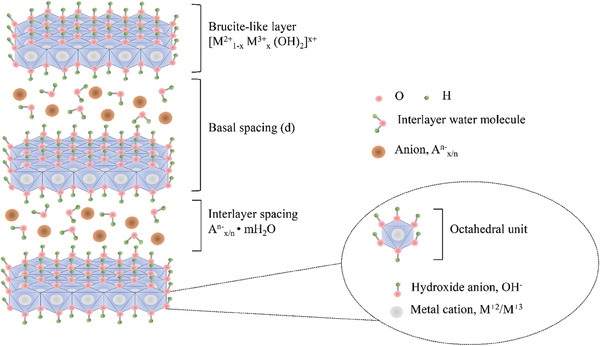
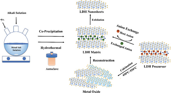
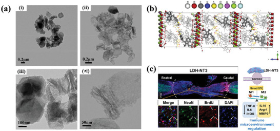
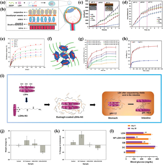
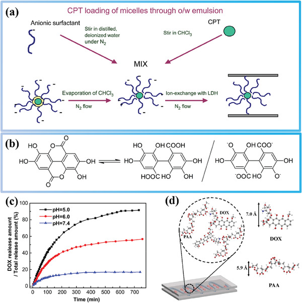
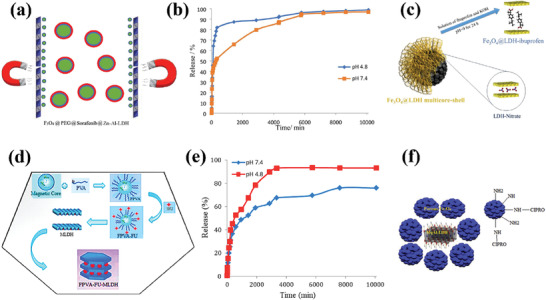
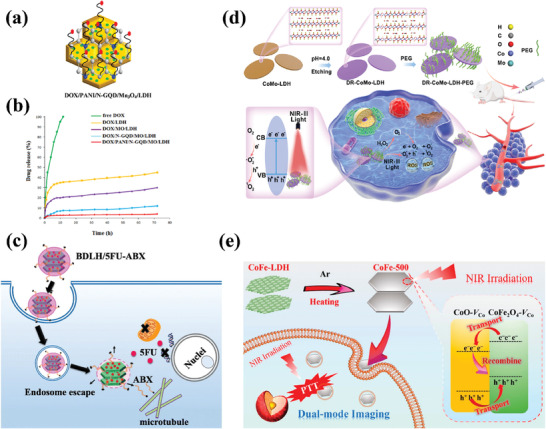
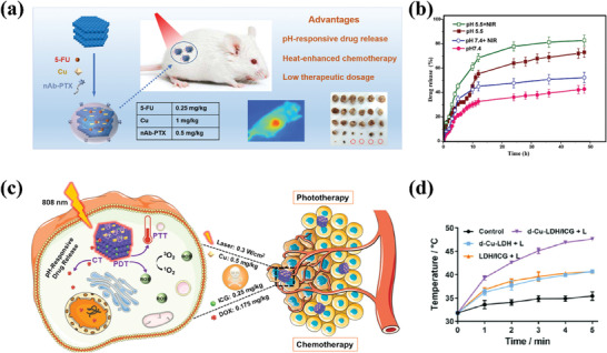
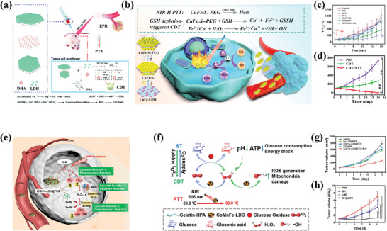
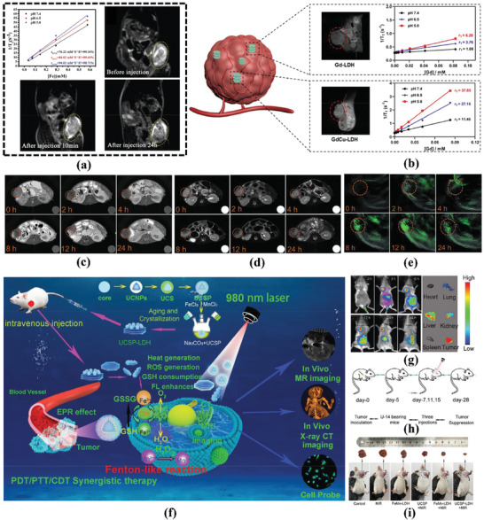
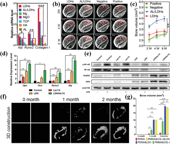
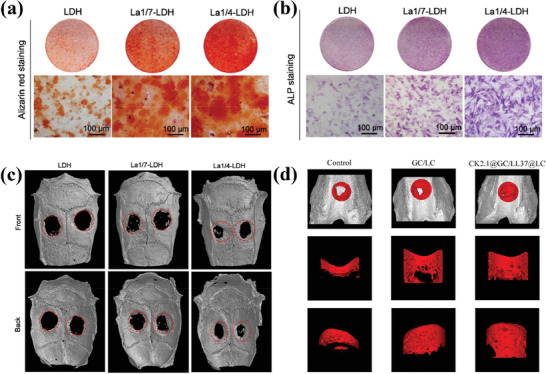
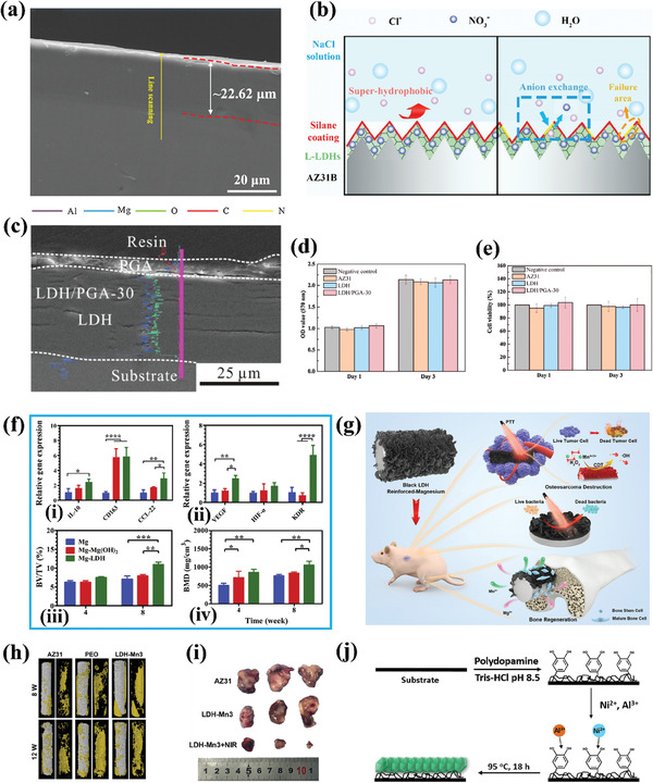
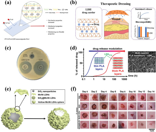
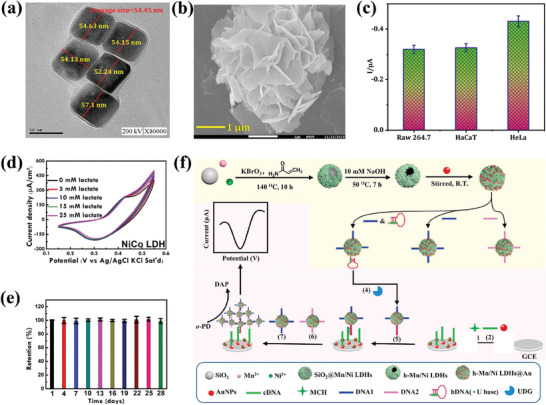
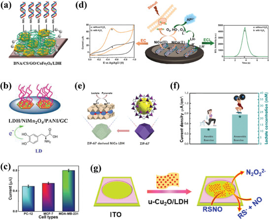
Layered double hydroxides (LDHs) have been widely studied for biomedical applications due to their excellent properties, such as good biocompatibility, degradability, interlayer ion exchangeability, high loading capacity, pH-responsive release, and large specific surface area. Furthermore, the flexibility in the structural composition and ease of surface modification of LDHs makes it possible to develop specifically functionalized LDHs to meet the needs of different applications. In this review, the recent advances of LDHs for biomedical applications, which include LDH-based drug delivery systems, LDHs for cancer diagnosis and therapy, tissue engineering, coatings, functional membranes, and biosensors, are comprehensively discussed. From these various biomedical research fields, it can be seen that there is great potential and possibility for the use of LDHs in biomedical applications. However, at the same time, it must be recognized that the actual clinical translation of LDHs is still very limited. Therefore, the current limitations of related research on LDHs are discussed by combining limited examples of actual clinical translation with requirements for clinical translation of biomaterials. Finally, an outlook on future research related to LDHs is provided.

Keywords: biomedical application; drug delivery; exchangeability; layered double hydroxides; nanotheranostics.


Conflict of interest statement

P.v.R also is a co‐founder, scientific advisor, and shareholder in BiomACS BV, a biomedical‐oriented screening company. The authors declare no other conflict of interest. The authors declare no competing financial interest.

Figures

Figure 1
Figure 1
Schematic diagram of LDHs structure.
Figure 2
Figure 2
Schematic diagram of common synthetic methods for LDHs.
Figure 3
Figure 3
a) Morphologies under TEM of MgAl‐NO3 L‐LDH‐MO after released in (i) PBS pH6.5, and (ii) PBS pH 5.2. Layered structures of MgAl‐NO3 L‐LDH‐MO under TEM after being released in (iii) PBS pH 6.5, and (iv) PBS pH 5.2. Reprinted with permission from Ref.[99] Copyright 2023, Wiley‐VCH. b) Final model of LDH‐Sulindac after molecular dynamics production run, view along a‐axis. Reprinted with permission from Ref.[101] Copyright 2020, Elsevier. c) Immunomodulatory layered double hydroxide nanoparticles enable neurogenesis by targeting transforming growth factor‐β receptor 2. Reprinted with permission from Ref.[102] Copyright 2021, American Chemical Society.
Figure 4
Figure 4
a) Schematic design of multifunctional CMCG‐GS‐DEXP‐LDH. b) The scheme of drug diffusion routes into the retina for the CMCG‐GS‐DEXP‐LDH hybrid nanocomposite eye drop. Reprinted with the permission from Ref.[104] Copyright 2020, Elsevier. c) Swelling profile of CMC/LDH(Cu/Al) bio‐nanocomposite hydrogel beads with different content of LDH in simulated gastrointestinal tract conditions and a digital photo of hydrogel bead in the dry and swelled state. d) AMX release behavior of CMC‐based hydrogel beads with different content of LDH(Cu/Al) wt.% in the simulated conditions with gastrointestinal tract passage (time and pH). Reprinted with the permission from Ref.[105] Copyright 2020, Elsevier. e) Release kinetics of diclofenac from Alg/LDH‐Dic beads at different pHs of release medium. Reprinted with the permission from Ref.[111] Copyright 2021, Elsevier. f) Schematic of DOX‐Cu MOF‐LDH. g) In vitro release profile of DOX from DOX‐Cu MOF‐LDH and Alg‐DOX‐Cu MOF‐LDH beads at different pH values (pH 5, 6.8, and 7.4). h) Swelling ratio of Alg‐DOX‐Cu MOF‐LDH at both pH 5 and 7.4. Reprinted with the permission from Ref.[112] Copyright 2023, Elsevier. i) Schematic illustrating the fabrication process of surface functionalized LDHs, and their substantial bile acid sequestering ability in the intestinal region. j) Changes in the weight of mice after 4 weeks of treatment with various tablet formulations of LDHs. k) Change in the cholesterol levels in mice after 4 weeks of treatment with various tablet formulations. Reprinted with the permission from Ref.[113] Copyright 2020, Elsevier. l) Blood glucose values of normoglycemic rats (N), diabetic control (DC), diabetic orally treated with GB (5 mg kg−1), NP LDH+GB, and LDH after 28 days of treatment. Reprinted with the permission from Ref.[114] Copyright 2023, Elsevier.
Figure 5
Figure 5
a) Schematic preparation process of nanobiohybrids as delivery vehicles for camptothecin (CPT). Reprinted with the permission from Ref.[115] Copyright 2004, Elsevier. b) Chemical equilibrium between EA and its opened form, 4,4′,5,5′,6,6′‐hexahydroxydiphenic acid, and its potential intercalated form. Reprinted with the permission from Ref.[117] Copyright 2020, MDPI. c) In vitro release profiles of LDH‐Gd/Au‐DOX under different pH values. Reprinted with the permission from Ref.[78] Copyright 2013, Elsevier. d) Schematic of the structure of DOX‐PAA‐LDH hybrids. Reprinted with the permission from Ref.[118] Copyright 2021, Elsevier.
Figure 6
Figure 6
a) Schematic the design of Fe3O4@PEG@Sorafenib@Zn‐Al‐LDH. Reprinted with the permission from Ref.[128] Copyright 2020, MDPI. b) The sorafenib release profiles from MPVASO‐ZLDH in phosphate‐buffered solutions at pH 4.8 and 7.4. Reprinted with the permission from Ref.[129] Copyright 2021, Elsevier. c) Schematic representation of the preparation of Fe3O4@LDH multicore–shell nanostructure before and after the intercalation of ibuprofen. Reprinted with the permission from Ref.[130] Copyright 2020, Springer. d) Schematic representation of the core–shell structure of magnetic FPVA‐FU‐MLDH. e) The cumulative release profiles of 5‐fluorouracil from its FPVA‐FU‐MLDH nanoparticles in phosphate‐buffered solution at pH 4.8 and pH 7.4. Reprinted with the permission from Ref.[131] Copyright 2019, MDPI. f) Schematic the design of MgAl‐LDH@PMN‐NH2. Reprinted with the permission from Ref.[132] Copyright 2020, Elsevier.
Figure 7
Figure 7
a) Schematic design of DOX/PANI/N‐GQD/Mn3O4/LDH. b) DOX release from the samples under different simulated physiological conditions of pH 7.4. Reprinted with the permission from Ref.[121] Copyright 2020, Elsevier. c) Schematic illustration of preparation and co‐delivery of 5FU‐ABX‐loaded albumin‐stabilized LDH nanoparticles (BLDH/5FU‐ABX) for colorectal cancer treatment. Reprinted with the permission from Ref.[136] Copyright 2021, Elsevier. d) Schematic illustration of the defect engineering of CoMo‐LDH nanosheets, surface modification with PEG, and its application in NIR‐III PDT. Reprinted with the permission from Ref.[149] Copyright 2020, American Chemical Society. e) Schematic Illustration of the Synthesis of CoFe‐500 from CoFe‐LDH, along with the Mode of Action of CoFe‐500 for NIR Light‐Driven PTT Guided by PA/MR/NIR Imaging. Reprinted with the permission from Ref.[151] Copyright 2022, Nature.
Figure 8
Figure 8
a) Schematic design of 5FU/Cu‐LDH@nAb‐PTX for synergistical chemo‐photo‐therapy. Reprinted with the permission from Ref.[137] Copyright 2021, Elsevier. b) Cumulative DOX release profile at pHs 5.5 and 7.4 in the absence and presence of NIR radiation. Reprinted with the permission from Ref.[135] Copyright 2020, Elsevier. c) Schematic Illustration of Multifunctional ICG/Cu‐LDH@BSA–DOX Nanomedicine. Reprinted with the permission from Ref.[120] Copyright 2021, American Chemical Society. d) Skin temperature profiles of tumor‐bearing mice in different groups under 808 nm laser irradiation (+L) at the power density of 0.23 W cm−2 (n  =  3). Reprinted with the permission from Ref.[143] Copyright 2020, Wiley.
Figure 9
Figure 9
a) Schematic illustration of the DHA delivery system of MnMgFe‐LDH toward efficient loading and mechanism of combined CDT/PTT treatment. Reprinted with the permission from Ref.[138] Copyright 2020, RSC. b) Schematic illustration of the preparation of CuFe2S3‐PEG NSs for efficient CDT/PTT. Reprinted with the permission from Ref.[144] Copyright 2021, Elsevier. c) Relative tumor volume of mice from the different groups treated under different conditions. Reprinted with the permission from Ref.[138] Copyright 2020, RSC. d) Tumor growth curves of mice with various treatments. Reprinted with the permission from Ref.[144] Copyright 2021, Elsevier. e) Scheme of ICG/Fe‐LDH@PEG mediated catalytic cascade reactions for self‐supplied H2O2 enhanced CDT. Reprinted with the permission from Ref.[145] Copyright 2022, Elsevier. f) Schematic illustration of the CoMnFe‐LDO composite hydrogel for synergetic tumor chemodynamic/starvation/photothermal therapy. Reprinted with the permission from Ref.[146] Copyright 2022, Elsevier. g) Tumor volume change of 4T1 tumor‐bearing mice after different treatments. Reprinted with the permission from Ref.[145] Copyright 2022, Elsevier. h) Tumor volume of tumor‐bearing mice treated with different groups under NIR irradiation. Reprinted with the permission from Ref.[146] Copyright 2022, Elsevier.
Figure 10
Figure 10
a) Enhanced relaxivity values after incubation with buffer solutions with different pH values at 37 °C for 24 h and MRI images of the 4T1 tumor model after the injection of Fe200‐LDH NPs (C(Fe2+) = 2.0 mmol−1) solution. Reprinted with the permission from Ref.[122] Copyright 2021, RSC. b) Schematic illustration of the in vivo MRI performance of Gd‐LDH and GdCu‐LDH nanoparticles. Reprinted with the permission from Ref.[173] Copyright 2023, RSC. c) In vivo T1‐weighted MR images and d) T2‐weighted MR images of the tumor‐bearing mice at various time points after i.v. injection of Ce6/CoMn‐LDH nanosheets (tumors are indicated by orange circles). e) PA imaging of the tumor‐bearing mice at various time points after i.v. injection of Ce6/CoMn‐LDH nanosheets. Reprinted with the permission from Ref.[150] Copyright 2020, RSC. f) Schematic illustration for the formation and transport of UCSP‐LDH in blood vessels, EPR‐mediated tumor accumulation, proposed PDT/PTT/CDT mechanism, and multiple imaging functions. g) In vivo fluorescence images of U14‐tumor‐bearing female mice taken after i.v. injection of the UCSP‐LDH nanocatalysts, in vitro fluorescence images of major organs and tumors at 12 h intervals post‐injection with the UCSP‐LDH nanocatalysts. h) Experimentation for the therapeutic model. i) Representative digital photographs of excised tumors in various treating groups of tumor‐bearing mice after 14 days of treatment. Reprinted with the permission from Ref.[147] Copyright 2020, Wiley.
Figure 11
Figure 11
a) Relative gene expression of MC3T3‐E1 cells for osteogenic markers (ALP, Runx2, and Collagen I) on day 7. b,c) Representative cross‐sectional views and quantitative analysis of regenerated bone in the femoral head. The scale bar is 5 mm (b). Reprinted with the permission from Ref.[218] Copyright 2020, RSC. d) Relative mRNA expression levels of osteogenic genes Opn, Ocn, Runx2, and ALP after treatment with let‐7d, LDH, or LDH/let‐7d for 7 days. e) Western blot analysis of RUNX2, OPN, and NF‐κB signaling. Reprinted with the permission from Ref.[219] Copyright 2021, American Chemical Society. f,g) 3D construction of newly formed bone and quantitative analysis. Note: The 3D reconstructed CT images, from left to right and from top to bottom, correspond to the PMMA group, PMMA&COL‐I&LDH group, PMMA&LDH group, and PMMA&COL‐I group, respectively. Reprinted with the permission from Ref.[220] Copyright 2021, American Chemical Society.
Figure 12
Figure 12
a) Alizarin red staining images and b) ALP staining images of the rBMSCs‐OVX as‐treated with La‐LDH and LDH scaffolds at days 21 and 7, respectively. c) Micro‐CT images of OVX‐rat cranial defects implanted with LDH, La1/7‐LDH, and La1/4‐LDH scaffolds at 12 weeks of post‐implantation. Reprinted with the permission from Ref.[222] Copyright 2021, PMC. d) Micro‐CT reconstruction of repaired tissue of osteochondral defect in vivo. Reprinted with the permission from Ref.[223] Copyright 2021, Elsevier.
Figure 13
Figure 13
a) Cross‐sectional morphologies of MgAl‐SDS‐LDHs film. Reprinted with the permission from Ref.[248] Copyright 2021, Elsevier. b) Anti‐corrosion mechanism of the composite (L‐LDHs‐OTS) coating. Reprinted with the permission from Ref.[249] Copyright 2021, American Chemical Society. c) Cross‐sectional morphologies and corresponding line scan images (inset) LDH/PGA‐30 coating. d) OD values and e) Cell viability of NIH 3T3 cultured in different extracts prepared with negative control, AZ31 substrate, LDH coated sample, and, LDH/PGA‐30 coated sample for 1 and 3 days. Reprinted with the permission from Ref.[250] Copyright 2020, Elsevier. f) (i)Expression of M2‐related genes of RAW264.7 cultured in the extracts for 3 days. (ii) Angiogenesis‐related gene expression of HUVECs after being cultured in MCM. Calculated bone volume/tissue volume (BV/TV) (iii) and trabecular bone mineral density (BMD) (iv). Reprinted with the permission from Ref.[255] Copyright 2021, Elsevier. g) Stepwise therapeutic strategy for osteosarcoma destruction, antibacterial, and followed bone regeneration. h) 3D reconstruction images of Micro‐CT results of the various samples after implantation in the femur of SD rat for 8 and 12 weeks, yellow section indicates new bone. i) Time‐dependent tumor‐growth curves of the mice after different treatments. Reprinted with the permission from Ref.[251] Copyright 2022, Elsevier. j) Scheme of preparation of NiAl‐LDHs film using PD as a platform. Reprinted with the permission from Ref.[257] Copyright 2017, Elsevier.
Figure 14
Figure 14
a) Schematic of PVA/PAA/LDH nanocomposite film. Reprinted with permission from Ref.[258] Copyright 2022, Elsevier. b) Schematic illustration of polymer/iron‐based LDH as a wound dressing. Reprinted with the permission from Ref.[261] Copyright 2020, MDPI. c) Photograph of in vitro antimicrobial activity evaluated by Agar disk diffusion assay corresponding to HS, Cip/HS, and (LDH‐Cip)/HS films (6 mm diameter) against Staphylococcus aureus. The dotted line indicates the total area of the swollen films. Reprinted with the permission from Ref.[262] Copyright 2020, Elsevier. d) Schematic tunable of PLA/LDH film with adjustable drug release rate prepared by electrospinning and electrospray technique. Reprinted with the permission from Ref.[263] Copyright 2020, Elsevier. e) The synthetic scheme of the hollow Mn/Ni LDHs structure. f) Photographs of different samples in the mice wound healing model with infection of S. aureus after different times. Reprinted with the permission from Ref.[275] Copyright 2020, Wiley.
Figure 15
Figure 15
a) TEM images FeMn LDHs‐12h. Reprinted with the permission from Ref.[294] Copyright 2020, RSC. b) FESEM images of NiMn LDH‐180 °C. c) Comparisons of current responses of NiMn LDH toward H2O2 secreted from Raw 264.7, HaCaT, and HeLa cells. Reprinted with the permission from Ref.[292] Copyright 2021, Elsevier. d) Cyclic voltammograms of the NiCo LDH modified SPE at a scan rate of 50 mV s−1 in 0.1 m NaOH with various lactate concentrations (0–25 mm). e) Amperometric response of NiCo LDH modified electrode toward 10 mm lactate for 28 days. Reprinted with the permission from Ref.[289] Copyright 2021, Elsevier. f) Schematic illustration of the proposed signal‐enhanced electrochemical biosensor for UDG detection based on h‐MnNi LDHs. Reprinted with the permission from Ref.[37] Copyright 2021, Elsevier.
Figure 16
Figure 16
a) Schematic the structure of DNA/GO/CoFe2O4/ZnAl‐LDH/FTO bioelectrode. Reprinted with the permission from Ref.[299] Copyright 2020, Elsevier. b) Schematic the structure of LDH/NiMn2O4/PANI/GC bioelectrode. Reprinted with the permission from Ref.[296] Copyright 2020, Elsevier. c) Amperometric current responses of CNTs@NiAl LDH modified electrode sensor for different cell lines. Reprinted with the permission from Ref.[295] Copyright 2022, Elsevier. d) Illustration for the Dual‐Mode Mechanism of Peroxidase‐Like N‐Doped Carbon Nanotubes Loaded with NiCo‐Layered Double‐Hydroxide Nanoflowers for H2O2 Determination. Reprinted with the permission from Ref.[293] Copyright 2022, American Chemistry Society. e) Schematic illustration of ZIF‐67 derived NiCo LDH for lactate detection. f) Amperometric response of ZIF‐67 derived NiCo LDH modified electrode sensor at applied voltage 0.55 V for different concentrations of lactate. Reprinted with the permission from Ref.[290] Copyright 2022, Elsevier. g) Schematic Illustration of electrochemical detection strategy of synthesized u‐Cu2O/LDH for RSNO. Reprinted with the permission from Ref.[297] Copyright 2022, Elsevier.

Similar articles

Cited by

References

    1. Murali A., Lokhande G., Deo K. A., Brokesh A., Gaharwar A. K., Mater. Today 2021, 50, 276. - PMC - PubMed
    1. Hu T., Mei X., Wang Y., Weng X., Liang R., Wei M., Sci. Bull. 2019, 64, 1707. - PubMed
    1. Chimene D., Alge D. L., Gaharwar A. K., Adv. Mater. 2015, 27, 7261. - PubMed
    1. Hochstetter C., J. Für Prakt. Chemie 1842, 27, 375.
    1. Mandarino J. A., Mineral. Rec. 1998, 29, 227.

MeSH terms

LinkOut - more resources