. 2024 Apr 10;15(4):e0041924.
doi: 10.1128/mbio.00419-24. Epub 2024 Mar 19.

Structural insight into Escherichia coli CsgA amyloid fibril assembly

Affiliations

Structural insight into Escherichia coli CsgA amyloid fibril assembly

Fan Bu et al. mBio. .

Abstract

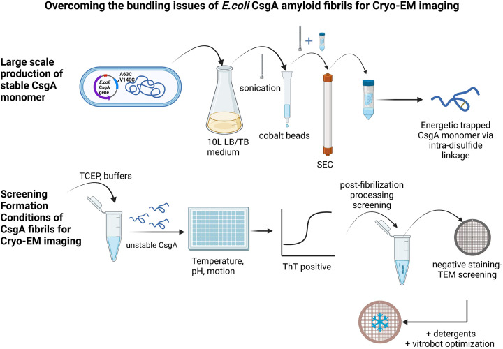
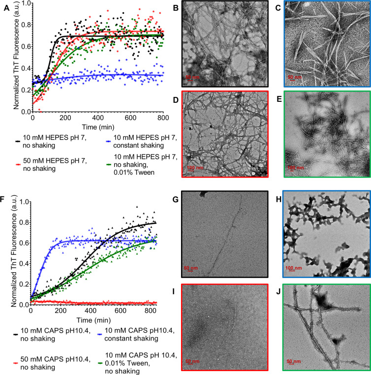
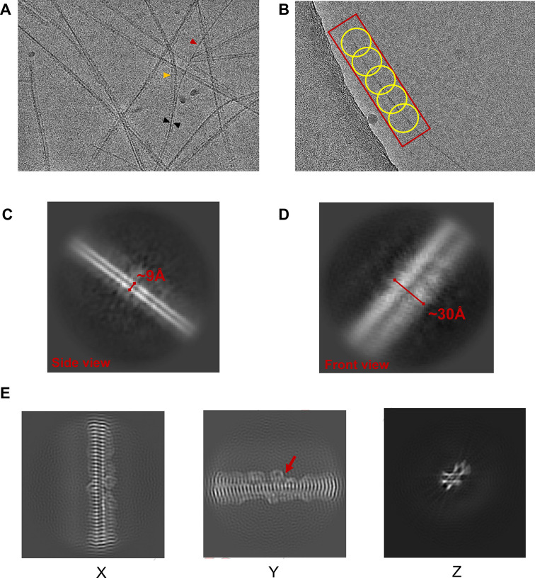
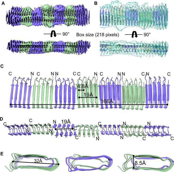
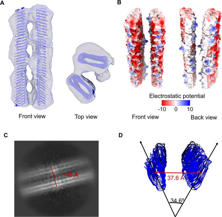
The discovery of functional amyloids in bacteria dates back several decades, and our understanding of the Escherichia coli curli biogenesis system has gradually expanded over time. However, due to its high aggregation propensity and intrinsically disordered nature, CsgA, the main structural component of curli fibrils, has eluded comprehensive structural characterization. Recent advancements in cryo-electron microscopy (cryo-EM) offer a promising tool to achieve high-resolution structural insights into E. coli CsgA fibrils. In this study, we outline an approach to addressing the colloidal instability challenges associated with CsgA, achieved through engineering and electrostatic repulsion. Then, we present the cryo-EM structure of CsgA fibrils at 3.62 Å resolution. This structure provides new insights into the cross-β structure of E. coli CsgA. Additionally, our study identifies two distinct spatial arrangements within several CsgA fibrils, a 2-CsgA-fibril pair and a 3-CsgA-fibril bundle, shedding light on the intricate hierarchy of the biofilm extracellular matrix and laying the foundation for precise manipulation of CsgA-derived biomaterials.IMPORTANCEThe visualization of the architecture of Escherichia coli CsgA amyloid fibril has been a longstanding research question, for which a high-resolution structure is still unavailable. CsgA serves as a major subunit of curli, the primary component of the extracellular matrix generated by bacteria. The support provided by this extracellular matrix enables bacterial biofilms to resist antibiotic treatment, significantly impacting human health. CsgA has been identified in members of Enterobacteriaceae, with pathogenic E. coli being the most well-known model system. Our novel insights into the structure of E. coli CsgA protofilaments form the basis for drug design targeting diseases associated with biofilms. Additionally, CsgA is widely researched in biomaterials due to its self-assembly characteristics. The resolved spatial arrangements of CsgA amyloids revealed in our study will further enhance the precision design of functional biomaterials. Therefore, our study uniquely contributes to the understanding of CsgA amyloids for both microbiology and material science.

Keywords: bacteria; biofilms; curli; electron microscopy; functional amyloids; protein aggregation; protein structure-function.


Conflict of interest statement

The authors declare no conflict of interest.

Figures

Fig 1
Fig 1
Schematic of how to overcome the bundling issues of E. coli CsgA fibrils for cryo-EM data collection. The optimization process involved adjusting the medium type, induction temperature, and induction time to achieve the highest yield of CsgA monomer. Then, various conditions during fibrillation (pH, salt, agitation, and additives) and de-bundling methods (heat, detergent, sonication, and buffer exchange) after assembly were tested to obtain de-bundled CsgA fibrils suitable for cryo-EM imaging. Lastly, data processing procedures were fine-tuned to generate good 2D classes and 3D density maps.
Fig 2
Fig 2
Effect of different growth conditions on CsgA fibrillation behavior. (A) The aggregation kinetics of CsgA in HEPES buffer were monitored by ThT fluorescence. The morphology of CsgA fibrils was characterized by negative-staining TEM. (B) 20 µM CsgA in 10 mM HEPES, pH 7, without shaking. (C) 20 µM CsgA in 10 mM HEPES, pH 7, with 200 rpm constant shaking. (D) 20 µM CsgA in 50 mM HEPES, pH 7, without shaking. (E) 20 µM CsgA in 10 mM HEPES, pH 7, without shaking + 0.01% Tween 20. (F) The aggregation kinetics of CsgA in CAPS buffer were monitored by ThT fluorescence. (G) 20 µM CsgA in 10 mM CAPS, pH 10.4, without shaking. (H) 20 µM CsgA in 10 mM CAPS, pH 10.4, with 200 rpm constant shaking. (I) 20 µM CsgA in 50 mM CAPS, pH 10.4, without shaking. (J) 20 µM CsgA in 10 mM CAPS, pH 10.4, without shaking + 0.01% Tween 20.
Fig 3
Fig 3
Two-dimensional representations of E. coli CsgA fibrils. (A) A representative cryo-EM image of CsgA fibrils obtained in 10 mM CAPS + 0.01% Tween 20, pH 10.4, buffer without shaking, and treated with post-fibrillation heating. The red and yellow arrows indicate a single fibril and 2-CsgA-fibril pair, respectively, derived from a 3-CsgA-fibril bundle (black arrow). (B) Sections of CsgA fibrils were picked using Blob picker with some overlapping between picks. Side (C) and front (D) views of a single CsgA fibril (representative 2D classes). (E) Central slice of the map in three orthogonal projections.
Fig 4
Fig 4
Cryo-EM structure of CsgA fibrils. Front and side views of the cryo-EM unsharpened map of CsgA fibrils (A) and with the structural model fitted (PDB: 8ENQ), presented as a ribbon cartoon (B). Front (C), side (D), and top (E) views of the CsgA fibril structural model and the beta-helical sizing of the fibril.
Fig 5
Fig 5
Predominant spatial organization of CsgA fibrils. (A) A 2-CsgA-fibril pair (front and top views). The CsgA model (PDB: 8ENQ) was docked into the class 3 cryo-EM map generated with a 260-pixel box size. (B) Electrostatic potential of a 2-CsgA-fibril pair uncovered the hydrophobic attraction between the side of two CsgA fibrils and the electrostatic repulsion (negative to negative charge) between the front of two CsgA fibrils. (C) A representative 2D class of 2-CsgA-fibril pair (side view). (D) The fibrils were tilted 34.6° relative to each other, and the distance between their centers was 37.6 Å. The angle and distance were measured in PyMoL.

References

    1. Romero P, Obradovic Z, Li X, Garner EC, Brown CJ, Dunker AK. 2001. Sequence complexity of disordered protein. Proteins 42:38–48. doi: 10.1002/1097-0134(20010101)42:1<38::AID-PROT50>3.0.CO;2-3 - DOI - PubMed
    1. Papoian GA. 2008. Proteins with weakly funneled energy landscapes challenge the classical structure–function paradigm. Proc Natl Acad Sci U S A 105:14237–14238. doi: 10.1073/pnas.0807977105 - DOI - PMC - PubMed
    1. Mukhopadhyay S. 2020. The dynamism of intrinsically disordered proteins: binding-induced folding, amyloid formation, and phase separation. J Phys Chem B 124:11541–11560. doi: 10.1021/acs.jpcb.0c07598 - DOI - PubMed
    1. Stephens AD, Kaminski Schierle GS. 2019. The role of water in amyloid aggregation kinetics. Curr Opin Struct Biol 58:115–123. doi: 10.1016/j.sbi.2019.06.001 - DOI - PubMed
    1. Gallat F-X, Laganowsky A, Wood K, Gabel F, van Eijck L, Wuttke J, Moulin M, Härtlein M, Eisenberg D, Colletier J-P, Zaccai G, Weik M. 2012. Dynamical coupling of intrinsically disordered proteins and their hydration water: comparison with folded soluble and membrane proteins. Biophys J 103:129–136. doi: 10.1016/j.bpj.2012.05.027 - DOI - PMC - PubMed

LinkOut - more resources