

Relationship Between Reactive Astrocytes, by [18F]SMBT-1 Imaging, with Amyloid-Beta, Tau, Glucose Metabolism, and TSPO in Mouse Models of Alzheimer's Disease
- PMID: 38502413
- PMCID: PMC11415417
- DOI: 10.1007/s12035-024-04106-7
Relationship Between Reactive Astrocytes, by [18F]SMBT-1 Imaging, with Amyloid-Beta, Tau, Glucose Metabolism, and TSPO in Mouse Models of Alzheimer's Disease
Abstract
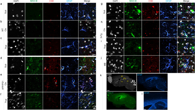
Reactive astrocytes play an important role in the development of Alzheimer's disease (AD). Here, we aimed to investigate the temporospatial relationships among monoamine oxidase-B, tau and amyloid-β (Aβ), translocator protein, and glucose metabolism by using multitracer imaging in AD transgenic mouse models. Positron emission tomography (PET) imaging with [18F]SMBT-1 (monoamine oxidase-B), [18F]florbetapir (Aβ), [18F]PM-PBB3 (tau), [18F]fluorodeoxyglucose (FDG), and [18F]DPA-714 (translocator protein) was carried out in 5- and 10-month-old APP/PS1, 11-month-old 3×Tg mice, and aged-matched wild-type mice. The brain regional referenced standard uptake value (SUVR) was computed with the cerebellum as the reference region. Immunofluorescence staining was performed on mouse brain tissue slices. [18F]SMBT-1 and [18F]florbetapir SUVRs were greater in the cortex and hippocampus of 10-month-old APP/PS1 mice than in those of 5-month-old APP/PS1 mice and wild-type mice. No significant difference in the regional [18F]FDG or [18F]DPA-714 SUVRs was observed in the brains of 5- or 10-month-old APP/PS1 mice or wild-type mice. No significant difference in the SUVRs of any tracer was observed between 11-month-old 3×Tg mice and age-matched wild-type mice. A positive correlation between the SUVRs of [18F]florbetapir and [18F]DPA-714 in the cortex and hippocampus was observed among the transgenic mice. Immunostaining validated the distribution of MAO-B and limited Aβ and tau pathology in 11-month-old 3×Tg mice; and Aβ deposits in brain tissue from 10-month-old APP/PS1 mice. In summary, these findings provide in vivo evidence that an increase in astrocyte [18F]SMBT-1 accompanies Aβ accumulation in APP/PS1 models of AD amyloidosis.
Keywords: Alzheimer’s disease; Amyloid-beta; Glia; MAO-B; PET; TSPO; Tau.
© 2024. The Author(s).
Conflict of interest statement
RMN is employee and shareholder of Neurimmune AG, Switzerland.
The other authors declare no competing interests.
Figures


Similar articles
-
In vivo reactive astrocyte imaging using [18F]SMBT-1 in tauopathy and familial Alzheimer's disease mouse models: A multi-tracer study.J Neurol Sci. 2024 Jul 15;462:123079. doi: 10.1016/j.jns.2024.123079. Epub 2024 Jun 4. J Neurol Sci. 2024. PMID: 38878650
-
(S)-[18F]THK5117 brain uptake is associated with Aβ plaques and MAO-B enzyme in a mouse model of Alzheimer's disease.Neuropharmacology. 2021 Sep 15;196:108676. doi: 10.1016/j.neuropharm.2021.108676. Epub 2021 Jun 30. Neuropharmacology. 2021. PMID: 34216585
-
Comparison of Monoamine Oxidase-A, Aβ Plaques, Tau, and Translocator Protein Levels in Postmortem Human Alzheimer's Disease Brain.Int J Mol Sci. 2023 Jun 28;24(13):10808. doi: 10.3390/ijms241310808. Int J Mol Sci. 2023. PMID: 37445985 Free PMC article.
-
[Development of SPECT Probes for In Vivo Imaging of β-Amyloid and Tau Aggregates in the Alzheimer's Disease Brain].Yakugaku Zasshi. 2017;137(11):1361-1365. doi: 10.1248/yakushi.17-00156. Yakugaku Zasshi. 2017. PMID: 29093372 Review. Japanese.
-
Integrating amyloid and tau imaging with proteomics and genomics in Alzheimer's disease.Cell Rep Med. 2024 Sep 17;5(9):101735. doi: 10.1016/j.xcrm.2024.101735. Cell Rep Med. 2024. PMID: 39293391 Free PMC article. Review.
Cited by
-
Spiral volumetric optoacoustic tomography of reduced oxygen saturation in the spinal cord of M83 mouse model of Parkinson's disease.Eur J Nucl Med Mol Imaging. 2025 Jan;52(2):427-443. doi: 10.1007/s00259-024-06938-w. Epub 2024 Oct 9. Eur J Nucl Med Mol Imaging. 2025. PMID: 39382580 Free PMC article.
-
Increased Cerebral Level of P2X7R in a Tauopathy Mouse Model by PET Using [18F]GSK1482160.ACS Chem Neurosci. 2024 Jun 5;15(11):2112-2120. doi: 10.1021/acschemneuro.4c00067. Epub 2024 May 22. ACS Chem Neurosci. 2024. PMID: 38776461 Free PMC article.
References
-
- Adlimoghaddam A, Snow WM, Stortz G, Perez C, Djordjevic J, Goertzen AL, Ko JH, Albensi BC (2019) Regional hypometabolism in the 3xTg mouse model of Alzheimer’s disease. Neurobiol Dis 127:264–277 - PubMed
-
- Andersen JV, Skotte NH, Christensen SK, Polli FS, Shabani M, Markussen KH, Haukedal H, Westi EW et al (2021) Hippocampal disruptions of synaptic and astrocyte metabolism are primary events of early amyloid pathology in the 5xFAD mouse model of Alzheimer’s disease. Cell Death Dis 12(11):954 - PMC - PubMed
-
- Barron AM, Ji B, Fujinaga M, Zhang MR, Suhara T, Sahara N, Aoki I, Tsukada H et al (2020) In vivo positron emission tomography imaging of mitochondrial abnormalities in a mouse model of tauopathy. Neurobiol Aging 94:140–148 - PubMed
MeSH terms
Substances
Grants and funding
- SCAHT-AP_22_01/Swiss Centre for Applied Human Toxicology
- 82272108/National Natural Science Foundation of China
- 81701732/National Natural Science Foundation of China
- 22ZR1409200/Natural Science Foundation of Shanghai Municipality
- 23Y11903200/Shanghai Science and Technology Innovation Action Plan Medical Innovation Research Project
LinkOut - more resources
Full Text Sources
Medical
Molecular Biology Databases
