. 2024 Mar 19;20(3):e1012101.
doi: 10.1371/journal.ppat.1012101. eCollection 2024 Mar.

Establishment of a lethal mouse model of emerging tick-borne orthonairovirus infections

Affiliations

Establishment of a lethal mouse model of emerging tick-borne orthonairovirus infections

Takuma Ariizumi et al. PLoS Pathog. .

Abstract

Emerging and reemerging tick-borne virus infections caused by orthonairoviruses (family Nairoviridae), which are genetically distinct from Crimean-Congo hemorrhagic fever virus, have been recently reported in East Asia. Here, we have established a mouse infection model using type-I/II interferon receptor-knockout mice (AG129 mice) both for a better understanding of the pathogenesis of these infections and validation of antiviral agents using Yezo virus (YEZV), a novel orthonairovirus causing febrile illnesses associated with tick bites in Japan and China. YEZV-inoculated AG129 mice developed hepatitis with body weight loss and died by 6 days post infection. Blood biochemistry tests showed elevated liver enzyme levels, similar to YEZV-infected human patients. AG129 mice treated with favipiravir survived lethal YEZV infection, demonstrating the anti-YEZV effect of this drug. The present mouse model will help us better understand the pathogenicity of the emerging tick-borne orthonairoviruses and the development of specific antiviral agents for their treatment.


Conflict of interest statement

The authors have declared that no competing interests exist.

Figures

Fig 1
Fig 1. YEZV infection in mouse models.
(A) Female 5-week-old C57BL/6 mice and BALB/c mice (n = 5) and sex-mixed 5-week-old AG129 mice (male: n = 4, female: n = 3) were inoculated intraperitoneally with 104 FFU of YEZV. Fourteen-day survival rates and body weight changes were compared between mock and YEZV infected groups. Relative body weight is shown as means with standard deviations. (B) AG129 mice were inoculated intraperitoneally with 104 FFU (male: n = 3, female: n = 3, red line), 103 FFU (male: n = 4, female: n = 3, yellow line), 102 FFU (male: n = 3, female: n = 3, green line), 10 FFU (male: n = 4, female: n = 3, blue line), and 1 FFU (male: n = 4, female: n = 3, gray line) of YEZV and monitored for 14-day survival rate and body weight change. Relative body weight was calculated and is shown as means with standard deviations. (C-E) AG129 mice were inoculated intraperitoneally with 104 FFU of YEZV and sacrificed for serum sampling at 2, 4, and 6 dpi (2 dpi: n = 5, 4 dpi: n = 5, 6 dpi: n = 3) for (C) blood biochemistry tests and quantification of (D) viral titer and (E) viral RNA in serum. Means with standard errors were shown with individual values as white circles; (C) Alanine transaminase (ALT), aspartate transaminase (AST), total bilirubin (TBIL), total protein (TP), albumin (ALB), globlin (GLOB) and glucose (Glu) values in serum, (D) virus titers in serum quantified by focus-formation assay, and (E) copy numbers of viral RNA quantified by RT-qPCR. Statistical analyses were performed using the Steel test, and the significant difference (p < 0.05) is indicated as an asterisk. (D) Dotted line indicated the lower limit of detection (L.O.D.) for the assay (0.4 FFU/μL).
Fig 2
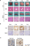
Fig 2. Histopathological findings in YEZV-infected AG129 mice.
AG129 mice inoculated intraperitoneally with 104 FFU of YEZV were sacrificed at 2, 4, and 6 dpi to collect organ samples. Mock-infected mice sacrificed at 6 dpi are shown as controls. Mouse organs were fixed in 10% phosphate-buffered formalin and (A) stained with hematoxylin-eosin (H&E) or (B and C) immunohistochemically stained for YEZV N. (A) Representative gross images and H&E-stained tissue images of the liver and spleen are shown. Each side of a square in the gross images is 5 mm. (B) Representative IHC images of the liver and spleen stained for YEZV N are shown with viral antigens in brown. (A and B) The scale bars on the histological images of the liver and spleen are 100 μm and 200 μm, respectively. (C) YEZV N protein (blue) and macrophage marker, Iba1 (brown) were stained by IHC double staining in the liver and spleen of YEZV-infected mice. The scale bars on IHC images of the liver and spleen tissues are 50 μm and 20 μm, respectively. Arrows indicate a cell in which both YEZV antigen and Iba1 were detected.
Fig 3
Fig 3. Viral titers and viral RNA in the liver and spleen of YEZV-infected AG129 mice.
Livers and spleens of YEZV-inoculated AG129 mice collected at 2, 4 and 6 dpi were used (2 dpi: n = 5, 4 dpi: n = 5, 6 dpi: n = 3) for analyzing (A) viral titers quantified by focus-formation assay and (B) the copy numbers of viral RNA (YEZV L-segment) quantified by RT-qPCR. Dotted lines indicated the lower limit of detection (L.O.D.) for the assay (4 FFU/mg). Plots lower than the L.O.D. are indicated by the half value of L.O.D. on the graph. White circles, thick bars, and thin lines indicate individual values, mean values and standard errors, respectively.
Fig 4
Fig 4. The effects of antivirals on YEZV infection in vitro.
Inhibitory effects of T-705 and ribavirin on YEZV replication in Vero cells were verified. Viral titers (white circles and solid lines) in the supernatant and cell viability (white triangles and dotted lines) at 6 dpi were compared with those in the absence of the chemical compounds.
Fig 5
Fig 5. Effects of antivirals on YEZV infection in vivo.
(A) The antiviral effects of T-705 in the AG129 mouse model were evaluated. AG129 mice intraperitoneally inoculated with 104 FFU of YEZV were orally dosed with T-705 (100 or 300 mg/kg/day) or vehicle using a stomach probe at 1 hour, 1, 2 and 3 dpi (represented by gray color on the figure). All mice were monitored for survival (left panel) and body weight changes (right panel) for 14 days. Fourteen-day survival rates and body weight changes were compared with the vehicle-treated group, 100 mg/kg/day of T-705-treated group and 300 mg/kg/day of T-705-treated group using a log-rank test. (B-E) Vehicle- or 300 mg/kg/day of T705-treated YEZV-infected AG129 mice were sacrificed at 2 and 4 dpi to collect blood and tissue samples. (B) Representative gross images and H&E-stained tissue images of the liver and spleen of mice are shown. Each side of a square in the gross images is 5 mm. The scale bars on the histological images of the liver and spleen are 100 μm and 500 μm respectively. (C) YEZV N proteins were detected by IHC in the liver and spleen of vehicle-treated and T-705-treated mice, and representative images are shown. The scale bars on the images of the liver and spleen tissues are 100 μm and 500 μm, respectively. (D) Viral titers and (E) the copy number of viral RNA (YEZV L-segment) in the liver and spleen of vehicle-treated and T-705-treated mice were quantified. Dotted lines indicated the lower limit of detection (L.O. D.) for the assay (4 FFU/mg of liver and spleen and 0.4 FFU/μL of serum). Plots lower than the L.O.D. are indicated by the half value of L.O.D. on the graph. Statistical differences in viral RNA copy number between the vehicle-treated group and T-705-group were examined by the Mann-Whitney U test, and the significant difference (p < 0.01) is indicated as double asterisks.

Similar articles

Cited by

References

    1. Kuhn JH, Adkins S, Alkhovsky SV, Avšič-Županc T, Ayllón MA, Bahl J, et al.. 2022 taxonomic update of phylum Negarnaviricota (Riboviria: Orthornavirae), including the large orders Bunyavirales and Mononegavirales. Arch Virol. 2022;167: 2857–2906. doi: 10.1007/s00705-022-05546-z - DOI - PMC - PubMed
    1. Hawman DW, Feldmann H. Crimean-Congo haemorrhagic fever virus. Nat Rev Microbiol. 2023; 1–15. - PMC - PubMed
    1. Krasteva S, Jara M, Frias-De-Diego A, Machado G. Nairobi Sheep Disease Virus: A Historical and Epidemiological Perspective. Front Vet Sci. 2020;7: 419. doi: 10.3389/fvets.2020.00419 - DOI - PMC - PubMed
    1. Burt FJ, Spencer DC, Leman PA, Patterson B, Swanepoel R. Investigation of tick-borne viruses as pathogens of humans in South Africa and evidence of Dugbe virus infection in a patient with prolonged thrombocytopenia. Epidemiol Infect. 1996;116: 353–361. doi: 10.1017/s0950268800052687 - DOI - PMC - PubMed
    1. Kalunda M, Mukwaya L G, Mukuye A, Lule M, Sekyalo E, Wright J, Casals J. Kasokero Virus: a New Human Pathogen from Bats (Rousettus Aegyptiacus) in Uganda. the American Journal of Tropical Medicine and Hygiene. 1986;35: 387–392. doi: 10.4269/ajtmh.1986.35.387 - DOI - PubMed

Publication types

Substances