Cardiac Magnetic Resonance to Predict Cardiac Mass Malignancy: The CMR Mass Score
- PMID: 38502734
- PMCID: PMC10949976
- DOI: 10.1161/CIRCIMAGING.123.016115
Cardiac Magnetic Resonance to Predict Cardiac Mass Malignancy: The CMR Mass Score
Erratum in
-
Correction to: Paolisso et al, "Cardiac Magnetic Resonance to Predict Cardiac Mass Malignancy: The CMR Mass Score".Circ Cardiovasc Imaging. 2024 Sep;17(9):e000084. doi: 10.1161/HCI.0000000000000084. Epub 2024 Sep 17. Circ Cardiovasc Imaging. 2024. PMID: 39288209 No abstract available.
Abstract
Background: Multimodality imaging is currently suggested for the noninvasive diagnosis of cardiac masses. The identification of cardiac masses' malignant nature is essential to guide proper treatment. We aimed to develop a cardiac magnetic resonance (CMR)-derived model including mass localization, morphology, and tissue characterization to predict malignancy (with histology as gold standard), to compare its accuracy versus the diagnostic echocardiographic mass score, and to evaluate its prognostic ability.
Methods: Observational cohort study of 167 consecutive patients undergoing comprehensive echocardiogram and CMR within 1-month time interval for suspected cardiac mass. A definitive diagnosis was achieved by histological examination or, in the case of cardiac thrombi, by histology or radiological resolution after adequate anticoagulation treatment. Logistic regression was performed to assess CMR-derived independent predictors of malignancy, which were included in a predictive model to derive the CMR mass score. Kaplan-Meier curves and Cox regression were used to investigate the prognostic ability of predictors.
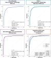
Results: In CMR, mass morphological features (non-left localization, sessile, polylobate, inhomogeneity, infiltration, and pericardial effusion) and mass tissue characterization features (first-pass perfusion and heterogeneity enhancement) were independent predictors of malignancy. The CMR mass score (range, 0-8 and cutoff, ≥5), including sessile appearance, polylobate shape, infiltration, pericardial effusion, first-pass contrast perfusion, and heterogeneity enhancement, showed excellent accuracy in predicting malignancy (areas under the curve, 0.976 [95% CI, 0.96-0.99]), significantly higher than diagnostic echocardiographic mass score (areas under the curve, 0.932; P=0.040). The agreement between the diagnostic echocardiographic mass and CMR mass scores was good (κ=0.66). A CMR mass score of ≥5 predicted a higher risk of all-cause death (P<0.001; hazard ratio, 5.70) at follow-up.
Conclusions: A CMR-derived model, including mass morphology and tissue characterization, showed excellent accuracy, superior to echocardiography, in predicting cardiac masses malignancy, with prognostic implications.
Keywords: cardiac magnetic resonance (CMR); cardiac masses; echocardiography; prognosis.
Conflict of interest statement
None.
Figures


Comment in
-
Benign or Malignant Cardiac Mass: Refining the Role of Cardiac Magnetic Resonance.Circ Cardiovasc Imaging. 2024 Mar;17(3):e016574. doi: 10.1161/CIRCIMAGING.124.016574. Epub 2024 Mar 19. Circ Cardiovasc Imaging. 2024. PMID: 38502737 No abstract available.
References
-
- D’Angelo EC, Paolisso P, Vitale G, Foà A, Bergamaschi L, Magnani I, Saturi G, Rinaldi A, Toniolo S, Renzulli M, et al. Diagnostic accuracy of cardiac computed tomography and 18-f fluorodeoxyglucose positron emission tomography in cardiac masses. JACC Cardiovasc Imaging. 2020;13:2400–2411. doi: 10.1016/j.jcmg.2020.03.021 - PubMed
-
- Lopez-Mattei JC, Lu Y. Multimodality imaging in cardiac masses: to standardize recommendations, the time is now! JACC Cardiovasc Imaging. 2020;13:2412–2414. doi: 10.1016/j.jcmg.2020.04.009 - PubMed
-
- Rahouma M, Arisha MJ, Elmously A, El-Sayed Ahmed MM, Spadaccio C, Mehta K, Baudo M, Kamel M, Mansor E, Ruan Y, et al. Cardiac tumors prevalence and mortality: a systematic review and meta-analysis. Int J Surg. 2020;76:178–189. doi: 10.1016/j.ijsu.2020.02.039 - PubMed
-
- Sultan I, Bianco V, Habertheuer A, Kilic A, Gleason TG, Aranda-Michel E, Harinstein ME, Martinez-Meehan D, Arnaoutakis G, Okusanya O. Long-term outcomes of primary cardiac malignancies: multi-institutional results from the national cancer database. J Am Coll Cardiol. 2020;75:2338–2347. doi: 10.1016/j.jacc.2020.03.041 - PubMed

