. 2024 Apr 4;111(4):761-777.
doi: 10.1016/j.ajhg.2024.02.014. Epub 2024 Mar 18.

Mono-allelic KCNB2 variants lead to a neurodevelopmental syndrome caused by altered channel inactivation

Affiliations

Mono-allelic KCNB2 variants lead to a neurodevelopmental syndrome caused by altered channel inactivation

Shreyas Bhat et al. Am J Hum Genet. .

Abstract

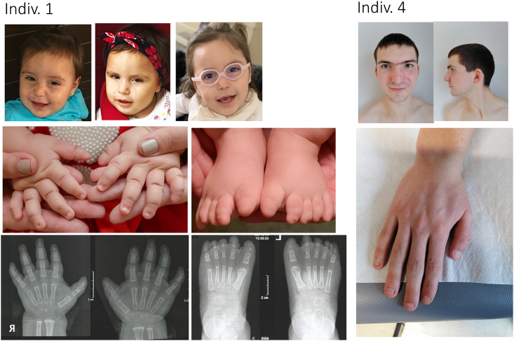
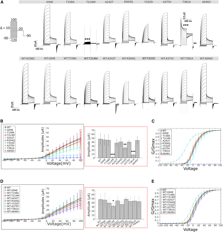
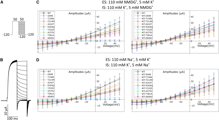
Ion channels mediate voltage fluxes or action potentials that are central to the functioning of excitable cells such as neurons. The KCNB family of voltage-gated potassium channels (Kv) consists of two members (KCNB1 and KCNB2) encoded by KCNB1 and KCNB2, respectively. These channels are major contributors to delayed rectifier potassium currents arising from the neuronal soma which modulate overall excitability of neurons. In this study, we identified several mono-allelic pathogenic missense variants in KCNB2, in individuals with a neurodevelopmental syndrome with epilepsy and autism in some individuals. Recurrent dysmorphisms included a broad forehead, synophrys, and digital anomalies. Additionally, we selected three variants where genetic transmission has not been assessed, from two epilepsy studies, for inclusion in our experiments. We characterized channel properties of these variants by expressing them in oocytes of Xenopus laevis and conducting cut-open oocyte voltage clamp electrophysiology. Our datasets indicate no significant change in absolute conductance and conductance-voltage relationships of most disease variants as compared to wild type (WT), when expressed either alone or co-expressed with WT-KCNB2. However, variants c.1141A>G (p.Thr381Ala) and c.641C>T (p.Thr214Met) show complete abrogation of currents when expressed alone with the former exhibiting a left shift in activation midpoint when expressed alone or with WT-KCNB2. The variants we studied, nevertheless, show collective features of increased inactivation shifted to hyperpolarized potentials. We suggest that the effects of the variants on channel inactivation result in hyper-excitability of neurons, which contributes to disease manifestations.

Keywords: KCNB2; channel inactivation, neurodevelopmental disorders, voltage-gated potassium channels; dysmorphism; epilepsy; global developmental delay.


Conflict of interest statement

Declaration of interests The authors declare no competing interests.

Figures

None
Graphical abstract
Figure 1
Figure 1
Photographs of two of the affected individuals Left: individual 1 through the years. Photos and X-rays of her hands and feet, illustrating nail hypoplasia and aplasia, and terminal phalanx hypoplasia (brachytelephalangia). Photos were taken at age 14 months; top right picture at 2 years 7 months. Right: individual 4, illustrating synophrys and nail hypoplasia. The proband was 18 years old at time of photo.
Figure 2
Figure 2
Schematic representation of mutations in Kv2.2 (A) Topology of Kv2.2 channel and schematic representation of distribution of KCNB2 variants. The variants are in the following regions: c.281G>A (p.Gly94Glu) and c.472A>G (p.Thr158Ala) in the N terminus; c.641C>T (p.Thr214Met) in the S1-S2 linker; c.724G>A (p.Ala242Thr) in the S2; c.827C>T (p.Pro276Leu) in the S3; c.911G>A (p.Arg304Gln) in the S4; c.994T>G (p.Tyr332Asp) in the S4-S5 linker; c.1124C>T (p.Ala375Val) and c.1141A>G (p.Thr381Ala) in the pore helix; and c.1937C>T (p.Ala646Val) in the C terminus. (B) Alignment of the variant Kv2.2 amino acids across different species. (C and D) Homology model and variant distribution of Kv2.2 as a tetramer (top view, C) and as a monomer (D) based on the known structure of the Kv1.2/2.1 chimera. The model was generated using alphafold2.
Figure 3
Figure 3
Activation properties of Kv2.2 variants (A) Activation currents from oocytes expressing WT-Kv2.2 and variant channels (expressed in the absence [top] and presence [below] of WT, see material and methods) were evoked by stepping from −90 mV to voltages ranging from −120 to +100 mV in 10 mV increments for 100 ms. This was followed by a voltage step to −20 mV for 100 ms and back to −90 mv for 5 s to allow recovery of the channels to deactivated states (protocol illustrated in the left corner). c.641C>T (p.Thr214Met) does not evoke any currents. c.994T>G (p.Tyr332Asp) show reduced currents. c.1141A>G (p.Thr381Ala) shows transient channel opening followed by a rapid inactivation of ionic currents. (B) Current-voltage (IV) relationship of Kv2.2 variants (left) and corresponding boxplots of maximal current amplitudes measured at +100 mV (right). p.Thr214Met, p.Tyr332Asp, and p.Thr381Ala show significant reduction in current amplitudes as compared to WT. (C) Conductance-voltage (GV) relationship of KCNB2 variants when expressed in oocytes alone. (D and E) IV (D) and GV (E) relationship of Kv2.2 variants when co-expressed with equal amounts of WT. WT:p.Thr214Met show significant reduction in current amplitudes as compared to WT alone. GV curves were best fitted by a sum of two Boltzmann relations of the form G/Gmax = Bottom + (Top1 − Bottom)/(1 + exp((V50(1) − X)/k1)) + (Top2 − Top1)/(1 + exp((V50(2) − X)/k2)). The fitting parameters (V50(1),V50(2), k1, k2) for the GV activation relationships have been compiled in Table 2. Values are provided as means ± SD from n > 6 oocytes per conditions from at least 2 independent experiments. Statistical significance was tested by Kruskal Wallis one-way analysis of variance followed by Dunn’s post-hoc test comparing amplitudes of the different variants to WT. p < 0.05, ∗∗p < 0.01, ∗∗∗p < 0.001.
Figure 4
Figure 4
Reversal potential of Kv2.2 variants (A) For calculating the reversal potentials of the Kv2.2 variants, currents were evoked by stepping from −120 mV to +50 mV for 100 ms followed by voltages ranging from −120 to +50 mV in 10 mV increments for 100 ms. This was followed by a voltage step to −120 mV for 20 ms and back to the holding potential of −90 mv for 5 s to allow recovery of the channels to deactivated states. The external and internal solutions used in these experiments contained K+ and either NMDG+ or Na+ in the concentrations mentioned in the figure. (B) Representative raw traces of the reversal potential protocol in an oocyte expressing WT-Kv2.2 in NMDG+ and K+ containing external and internal solutions. (C and D) The resulting IV curves for this protocol of Kv2.2 variants alone (left) or co-injected with WT (right) in solutions containing either NMDG+ and K+ (C) or Na+ and K+ (D) are shown. The variants do not appear to alter the K+ selectivity of the channel pore (X-intercept data compiled with 95% C.I. in Table S3). Mean ± SD are shown.
Figure 5
Figure 5
Inactivation features of currents by KCNB2 variants (A) Protocol to measure voltage-dependent inactivation in Kv2.2 variants. To measure inactivation, currents from oocytes expressing Kv2.2 variant channels were evoked by stepping from −90 mV to voltages ranging from −120 to +40 mV in 10 mV increments for 20 s. This was followed by a voltage step to +60 mV for 100 ms and back to −120 mv for 5 s to allow recovery of the channels from inactivation. (B and C) The raw traces from voltage steps highlighted in thickened line (−90 mV → 40 mV → 60 mV → 120 mV) are highlighted in (B) from oocytes expressing the individual variants alone or in (C) from oocytes expressing both WT and a specific variant. c.641C>T (p.Thr214Met) was excluded in (B) because of lack of any currents evoked by this variant. Currents evoked by c.1141A>G (p.Thr381Ala) are represented and further explained below. (D and E) The inactivation current-voltage (IV) relationship of these recordings are plotted. The IV relationship was best fitted by a sum of two Boltzmann relations of the form I/Imax = Top + (Bottom1 − Top)/(1 + exp((V50(1) − X)/k1)) + (Bottom2 − Bottom1)/(1 + exp((V50(2) − X)/k2)). The fitting parameters (V50(1),V50(2), k1, k2) for the IV relationships have been compiled in Table 3. Mean ± SD are shown. (F) The fitting function mentioned above also calculates the parameter “Bottom2,” which describes the extent of inactivation in these variants expressed either alone (top) or with WT (below). The differences in extent of inactivation between the variants and WT (red dashed lines) were tested for significance using the Kruskal Wallis one-way analysis of variance followed by Dunn’s post-hoc test comparing amplitudes of the different variants to WT-KCNB2. p ≤ 0.05, ∗∗p ≤ 0.01, ∗∗∗p ≤ 0.001. Values are provided as means ± SD from n > 6 oocytes per conditions from at least 2 independent experiments. (G) Representative current trace of an oocyte expressing only the p.Thr381Ala variant (blue trace) or only WT (black trace). p.Thr381Ala-expressing oocytes show diminished currents (blue trace) in the inactivation protocol as compared to WT, in a manner like the activation protocol in Figures 2 and 3. (H) Raw traces of the inactivation protocol of residual currents of the p.Thr381Ala variant. (I) The IV relationship of the p.Thr381Ala variant (blue line) shows recovery of inactivation with increasing voltages as opposed to WT (dashed fit representing the fit to WT IV shown in D and E).Mean ± SD are shown.

References

    1. Misonou H., Trimmer J.S. Determinants of voltage-gated potassium channel surface expression and localization in Mammalian neurons. Crit. Rev. Biochem. Mol. Biol. 2004;39:125–145. doi: 10.1080/10409230490475417. - DOI - PubMed
    1. Johnson B., Leek A.N., Tamkun M.M. Kv2 channels create endoplasmic reticulum/plasma membrane junctions: a brief history of Kv2 channel subcellular localization. Channels. 2019;13:88–101. doi: 10.1080/19336950.2019.1568824. - DOI - PMC - PubMed
    1. Kihira Y., Hermanstyne T.O., Misonou H. Formation of heteromeric Kv2 channels in mammalian brain neurons. J. Biol. Chem. 2010;285:15048–15055. doi: 10.1074/jbc.M109.074260. - DOI - PMC - PubMed
    1. Trimmer J.S. Immunological identification and characterization of a delayed rectifier K+ channel polypeptide in rat brain. Proc. Natl. Acad. Sci. USA. 1991;88:10764–10768. doi: 10.1073/pnas.88.23.10764. - DOI - PMC - PubMed
    1. Murakoshi H., Trimmer J.S. Identification of the Kv2.1 K+ channel as a major component of the delayed rectifier K+ current in rat hippocampal neurons. J. Neurosci. 1999;19:1728–1735. doi: 10.1523/JNEUROSCI.19-05-01728.1999. - DOI - PMC - PubMed

Publication types

Substances