

Sialic acid in the regulation of blood cell production, differentiation and turnover
- PMID: 38503445
- PMCID: PMC11223974
- DOI: 10.1111/imm.13780
Sialic acid in the regulation of blood cell production, differentiation and turnover
Abstract
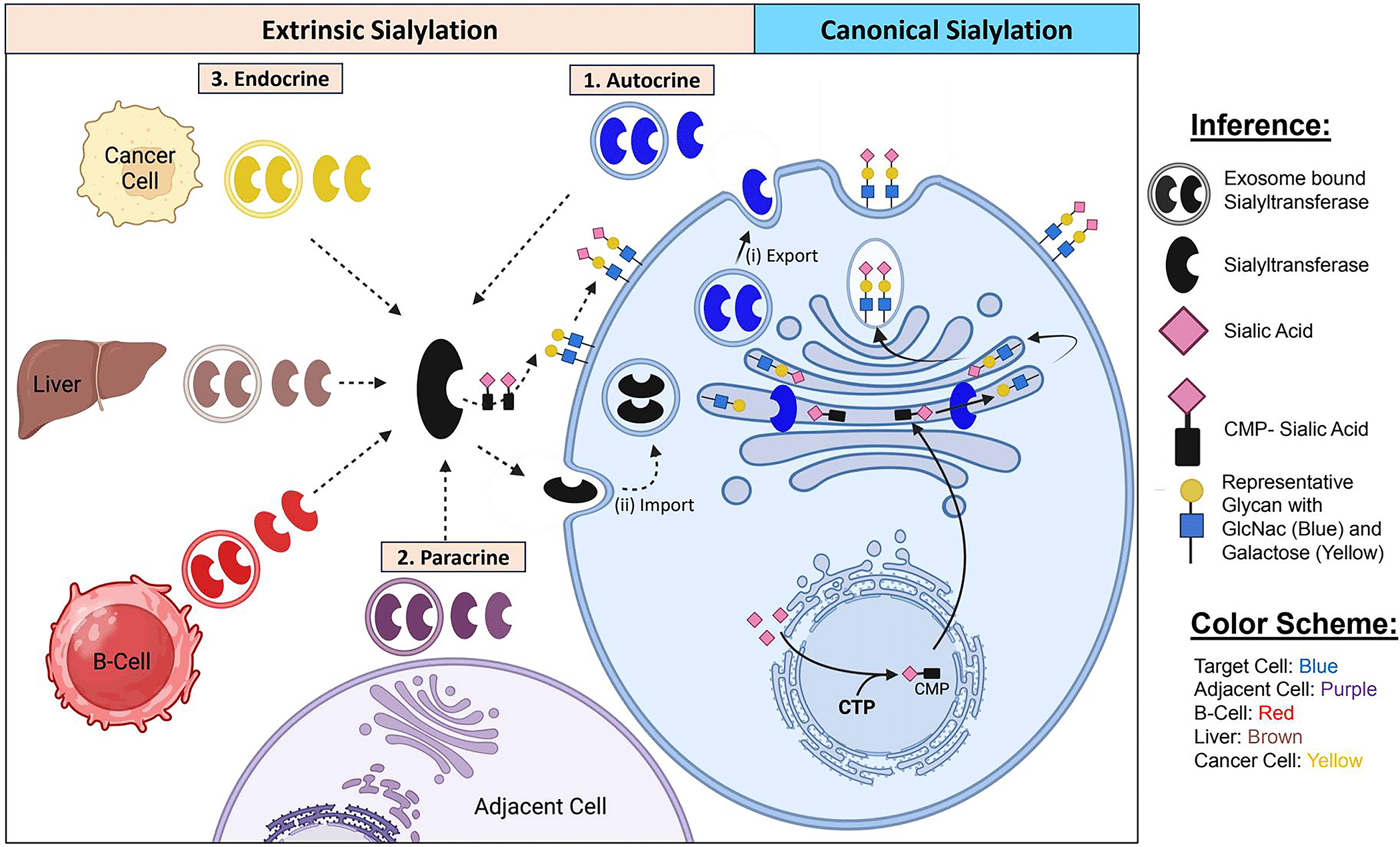
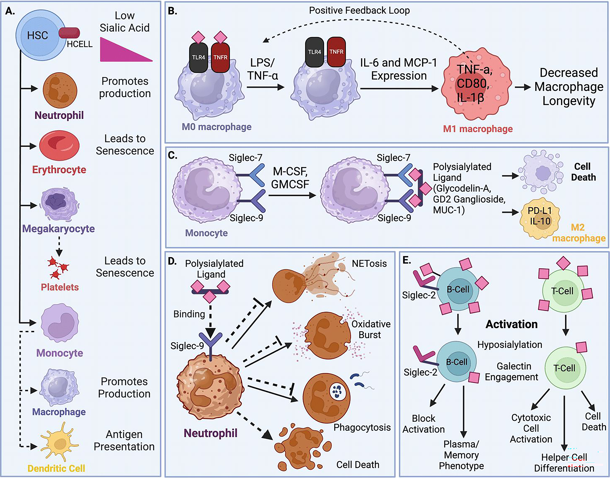
Sialic acid is a unique sugar moiety that resides in the distal and most accessible position of the glycans on mammalian cell surface and extracellular glycoproteins and glycolipids. The potential for sialic acid to obscure underlying structures has long been postulated, but the means by which such structural changes directly affect biological processes continues to be elucidated. Here, we appraise the growing body of literature detailing the importance of sialic acid for the generation, differentiation, function and death of haematopoietic cells. We conclude that sialylation is a critical post-translational modification utilized in haematopoiesis to meet the dynamic needs of the organism by enforcing rapid changes in availability of lineage-specific cell types. Though long thought to be generated only cell-autonomously within the intracellular ER-Golgi secretory apparatus, emerging data also demonstrate previously unexpected diversity in the mechanisms of sialylation. Emphasis is afforded to the mechanism of extrinsic sialylation, whereby extracellular enzymes remodel cell surface and extracellular glycans, supported by charged sugar donor molecules from activated platelets.
Keywords: bone marrow; cell death; extrinsic sialylation; galactose; galectin; haematopoiesis; lectins; sialic acid; sialyltransferase; siglec.
© 2024 The Authors. Immunology published by John Wiley & Sons Ltd.
Conflict of interest statement
Figures
Similar articles
-
Short-Term Memory Impairment.2024 Jun 8. In: StatPearls [Internet]. Treasure Island (FL): StatPearls Publishing; 2025 Jan–. 2024 Jun 8. In: StatPearls [Internet]. Treasure Island (FL): StatPearls Publishing; 2025 Jan–. PMID: 31424720 Free Books & Documents.
-
Sialylated glycoproteins bind to Siglec-9 in a cis manner on platelets to suppress platelet activation.J Thromb Haemost. 2025 Jul;23(7):2270-2283. doi: 10.1016/j.jtha.2025.03.027. Epub 2025 Apr 7. J Thromb Haemost. 2025. PMID: 40204021
-
α2,6-linked sialic acid serves as a high-affinity receptor for cancer oncolytic virotherapy with Newcastle disease virus.J Cancer Res Clin Oncol. 2017 Nov;143(11):2171-2181. doi: 10.1007/s00432-017-2470-y. Epub 2017 Jul 7. J Cancer Res Clin Oncol. 2017. PMID: 28687873 Free PMC article.
-
Management of urinary stones by experts in stone disease (ESD 2025).Arch Ital Urol Androl. 2025 Jun 30;97(2):14085. doi: 10.4081/aiua.2025.14085. Epub 2025 Jun 30. Arch Ital Urol Androl. 2025. PMID: 40583613 Review.
-
The Tumour Glyco-Code: Sialylation as a Mediator of Stromal Cell Immunosuppression in the Tumour Microenvironment.Eur J Immunol. 2025 Jul;55(7):e70000. doi: 10.1002/eji.70000. Eur J Immunol. 2025. PMID: 40667828 Free PMC article. Review.
Cited by
-
Insights on the Role of Sialic Acids in Acute Lymphoblastic Leukemia in Children.Int J Mol Sci. 2025 Mar 1;26(5):2233. doi: 10.3390/ijms26052233. Int J Mol Sci. 2025. PMID: 40076855 Free PMC article. Review.
-
Targeting Non-Eosinophilic Immunological Pathways in COPD and AECOPD: Current Insights and Therapeutic Strategies.Int J Chron Obstruct Pulmon Dis. 2025 Mar 5;20:511-532. doi: 10.2147/COPD.S506616. eCollection 2025. Int J Chron Obstruct Pulmon Dis. 2025. PMID: 40066199 Free PMC article. Review.
-
When a negative (charge) is not a positive: sialylation and its role in cancer mechanics and progression.Front Oncol. 2024 Nov 19;14:1487306. doi: 10.3389/fonc.2024.1487306. eCollection 2024. Front Oncol. 2024. PMID: 39628991 Free PMC article. Review.
References
Publication types
MeSH terms
Substances
Grants and funding
LinkOut - more resources
Full Text Sources
