. 2024 Mar 19;15(1):2459.
doi: 10.1038/s41467-024-46760-4.

VCF1 is a p97/VCP cofactor promoting recognition of ubiquitylated p97-UFD1-NPL4 substrates

Affiliations

VCF1 is a p97/VCP cofactor promoting recognition of ubiquitylated p97-UFD1-NPL4 substrates

Ann Schirin Mirsanaye et al. Nat Commun. .

Abstract

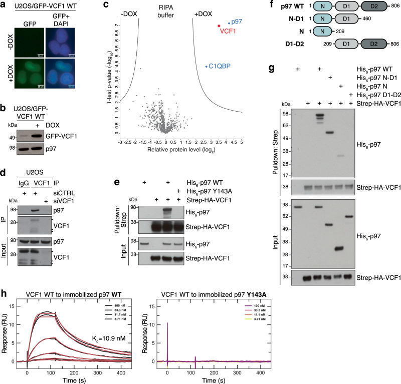
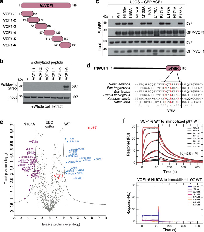
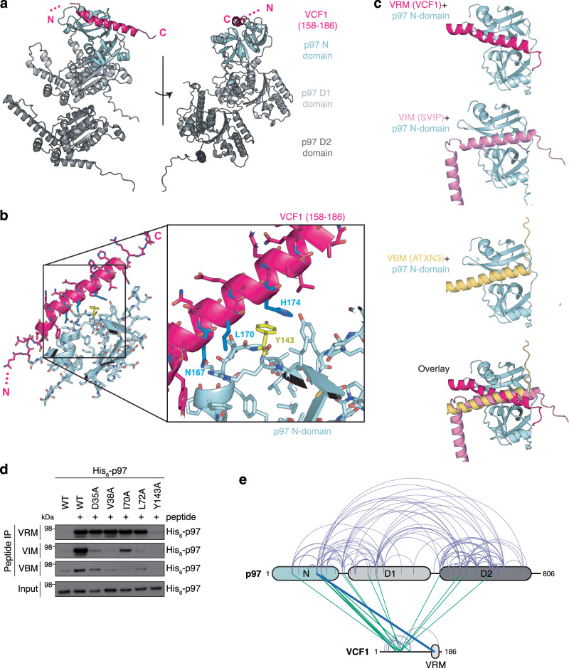
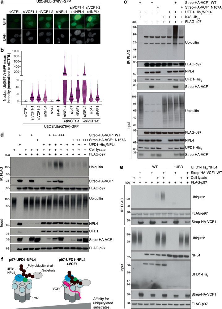
The hexameric AAA+ ATPase p97/VCP functions as an essential mediator of ubiquitin-dependent cellular processes, extracting ubiquitylated proteins from macromolecular complexes or membranes by catalyzing their unfolding. p97 is directed to ubiquitylated client proteins via multiple cofactors, most of which interact with the p97 N-domain. Here, we discover that FAM104A, a protein of unknown function also named VCF1 (VCP/p97 nuclear Cofactor Family member 1), acts as a p97 cofactor in human cells. Detailed structure-function studies reveal that VCF1 directly binds p97 via a conserved α-helical motif that recognizes the p97 N-domain with unusually high affinity, exceeding that of other cofactors. We show that VCF1 engages in joint p97 complex formation with the heterodimeric primary p97 cofactor UFD1-NPL4 and promotes p97-UFD1-NPL4-dependent proteasomal degradation of ubiquitylated substrates in cells. Mechanistically, VCF1 indirectly stimulates UFD1-NPL4 interactions with ubiquitin conjugates via its binding to p97 but has no intrinsic affinity for ubiquitin. Collectively, our findings establish VCF1 as an unconventional p97 cofactor that promotes p97-dependent protein turnover by facilitating p97-UFD1-NPL4 recruitment to ubiquitylated targets.


Conflict of interest statement

The authors declare no competing interests.

Figures

Fig. 1
Fig. 1. VCF1 is a p97-binding protein that interacts tightly with the p97 N-domain.
a Representative images of U2OS/GFP-VCF1 WT cells treated or not with Doxycycline (DOX) for 16 h. Scale bars, 10 μM. b Immunoblot analysis of U2OS/GFP-VCF1 WT cells treated as in (a). c Mass spectrometry analysis of VCF1-interacting proteins. U2OS/GFP-VCF1 WT cells were treated or not with DOX for 16 h, subjected to GFP immunoprecipitation (IP) in partially denaturing RIPA buffer and analyzed by mass spectrometry. Volcano plot shows enrichment of individual proteins (+DOX/-DOX ratio) plotted against the P value (Supplementary Data 1). Dashed lines indicate the significance thresholds (two-sided t test, FDR < 0.05, s0 = 1). d Immunoblot analysis of VCF1 or pre-immune serum (IgG) IPs from whole cell lysates of U2OS cells transfected with non-targeting control (CTRL) or VCF1 siRNAs. Three specific bands corresponding to different VCF1 isoforms are detected by the VCF1 antibody, with the slower-migrating major band representing the 186-amino acid protein (isoform 1). e Immunoblot analysis of in vitro binding reactions in RIPA buffer containing purified His6-p97 and Strep-HA-VCF1 proteins that were subjected to StrepTactin (Strep) pulldown. f Schematic showing recombinant His6-p97 proteins used in (g). g As in (e), but using recombinant His6-p97 proteins shown in (f). Note that less His6-p97 N protein than His6-p97 WT and His6-p97 N-D1 co-purifies with Strep-HA-VCF1, since the isolated p97 N domain does not form hexamers, unlike p97 WT and p97 N-D1. h Surface Plasmon Resonance (SPR) sensorgrams for the interaction between recombinant Strep-HA-VCF1 WT and immobilized His6-p97 proteins. For His6-p97 WT (left panel), black traces represent the experimental data and red traces correspond to the global fit of the two replicates according to a 1:1 interaction model. For the His6-p97 Y143A mutant (right panel), no dose-dependent response was observed. Data information: Data are representative of three (a, b, d, e, g, h) independent experiments with similar outcome. Source data are provided as a Source Data file.
Fig. 2
Fig. 2. VCF1 binds p97 via an α-helical motif (VRM).
a Schematic of VCF1 peptides used in (b). b Biotinylated VCF1 peptides in (a) were incubated with whole cell extracts of U2OS cells, subjected to StrepTactin (Strep) pulldown in RIPA buffer and analyzed by immunoblotting. c Immunoblot analysis of GFP IPs from U2OS cells transfected with indicated GFP-VCF1 expression constructs. d Sequence alignment showing conservation of the p97-binding motif (VRM) in selected vertebrate VCF1 proteins, with residues essential for p97 binding highlighted in red. e Comparison of VCF1 WT and N167A interactomes. U2OS/GFP-VCF1 WT or N167A cells treated with DOX for 16 h were subjected to GFP IP in non-denaturing EBC buffer and analyzed by mass spectrometry. Volcano plot shows enrichment of individual proteins (WT/N167A ratio) plotted against the P value (Supplementary Data 3). Dashed lines indicate the significance thresholds (two-sided t test, FDR < 0.01, s0 = 2). f SPR sensorgrams for the interaction between VCF1-6 peptide (a) and immobilized recombinant His6-p97. For the VCF1-6 WT peptide (left panel), black traces represent the experimental data and red traces correspond to the global fit of the two replicates according to a 1:1 interaction model. For the VCF1-6 N167A peptide (right panel), no dose-dependent response was observed. Data information: Data are representative of two (b, c, f) independent experiments with similar outcome. Source data are provided as a Source Data file.
Fig. 3
Fig. 3. Structural modeling of VCF1-p97 complex formation.
a AlphaFold-Multimer model of the complex between full-length VCF1 and a p97 monomer (colored by domains), predicting that the C-terminus of the otherwise highly unstructured VCF1 forms an α-helix (pink, residues 158−186) that interacts with the groove of the bipartite p97 N-domain (left panel). b Closeup view of the VCF1-p97 binding interface predicted by AlphaFold-Multimer (a), showing that it is involves Y143 (yellow) in the p97 N-domain and residues N167, L170 and H174 (blue) in VCF1 (right panel). c Comparison of AlphaFold-Multimer models of the complexes formed between the p97 monomer (N-domain in pale cyan) and VCF1 (pink), SVIP (rosa) or ATXN3 (yellow). d Biotinylated peptides spanning the VRM in VCF1, VIM in SVIP or VBM in ATXN3 immobilized on StrepTactin agarose were incubated with purified WT or mutant His6-p97 proteins, washed extensively in RIPA buffer and analyzed by immunoblotting. e Schematic visualization of all heteromeric VCF1-p97 crosslinks (green) identified by mass spectrometry analysis of purified FLAG-p97 and Strep-HA-VCF1 proteins incubated with the crosslinker disuccinimidyl dibutyric urea (DSBU) (Supplementary Fig. 3g). Self-crosslinks are shown in light purple. All visualized crosslinks scored >50 using MeroX, with a score of at least 19.4 required to ensure an FDR < 0.01 via decoy analysis. The line highlighted in blue visualizes a crosslink (with MeroX score of 116) between the p97 N-domain (K136) and the VCF1 VRM (Y163) (Supplementary Fig. 3f). Data information: Data are representative of two (d) independent experiments with similar outcome. Source data are provided as a Source Data file.
Fig. 4
Fig. 4. VCF1 and UFD1-NPL4 form a joint complex with p97.
a Mass photometry-based mass distribution of His6-p97 (blue) and His6-p97 in the presence of saturating concentrations of full-length Strep-HA-VCF1 WT (magenta) (left panel) or N167A mutant (gray) (right panel). The mass photometry measurements show major species with molecular weights of 535 kDa (corresponding to a p97 hexamer) and 591 kDa (corresponding to a p97 hexamer bound by 2−3 VCF1 molecules). b Binding reactions containing indicated combinations of purified Strep-HA-VCF1, His6-p97 and GST-UFD1-NPL4 proteins were subjected to Glutathione (GST) pulldown and analyzed by immunoblotting. c As in (b), except reactions containing indicated combinations of purified Strep-HA-VCF1, His6-p97 and UFD1-His6-NPL4 proteins were subjected to StrepTactin (Strep) pulldown. d Purified VCF1 was incubated alone or with equimolar concentrations of p97 and UFD1-NPL4 and then separated by size-exclusion chromatography (SEC). Immunoblot analysis of indicated fractions shows comigration of the p97-VCF1-UFD1-NPL4 complex. Data information: Data are representative of three (b, c) and two (a, d) independent experiments with similar outcome. Source data are provided as a Source Data file.
Fig. 5
Fig. 5. VCF1 stimulates p97-UFD1-NPL4 recruitment to ubiquitylated substrates to facilitate their degradation.
a Representative images of U2OS/Ub(G76V)-GFP cells transfected with indicated siRNAs. Scale bar, 10 μM. b U2OS/Ub(G76V)-GFP cells transfected with indicated siRNAs were subjected to quantitative image-based cytometry (QIBC) analysis of Ub(G76V)-GFP expression (solid lines, median; dashed lines, quartiles; >10,000 cells analyzed per condition). c Immunoblot analysis of in vitro binding reactions containing indicated combinations of purified FLAG-p97, Strep-HA-VCF1, UFD1-His6-NPL4 complex and K48-linked ubiquitin chains (K48-Ub2-7) that were subjected to FLAG IP. d As in (c), except that in vitro binding reactions containing purified proteins were incubated with whole cell extracts of U2OS cells where indicated and subjected to FLAG IP. e As in (d), using WT or ubiquitin binding-deficient (*UBD) UFD1-His6-NPL4 complex. f Model of VCF1-dependent stimulation of p97-UFD1-NPL4 binding to ubiquitylated client proteins. See main text for details. Data information: Data are representative of three (a, b, d) and two (c, e) independent experiments with similar outcome. Source data are provided as a Source Data file.

References

    1. van den Boom J, Meyer H. VCP/p97-mediated unfolding as a principle in protein homeostasis and signaling. Mol Cell. 2018;69:182–194. doi: 10.1016/j.molcel.2017.10.028. - DOI - PubMed
    1. Ye Y, Tang WK, Zhang T, Xia D. A mighty “Protein Extractor” of the cell: structure and function of the p97/CDC48 ATPase. Front. Mol. Biosci. 2017;4:39. doi: 10.3389/fmolb.2017.00039. - DOI - PMC - PubMed
    1. Twomey EC, et al. Substrate processing by the Cdc48 ATPase complex is initiated by ubiquitin unfolding. Science. 2019;365:eaax1033. doi: 10.1126/science.aax1033. - DOI - PMC - PubMed
    1. Bodnar NO, Rapoport TA. Molecular mechanism of substrate processing by the Cdc48 ATPase complex. Cell. 2017;169:722–735.e9. doi: 10.1016/j.cell.2017.04.020. - DOI - PMC - PubMed
    1. Weith M, et al. Ubiquitin-independent disassembly by a p97 AAA-ATPase complex drives PP1 holoenzyme formation. Mol. Cell. 2018;72:766–777.e6. doi: 10.1016/j.molcel.2018.09.020. - DOI - PubMed

MeSH terms