. 2024 Mar 19;15(1):2432.
doi: 10.1038/s41467-024-46761-3.

RAPP-containing arrest peptides induce translational stalling by short circuiting the ribosomal peptidyltransferase activity

Affiliations

RAPP-containing arrest peptides induce translational stalling by short circuiting the ribosomal peptidyltransferase activity

Martino Morici et al. Nat Commun. .

Erratum in

Abstract

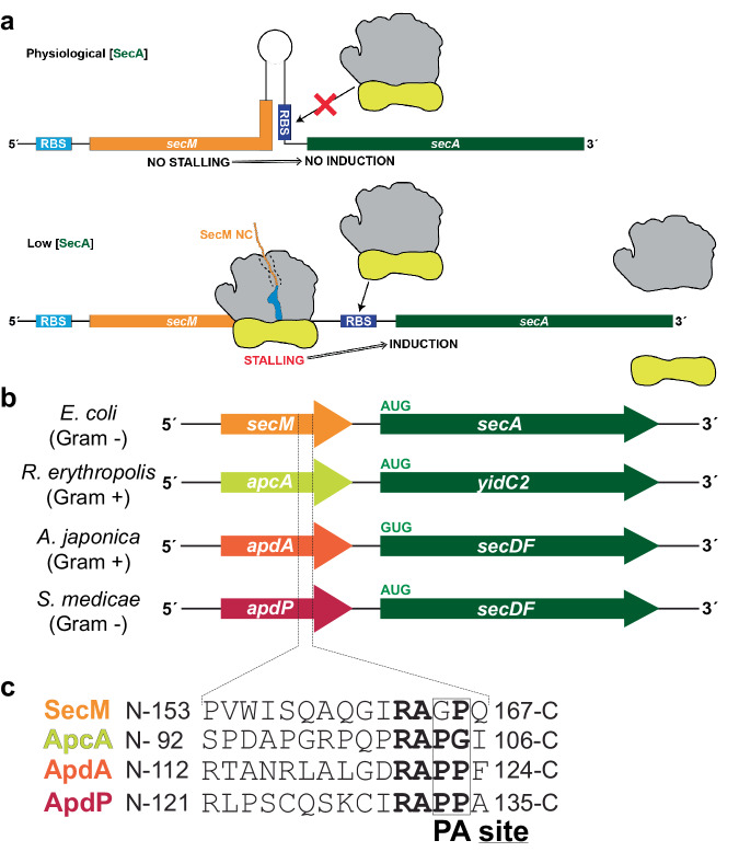
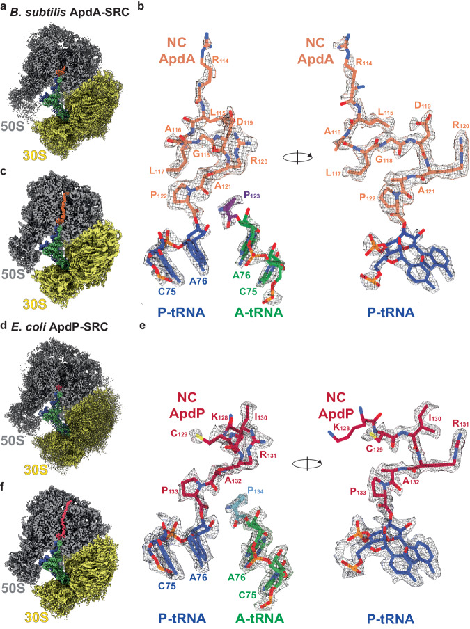
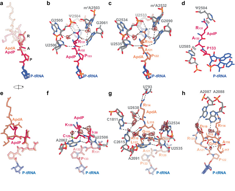
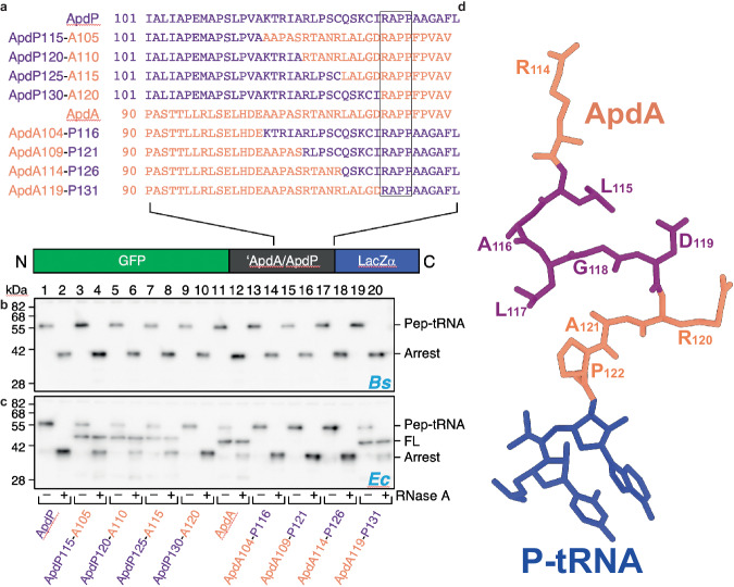
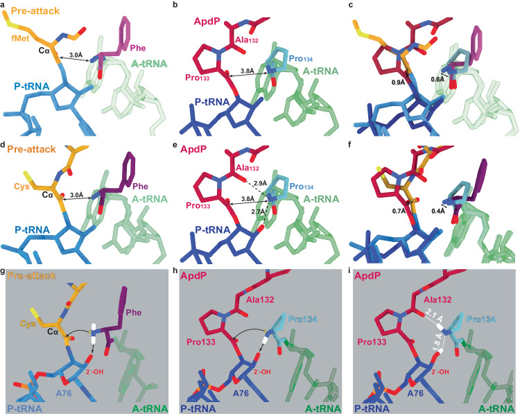
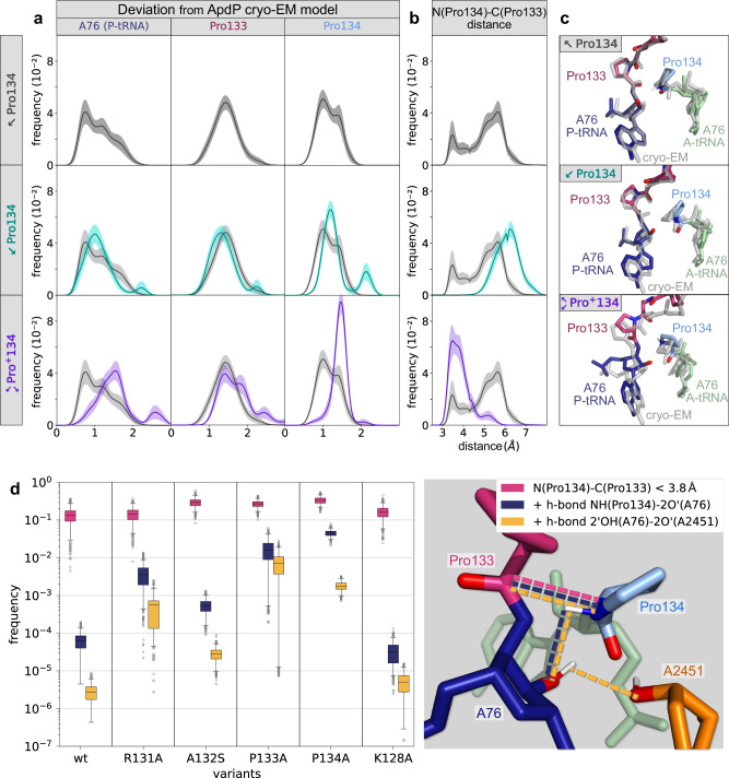
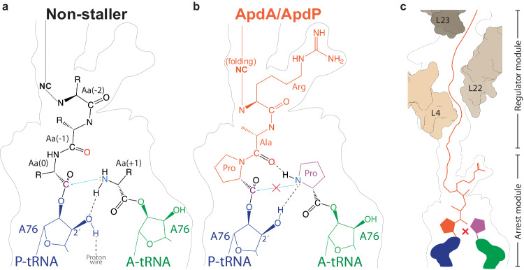
Arrest peptides containing RAPP (ArgAlaProPro) motifs have been discovered in both Gram-positive and Gram-negative bacteria, where they are thought to regulate expression of important protein localization machinery components. Here we determine cryo-EM structures of ribosomes stalled on RAPP arrest motifs in both Bacillus subtilis and Escherichia coli. Together with molecular dynamics simulations, our structures reveal that the RAPP motifs allow full accommodation of the A-site tRNA, but prevent the subsequent peptide bond from forming. Our data support a model where the RAP in the P-site interacts and stabilizes a single hydrogen atom on the Pro-tRNA in the A-site, thereby preventing an optimal geometry for the nucleophilic attack required for peptide bond formation to occur. This mechanism to short circuit the ribosomal peptidyltransferase activity is likely to operate for the majority of other RAPP-like arrest peptides found across diverse bacterial phylogenies.


Conflict of interest statement

The authors declare no competing interests.

Figures

Fig. 1
Fig. 1. Arrangement of bacterial regulatory operons.
a Schematic representation for the regulation of SecA by SecM. Upper panel: When SecA levels are high, the pulling force of SecA on the SecM nascent chain prevents stalling, and therefore the ribosome binding site (RBS) of the downstream secA gene is sequestered in a stem-loop structure, preventing SecA expression. Lower panel: When SecA levels are low, ribosomes stall during translation of SecM, leading to mRNA rearrangements that expose the RBS of the secA gene, leading to the expression of SecA. b Examples of bacterial operons containing regulatory upstream open reading frames (uORFs), including secM-secA in Gram-negative γ-proteobacteria, such as Escherichia coli, apcA-yidC2 from Gram-positive actinomycetes, such as Rhodococcus erythropolis, apdA-secDF from Gram-positive actinomycetes, such as Amycolatopsis japonica, and apdP-secDF from Gram-negative α-proteobacteria, such as Sinorhizobium medicae. c Amino acid sequences for the SecM, ApcA, ApdA, and ApdP arrest peptides, aligned based on stalling site during translation, with A- and P-site positions indicated. Conserved residues around the stalling sites are highlighted in bold.
Fig. 2
Fig. 2. Cryo-EM structures of ApdA- and ApdP-SRCs.
a Cryo-EM map of the post-processed B. subtilis ApdA-SRC with a transverse section of the 50S (gray) to reveal density for the nascent chain (orange), P-tRNA (blue), A-tRNA (green); 30S (yellow). b Two views showing the cryo-EM map density for A- and P-site tRNAs of the post-processed B. subtilis ApdA-SRC. The P-site tRNA (blue) bears the ApdA nascent chain (orange), whereas the A-site tRNA (green) carries proline (purple). c as (a), but cryo-EM map of 3D-refined B. subtilis ApdA-SRC. d Cryo-EM map of the post-processed E. coli ApdP-SRC with a transverse section of the 50S (gray) to reveal density for the nascent chain (red), P-tRNA (blue), A-tRNA (green); 30S (yellow). e Two views showing cryo-EM map density for A- and P-site tRNAs of the post-processed E. coli ApdP-SRC. The P-site tRNA (blue) bears the ApdP nascent chain (red), whereas the A-site tRNA (green) carries proline (cyan). f as (d), but cryo-EM map of 3D-refined E. coli ApdP-SRC.
Fig. 3
Fig. 3. Interaction of ApdA and ApdP NCs within the ribosomal tunnel.
a Overlay (aligned on the basis of the 23S rRNA) of the molecular models for ApdA (orange), ApdP (red), and P-tRNA (blue) with a focus on the RAP motif. b, c Interactions of RAP motif of the b ApdP (red) and c ApdA (orange) nascent chains with the exit tunnel nucleotides (gray) of the b E. coli and c B. subtilis 23S rRNA, respectively. Potential hydrogen bonds are shown as dashed lines and water molecules as red spheres (with meshed density). d Rotated view of (b) showing the stacking interactions (depicted as three parallel lines) between the conserved RAP motif of ApdP (red) and 23S rRNA nucleotides (gray). In (b) and (d), ψ indicates the presence of pseudouridine at position 2504 in E. coli. e Rotated view of (a) with a focus on residues N-terminal to the RAP motif. fh Interactions (f) ApdP (red) and (g, h) ApdA (orange) nascent chains with the exit tunnel nucleotides (gray) of the f E. coli and g, h B. subtilis 23S rRNA, respectively. Potential hydrogen bonds are shown as dashed lines and water molecules as red spheres (with meshed density).
Fig. 4
Fig. 4. Species-specific stalling of chimeric ApdA-ApdP constructs.
a Sequences of different chimeras between ApdA (orange) and ApdP (purple) (with the common RAPP motif boxed) cloned in the GFP-LacZα construct shown at the bottom. b, c Western blot against GFP showing the outcome of the stalling assay in b B. subtilis and c E. coli in vitro translation system for the chimeras listed in (a); each reaction was loaded before (−) and after (+) RNase A treatment. Bands corresponding to peptidyl-tRNA (Pep-tRNA), full-length peptide (FL), and truncated peptide arising due to the stalling (Arrest) are indicated. Experiments were performed in two independent experiments with similar results. Source data are provided as a Source Data file. d Structure of ApdA (orange) with residues colored purple that enhanced stalling on E. coli ribosomes when substituted with the corresponding ApdP residues.
Fig. 5
Fig. 5. ApdA/ApdP stabilize the pre-attack state at the PTC.
a View of the PTC of a pre-attack state (PDB ID 1VY4), showing a fMet-NH-tRNA (gold/blue) at the P-site and a phenylyl-NH-tRNA (purple/green) at the A-site. The distance (3.0 Å) between the attacking amine of the A-tRNA and the carbonyl-carbon of the P-tRNA is arrowed. b Same view as (a), but for the ApdP-SRC with ApdP-tRNA (red/dark blue) in the P-site and Pro-tRNA (cyan/green) in the A-site. c Overlay of (a, b) (aligned on the basis of the 23S rRNA) highlighting the difference in the distance between the attacking nitrogen groups of A-site aminoacyl moiety and the carbonyl carbons at the P-site. d View of the PTC of a pre-attack state (PDB ID 8CVK), showing a tripeptidyl-NH-tRNA (gold/blue) at the P-site and a phenyl-NH-tRNA (purple/green) at the A-site. The distance (3.0 Å) between the attacking nitrogen of the A-tRNA and the carbonyl-carbon of the P-tRNA is arrowed. e Same view as (d), but for the ApdP-SRC with ApdP-tRNA (red/dark blue) in the P-site and Pro-tRNA (cyan/green) in the A-site. f Overlay of (d, e) (aligned on the basis of the 23S rRNA) highlighting the difference in the distance between the attacking nitrogen groups at the A-site and the carbonyl carbons at the P-site. g Schematic view of the PTC of a pre-attack from (d), but with hydrogen atoms (white) modeled in silico for the α-amino group of the Phe moiety in the A-site. The yellow spheres indicate the lone pair electrons that make the nucleophilic attack (arrowed) on the carbonyl-carbon of the fMet moiety on the P-site tRNA. h Same schematic as (b), but with the hydrogen atom (white) modeled in silico towards the 2’OH of A76 of the P-site tRNA, which would allow a nucleophilic attack (arrowed) on the carbonyl-carbon of the Pro133 moiety attached to the P-site tRNA. i Same schematic as (h) but with the hydrogen atom (white) modeled toward the carbonyl of Ala132, a conformation that would prohibit any nucleophilic attack.
Fig. 6
Fig. 6. MD simulations of ApdP in the ribosome.
a, b For different protonation states (Pro134, Pro134, Pro+134), histograms of deviations (rmsd) from the cryo-EM model (ribose ring of A76, Pro134, and Pro133) (a) and of the distances between the α-amino N of Pro134 and the carbonyl C of Pro133 (b) are shown. Uncharged protonation states with the N-H pointing either towards the O (Ala132) or towards O2’ (A76) are denoted by Pro134 and Pro134, respectively. Pro+134 denotes the charged state with both hydrogens. Lines and error bars in (a) and (b) were obtained from the mean and standard deviations of 10,000 bootstraps of 20 independent simulations. c From the MD simulations of each protonation state, structures corresponding to the most probable rmsd values are shown (colored) and compared with the stalled cryo-EM structure (grey). d Frequencies of the conformations fulfilling three conditions required for peptide bond formation. Frequencies of N(Pro134)-C(Pro133) distances lower than 3.8 Å (proximity requirement, magenta). Frequency of conformations which, in addition to the first condition, contain an N-H(Pro134)−2’O(A76) hydrogen bond (blue). Frequency of the conformations that additionally contain the 2’OH(A76)−2’O(A2451) hydrogen bond (yellow). The box plots were obtained by bootstrapping 10,000 samples of 20 independent simulations for each variant. The boxes extend from the first to third quartiles. Whiskers display a 95% confidence interval. Points out of the confidence interval are shown (grey circles). Source data can be obtained from Zenodo (10.5281/zenodo.10426362).
Fig. 7
Fig. 7. Model for ApdA/ApdP-mediated translational stalling.
a, b Schematic representations of the PTC for a canonical non-stalling nascent polypeptide chains, where the lone pair electrons on the α-amino group of the aminoacyl moiety attached to the A-site tRNA makes a nucleophilic attack (blue arrow) on the carbonyl-carbon of the peptidyl-tRNA in the P-site. The nucleophilicity of the α-amino group is increased by the extraction of a proton by the 2’ OH of ribose of A76 of the P-tRNA. b RAPP-mediated translation stalling by ApdA or ApdP, where the nucleophilic attack of the nitrogen of the A-site Pro on the carbonyl-carbon of the peptidyl-tRNA cannot occur because (i) the hydrogen of the nitrogen of Pro is involved in a hydrogen bond with the carbonyl-oxygen of Ala of the RAP motif in the P-site, and (ii) the 2’O of the ribose of A76 donates hydrogen to form a hydrogen bond with the lone pair electron, rather than extracting the proton as required for peptide bond formation. c ApdA and ApdP stalling is strongly driven by the RAPP arrest module; however, the N-terminal regulator module also contributes by fine-tuning the stalling efficiency.

References

    1. Wilson DN, Beckmann R. The ribosomal tunnel as a functional environment for nascent polypeptide folding and translational stalling. Curr. Opin. Struct. Biol. 2011;21:1–10. doi: 10.1016/j.sbi.2011.01.007. - DOI - PubMed
    1. Ito K, Chiba S. Arrest peptides: cis-acting modulators of translation. Annu Rev. Biochem. 2013;82:171–202. doi: 10.1146/annurev-biochem-080211-105026. - DOI - PubMed
    1. Wilson DN, Arenz S, Beckmann R. Translation regulation via nascent polypeptide-mediated ribosome stalling. Curr. Opin. Struct. Biol. 2016;37:123–133. doi: 10.1016/j.sbi.2016.01.008. - DOI - PubMed
    1. Dever TE, Ivanov IP, Sachs MS. Conserved upstream open reading frame nascent peptides that control translation. Annu. Rev. Genet. 2020;54:237–264. doi: 10.1146/annurev-genet-112618-043822. - DOI - PMC - PubMed
    1. Ramu H, Mankin A, Vazquez-Laslop N. Programmed drug-dependent ribosome stalling. Mol. Microbiol. 2009;71:811–824. doi: 10.1111/j.1365-2958.2008.06576.x. - DOI - PubMed

MeSH terms