. 2024 Mar 19;15(1):2431.
doi: 10.1038/s41467-024-46762-2.

The SecM arrest peptide traps a pre-peptide bond formation state of the ribosome

Affiliations

The SecM arrest peptide traps a pre-peptide bond formation state of the ribosome

Felix Gersteuer et al. Nat Commun. .

Erratum in

Abstract

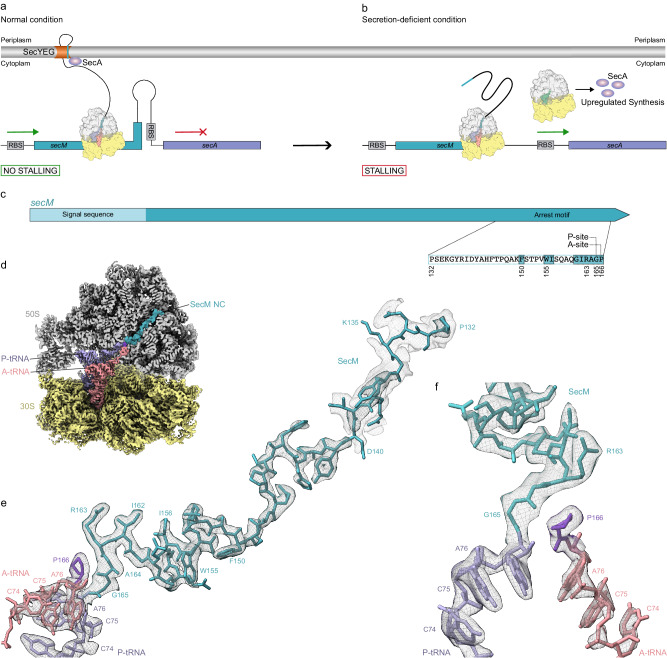
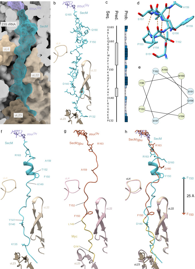
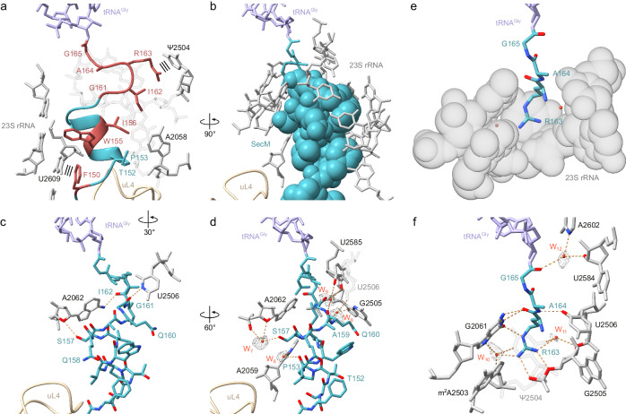
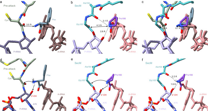
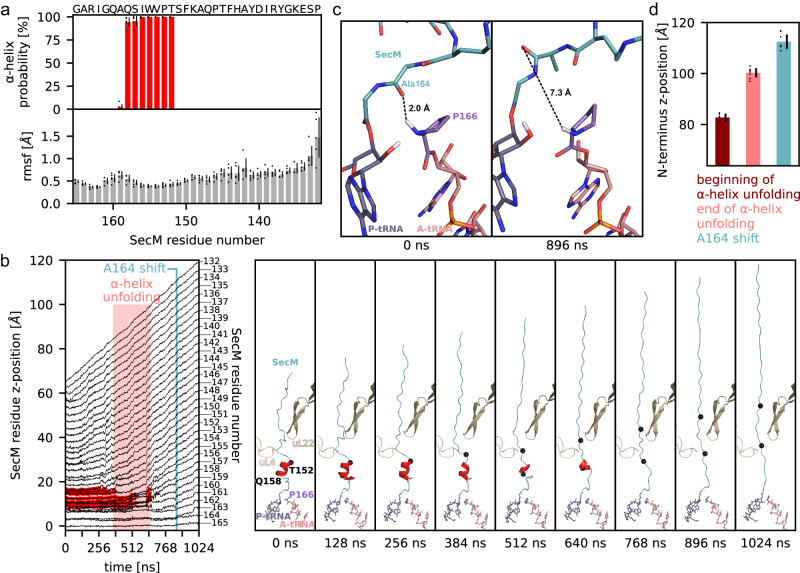
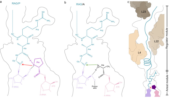
Nascent polypeptide chains can induce translational stalling to regulate gene expression. This is exemplified by the E. coli secretion monitor (SecM) arrest peptide that induces translational stalling to regulate expression of the downstream encoded SecA, an ATPase that co-operates with the SecYEG translocon to facilitate insertion of proteins into or through the cytoplasmic membrane. Here we present the structure of a ribosome stalled during translation of the full-length E. coli SecM arrest peptide at 2.0 Å resolution. The structure reveals that SecM arrests translation by stabilizing the Pro-tRNA in the A-site, but in a manner that prevents peptide bond formation with the SecM-peptidyl-tRNA in the P-site. By employing molecular dynamic simulations, we also provide insight into how a pulling force on the SecM nascent chain can relieve the SecM-mediated translation arrest. Collectively, the mechanisms determined here for SecM arrest and relief are also likely to be applicable for a variety of other arrest peptides that regulate components of the protein localization machinery identified across a wide range of bacteria lineages.


Conflict of interest statement

The authors declare no competing interests.

Figures

Fig. 1
Fig. 1. Regulation of SecM and cryo-EM structure of SecM-SRC.
a Schematic representation of secM-secA mRNA illustrating the stem-loop structure at the stalling site of secM leader peptide (teal) that sequesters the ribosome-binding site (RBS) of the secA gene (lavender) thereby preventing secA translation. By SecYEG-mediated SecM translocation, SecM-induced stalling is relieved. b Upon stalling of the ribosome at the stalling site of secM (teal) the ribosome-binding site of the secA gene (lavender) becomes accessible and translation of secA starts. c Schematic representation of the SecM gene used in the SRC formation with SecM signal sequence and arrest motif as well as functionally relevant amino acids, A- and P-site of the arrest motif indicated. d Cryo-EM map of the 3D-refined E. coli SecM-SRC with transverse section of the 50S (grey) to reveal density for the nascent chain (teal), P-tRNA (lavender), proline 166 of SecM (grape), A-tRNA (salmon) and 30S (yellow). e, f Two views showing the cryo-EM map density (black mesh) for A- and P-site tRNA as well as the attached nascent chain and proline of the 3D refined E. coli SecM-SRC. The P-site tRNA (lavender) bears the SecM nascent chain (teal), whereas the A-site tRNA (salmon) carries proline (grape). Additional density at lower threshold for N-terminal part of nascent chain (grey mesh) in (e).
Fig. 2
Fig. 2. Formation of an α-helix inside the NPET by the SecM peptide.
a Transverse section of the NPET shown as surface (grey) with P-tRNA (lavender) and SecM (teal) in relation to uL4 (light gold), uL22 (gold) and uL23 (dark gold) in surface representation. b Cryo-EM density (transparent teal, threshold 0.008/∼2.6 σ) and model of SecM (teal) attached to the P-tRNA (lavender) in relation to uL4 (light gold), uL22 (gold) and uL23 (dark gold). c Secondary structure prediction (Pred.) and probability (Prob.) of the SecM (Seq.) inside the NPET determined using PSIPRED. d Helix region of SecM (teal) inside the NPET and potential hydrogen bonds shown as dashed orange lines. e Downward cross-sectional view of the SecM helix axis with non-polar amino acids coloured in yellow and polar amino acids coloured in blue. f Structure of SecM (teal) in ribbon representation attached to the P-tRNA (lavender) in relation to uL4 (light gold), uL22 (gold) and uL23 (dark gold). g Myc-SecM (PDB ID 3JBU) attached to the P-tRNA (tangerine/yellow) in relation to uL4 (light rose), uL22 (rose) and uL23 (dark rose). h Overlay (aligned on basis of 23S rRNA) of (f) SecM and (g) SecM3jbu with distance between F150 position from the two models arrowed.
Fig. 3
Fig. 3. Interactions of SecM with components of the NPET.
Interactions of a N-terminal and b, c middle part of SecM (teal) inside the NPET with 23S rRNA (grey), uL4 (light gold), uL22 (gold) and uL23 (dark gold). In (a) and (b) direct interactions are shown whereas in (c) water-mediated interactions for the middle part of SecM (teal) are indicated. Potential hydrogen bonds are shown as dashed orange lines, stacking interactions as three parallel lines and water molecules as red spheres with meshed density.
Fig. 4
Fig. 4. Interactions of SecM stalling motif with components of the NPET.
a SecM arrest peptide (teal) with surrounding 23S rRNA (grey) and residues of the SecM stalling motif highlighted in red. Stacking interactions depicted as three parallel lines. b Space filling representation of SecM stalling motif (teal) from F150 to I162 with surrounding 23S rRNA (grey). c Direct interactions of SecM stalling motif (teal) from F150 to I162 with 23S rRNA (grey). d Water-mediated interactions of SecM stalling motif (teal) from F150 to I162 with 23S rRNA (grey). e Space filling representation of a distinct pocket of the NPET formed by 23S rRNA (grey) and entering SecM 163RAG165 (teal). f Direct and water-mediated interactions of SecM 163RAG165 (teal) with 23S rRNA (grey). Potential hydrogen bonds are shown as dashed orange lines and water molecules as red spheres (with meshed density).
Fig. 5
Fig. 5. Model for SecM-mediated PTC arrangement leading to translational stalling.
a View of the PTC of a pre-attack state (PDB ID 8CVK), showing a tripeptidyl-NH-tRNA (green/dark lavender) at the P-site and a phenyl-NH-tRNA (slate blue/brown) at the A-site. The distance between the attacking amine of the A-tRNA and the carbonyl carbon of the P-site is indicated by a double arrow. b Same view as (a), but for SecM-SRC with SecM-tRNA (teal/lavender) in the P-site and Pro-tRNA (grape/salmon) in the A-site. c Overlay of (a) and (b) (aligned on the basis of 23S rRNA) highlighting the difference in the distance between the attacking amino groups at the A-site and the carbonyl carbon at the P-site. d Schematic view of the PTC of a pre-attack from (a), but with hydrogen atoms (white) modelled in silico for the amino group of the phenylalanine in the A-site. Black spheres indicate the lone pair electrons that make the nucleophilic attack (arrowed) on the carbonyl carbon of the cysteine attached to the P-site tRNA. e Same schematic as (b), but with hydrogen atom (white) modelled in silico towards the 2′OH of A76 of the P-site tRNA, which would allow a nucleophilic attack (arrowed) on the carbonyl carbon of the Gly165 attached to the P-site tRNA. f Same schematic as (e) but with the hydrogen atom (white) modelled towards the carbonyl of Ala164, a conformation that would prohibit any nucleophilic attack.
Fig. 6
Fig. 6. MD simulations of the stalling release by pulling on N-terminus.
a Probability of SecM residues being in an α-helix and their root mean square fluctuations in the absence of a pulling force. Mean (bars) and standard deviations (black lines) are shown for 5 independent simulations (circles). b Left panel: For one pulling simulation (length 1024 ns), positions of SecM residues along the tunnel axis are shown as a function of time. Initial G165 position is set to zero. Residues in α-helix secondary structure are highlighted in red. Unfolding of α-helix and beginning of A164 shift are indicated by light red rectangle and teal vertical line, respectively. Right panel: intermediate structures at indicated times. c Conformation of the PTC before pulling and after the A164 shift. Distance between Pro166 and A164 carbonyl oxygen. d Mean and standard deviation of N-terminus position at beginning (dark red) and end of α-helix unfolding (light red) as well as A164 shift (teal) are shown. Mean (bars) and standard deviations (black lines) are shown for 8 independent simulations (circles). DSSP was used to assign α-helices. Source data be obtained from Zenodo (10.5281/zenodo.10492465).
Fig. 7
Fig. 7. Model for SecM-mediated translational arrest.
Schematic representation of the (a) RAG/P arrest module from SecM and (b) non-stalling RAG/A motif in the PTC. c SecM stalling is strongly driven by the RAG-P arrest module, however, the N-terminal regulator module also modulates and fine-tunes the stalling efficiency.

Similar articles

Cited by

References

    1. Wilson DN, Beckmann R. The ribosomal tunnel as a functional environment for nascent polypeptide folding and translational stalling. Curr. Opin. Struct. Biol. 2011;21:1–10. doi: 10.1016/j.sbi.2011.01.007. - DOI - PubMed
    1. Ito K, Chiba S. Arrest peptides: cis-acting modulators of translation. Annu. Rev. Biochem. 2013;82:171–202. doi: 10.1146/annurev-biochem-080211-105026. - DOI - PubMed
    1. Wilson DN, Arenz S, Beckmann R. Translation regulation via nascent polypeptide-mediated ribosome stalling. Curr. Opin. Struct. Biol. 2016;37:123–133. doi: 10.1016/j.sbi.2016.01.008. - DOI - PubMed
    1. Dever TE, Ivanov IP, Sachs MS. Conserved upstream open reading frame nascent peptides that control translation. Annu. Rev. Genet. 2020;54:237–264. doi: 10.1146/annurev-genet-112618-043822. - DOI - PMC - PubMed
    1. Nakatogawa H, Ito K. Secretion monitor, SecM, undergoes self-translation arrest in the cytosol. Mol. Cell. 2001;7:185–192. doi: 10.1016/S1097-2765(01)00166-6. - DOI - PubMed

MeSH terms