

Blunted brain responses to neutral faces in healthy first-degree relatives of patients with schizophrenia: an image-based fMRI meta-analysis
- PMID: 38503766
- PMCID: PMC10951276
- DOI: 10.1038/s41537-024-00452-6
Blunted brain responses to neutral faces in healthy first-degree relatives of patients with schizophrenia: an image-based fMRI meta-analysis
Abstract
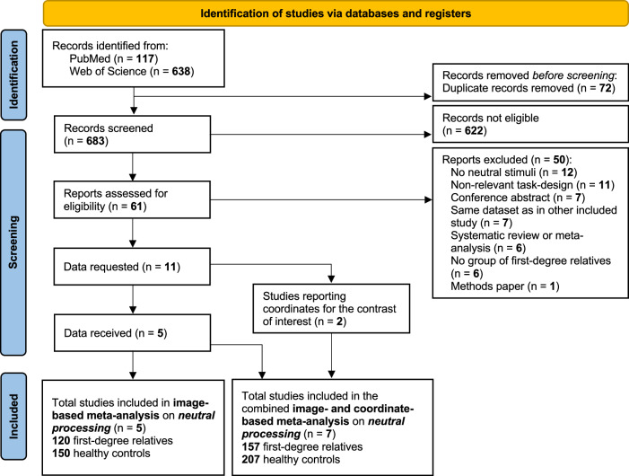
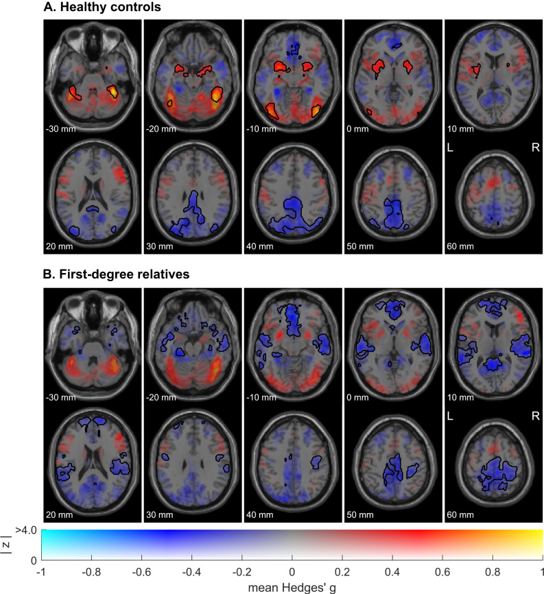
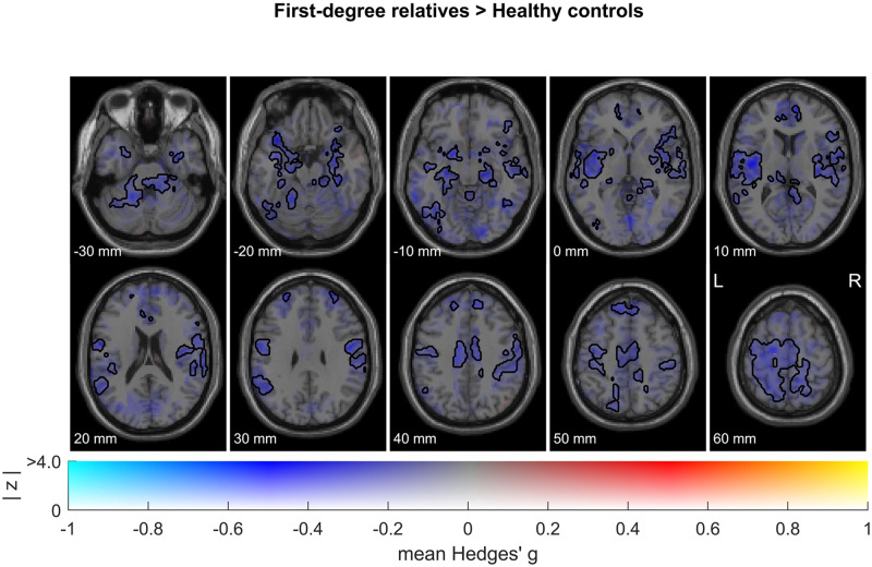
Schizophrenia is characterized by the misattribution of emotional significance to neutral faces, accompanied by overactivations of the limbic system. To understand the disorder's genetic and environmental contributors, investigating healthy first-degree relatives is crucial. However, inconsistent findings exist regarding their ability to recognize neutral faces, with limited research exploring the cerebral correlates of neutral face processing in this population. Thus, we here investigated brain responses to neutral face processing in healthy first-degree relatives through an image-based meta-analysis of functional magnetic resonance imaging studies. We included unthresholded group-level T-maps from 5 studies comprising a total of 120 first-degree relatives and 150 healthy controls. In sensitivity analyses, we ran a combined image- and coordinate-based meta-analysis including 7 studies (157 first-degree relatives, 207 healthy controls) aiming at testing the robustness of the results in a larger sample of studies. Our findings revealed a pattern of decreased brain responses to neutral faces in relatives compared with healthy controls, particularly in limbic areas such as the bilateral amygdala, hippocampus, and insula. The same pattern was observed in sensitivity analyses. These results contrast with the overactivations observed in patients, potentially suggesting that this trait could serve as a protective factor in healthy relatives. However, further research is necessary to test this hypothesis.
© 2024. The Author(s).
Conflict of interest statement
G.B. has received lecture fees from Lundbeck. E.F. has received consultancy fees from Bristol Myers Squibb, Janssen-Cilag, Lilly, Lundbeck, Otsuka Pharmaceutical Co., Ltd., Recordatti, and Sanofi and has lectured for AbbVie, AstraZeneca, Bristol Myers Squibb, Janssen-Cilag, Lundbeck, Otsuka, MSD, and Sanofi. M.J.S. is a full-time employee of Winterlight Labs. All other authors report no biomedical financial interest or potential conflicts of interest.
Figures
References
Publication types
Grants and funding
LinkOut - more resources
Full Text Sources
Miscellaneous
