. 2024 Aug;20(8):981-990.
doi: 10.1038/s41589-024-01578-5. Epub 2024 Mar 19.

Design of amyloidogenic peptide traps

Affiliations

Design of amyloidogenic peptide traps

Danny D Sahtoe et al. Nat Chem Biol. 2024 Aug.

Abstract

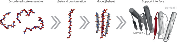
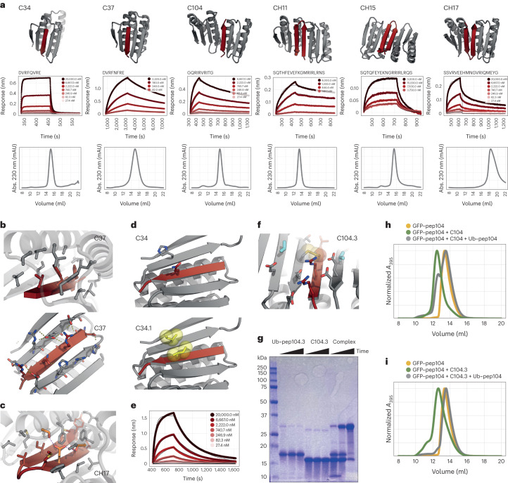
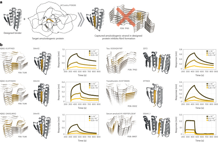
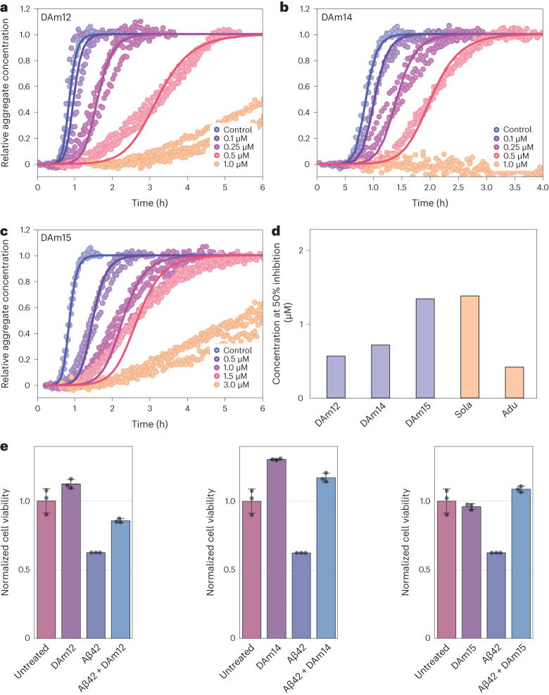
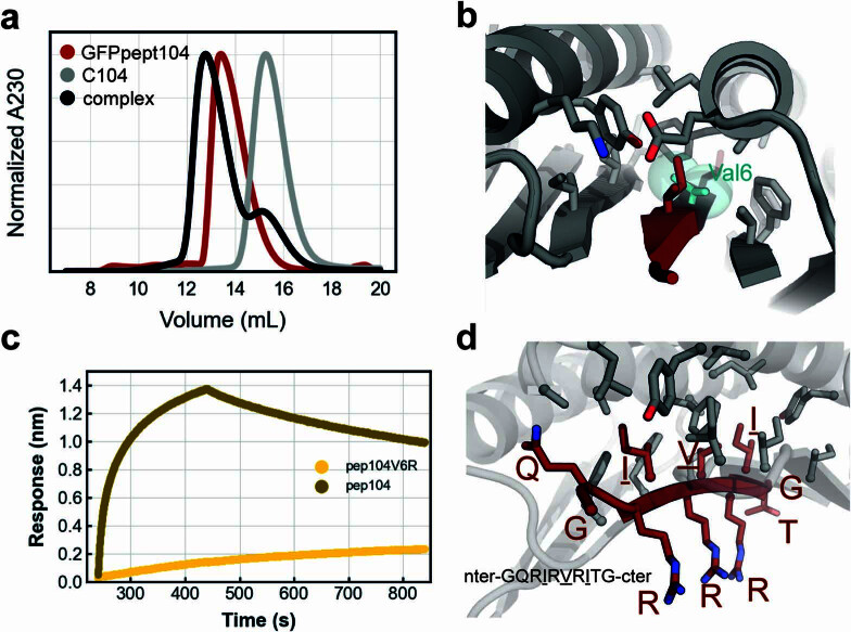
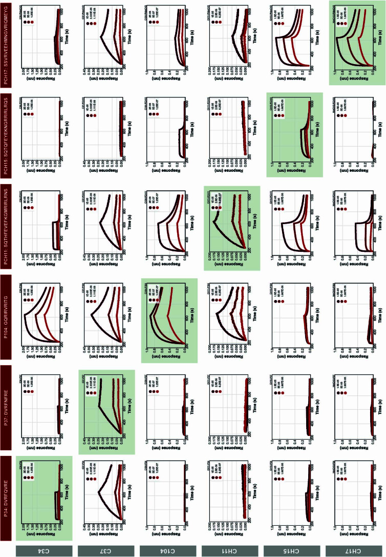
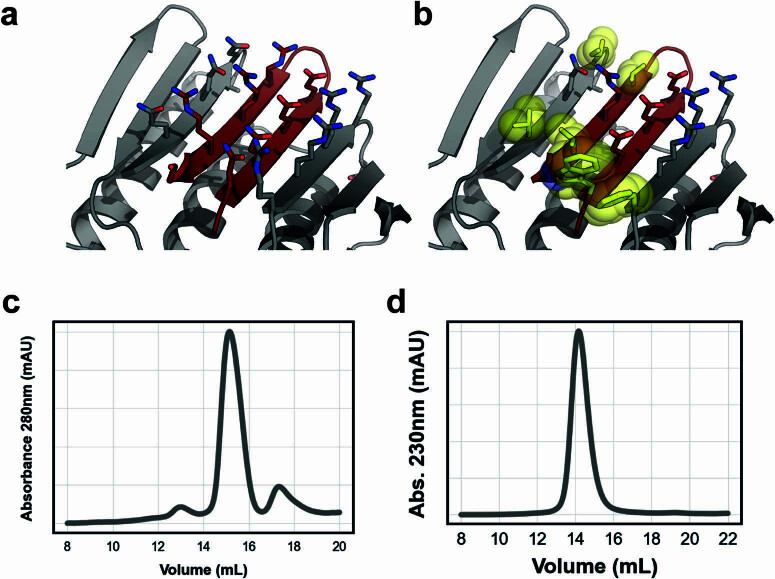
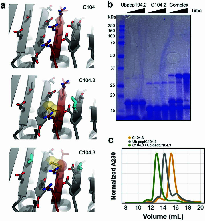
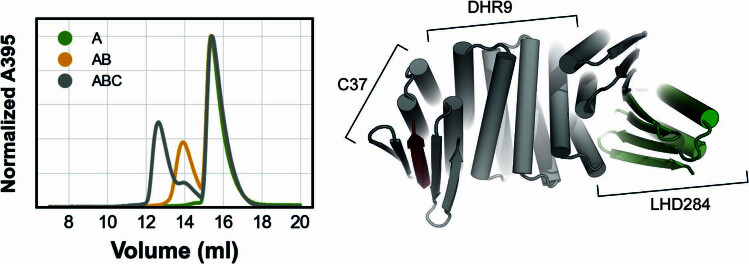
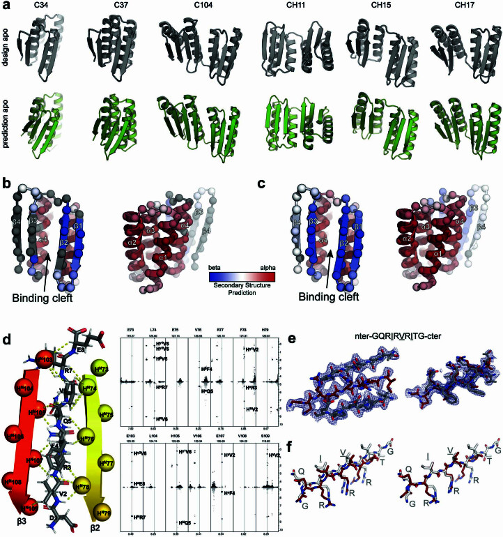
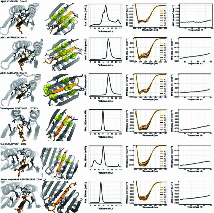
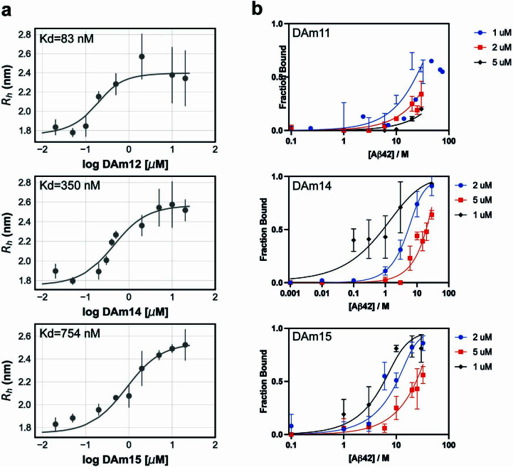
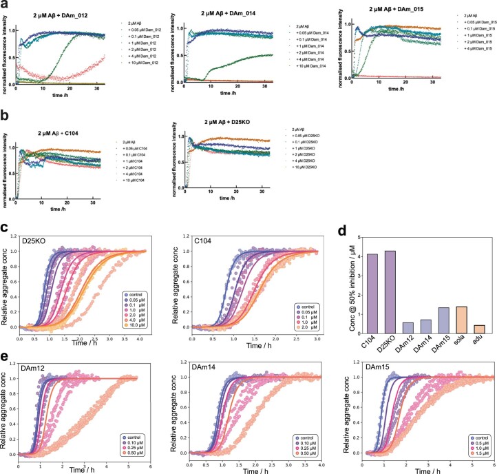
Segments of proteins with high β-strand propensity can self-associate to form amyloid fibrils implicated in many diseases. We describe a general approach to bind such segments in β-strand and β-hairpin conformations using de novo designed scaffolds that contain deep peptide-binding clefts. The designs bind their cognate peptides in vitro with nanomolar affinities. The crystal structure of a designed protein-peptide complex is close to the design model, and NMR characterization reveals how the peptide-binding cleft is protected in the apo state. We use the approach to design binders to the amyloid-forming proteins transthyretin, tau, serum amyloid A1 and amyloid β1-42 (Aβ42). The Aβ binders block the assembly of Aβ fibrils as effectively as the most potent of the clinically tested antibodies to date and protect cells from toxic Aβ42 species.


Conflict of interest statement

D.D.S., H.L.H. and D.B. are inventors on a PCT patent (PCT/US2024/010806) filed by the University of Washington that covers the use of the designed proteins and their variants described in this work. The remaining authors declare no competing interests.

Figures

Fig. 1
Fig. 1. Design approach for binding disordered protein fragments.
Intrinsically disordered regions of proteins and peptides have large conformational freedom but may be forced into predefined conformations such as β-strands that can be efficiently targeted by modeling a β-sheet that hydrogen bonds with the peptide. The interaction is stabilized by additional secondary structure elements supported by a two-domain single-chain protein flanking each side of the target peptide.
Fig. 2
Fig. 2. Characterization of designed peptide binders.
a, Design models for peptide binders (binder, gray; peptide, dark red). BLI traces with kinetic fits and SEC (S75 Increase 10/300) chromatograms of the purified binders are shown below the corresponding models. mAU, milli absorbance units. b, Detailed views of the solvent-exposed interface (bottom) and the buried interface (top) of C37. C-α atoms as spheres. c, Detailed view of the buried part of the interface of hairpin binder CH17 with the designed hydrogen-bond network depicted in orange sticks. d, Models of parent design C34 (top) and C34.1 (bottom) where a hydrophobic interaction pair (yellow sticks/spheres) is introduced to improve affinity. e, BLI traces of C34.1 binding to its peptide immobilized on biosensors. f, View of the designed interface disulfide on C104.3 (disulfide in spheres and sticks; additional redesigned residues in cyan). g, Representative nonreducing SDS−PAGE gel showing disulfide formation (time points of 0 min, 90 min and overnight). The experiment was reproduced twice with two independent protein preparations. Ub, ubiquitin. h, SEC traces of preformed noncovalent C104 complex + GFP−pep104. i, SEC traces of preformed covalent disulfide-linked C104.3 complex + GFP−pep104. Source data
Fig. 3
Fig. 3. Designed peptide−binder pairs function in mammalian cells.
a, Representative fluorescence microscopy images of mScartlet−CH15.1 localization to membranes in HeLa cells. Scale bars, 10 μm. PH, Pleckstrin homology domain. b, Representative fluorescence microscopy images of mScartlet−CH15.1 localizing to designed intracellular GFP-positive protein puncta in HeLa cells. Scale bars, 10 μm. Results were reproduced in two independent experiments. Source data
Fig. 4
Fig. 4. Structural characterization.
a, NMR spectra of 15N-labeled C34 in the absence (top) and presence (bottom) of tenfold excess target peptide, 25 °C. b, Secondary structure propensity as a function of residue, based on backbone 1H, 13C and 15N chemical shifts recorded at 50 °C using the SSP program. SSP scores for the apo form are shown with open circles, while those for the peptide-bound state are indicated with bars. The putative secondary structure of the designed protein is indicated above the plot. Positive values of SSP indicate α-helical structure, while negative values denote β-strands. c, 15N transverse relaxation rates as a function of residue. Low values, such as those in putative β4, indicate rapid timescale dynamics and are consistent with poorly formed structure. d, Designed model of C34. e, Left, overlay of the design model of a surface-redesigned version of C104 (gray) and the crystal structure (colors). Right; detailed interface view of the design (gray) and crystal structure (colors) with Ile8 shift indicated by the orange dashed arrow. f, Binding of CH15.1 to its hairpin peptide (left) or to the individual N-terminal strand (middle) or C-terminal strand (right) of the hairpin in BLI. Source data
Fig. 5
Fig. 5. Design of amyloid peptide traps.
a, Schematic illustration showing that designed amyloidogenic peptide binders (left, gray) can bind amyloidogenic sequences (yellow ribbon) that otherwise form amyloid fibrils through strand−strand interactions (right) and block fibril formation. AF2, AlphaFold2. b, Models of designs (middle column, designed binder in gray and target peptide in yellow) that bind amyloidogenic fragments (left column) from five different amyloid-forming proteins in BLI experiments (right column, legend in molar units). Source data
Fig. 6
Fig. 6. Inhibition of fibril formation.
ac, Aβ42 binders DAm12 (a), DAm14 (b) and DAm15 (c) strongly inhibit fibril formation at submicromolar concentrations in a ThT aggregation assay. Points are ThT fluorescence measurements; solid lines are fits of the kinetics expected when inhibitor binds Aβ42 monomer with the above-measured affinity and also inhibits secondary nucleation by direct interactions with the aggregates. d, Comparison of the Aβ42 aggregation inhibitory potential of the designed binders and clinical antibodies based on the concentration of inhibitor at which the aggregation reaction has been slowed by a fixed amount (that is, the half-time of aggregation (t½) is increased by 50%). Lower values indicate higher potency. The values for the clinical antibodies solanezumab (Sola) and aducanumab (Adu) are from ref. . e, DAm12, DAm14 and DAm15 protect neuroblastoma cells from Aβ42 toxicity. Cell viability was measured using the MTS assay at the aggregation half-time (where 50% of available Aβ42 protein has converted into aggregates and the highest concentration of cytotoxic oligomers is observed), in the presence of 1 µM Aβ42 and 2 µM of the designed binder. Data are presented as mean values ± s.d. (n = 3 individual wells as replicates). Source data
Extended Data Fig. 1
Extended Data Fig. 1. Designed peptide binder controls.
a, SEC binding assay showing that a fusion protein between GFP and 104 peptide binds to the C104 design on a S75 increase 10/300. b, Close-up view of the buried part of the C104 interface with Val6 shown in cyan sticks and spheres. Binder in gray and peptide in dark red. c, Biolayer interferometry trace of C104 binding to base peptide 104 and to a peptide with a V6R substitution. d, Interface close up view of C104 highlighting the hydrophobic-hydrophilic pattern of the peptide. Buried residues single letter amino acid identifiers are underlined. Source data
Extended Data Fig. 2
Extended Data Fig. 2. Specificity profile of peptide binder designs in BLI.
Peptides were immobilized onto octet biosensors at equal densities and incubated with all designs in separate experiments at three different binder concentrations. The on-target interactions are indicated with a light green background. The experiment was done for each different peptide from the base designs (Fig. 2a). Source data
Extended Data Fig. 3
Extended Data Fig. 3. Computational affinity maturation by introducing solvent exposed hydrophobic interaction pairs.
a, View of the solvent exposed interface of CH15 (binder gray, peptide dark red). b, View of the redesigned CH15.1 interface. Hydrophobic interaction pairs introduced to the base CH15 scaffold to improve affinity are highlighted in yellow sticks and spheres. Superdex 75 Increase 10/300 GL SEC traces of purified C34.1. (c) and CH15.1 (d). Source data
Extended Data Fig. 4
Extended Data Fig. 4. Disulfide functionalization of C104.
a, Close-up of C104 surface exposed interface (top) and of the disulfide bridge variants C104.2 (middle) and C104.3 (bottom). Disulfide bonds are highlighted with spheres while additional redesigned residues to optimally accommodate the disulfide bridges are highlighted in cyan thicker sticks. Designed binder in gray and peptide in dark red. b, Representative coommassie stained non-reducing SDS-PAGE gel monitoring disulfide bridge formation of C104.2. Time points are t=0, t=90min and t=overnight. Experiment was reproduced twice with 2 independent protein preparations c, Superdex 75 increase 10/300 GL SEC binding assay confirming that the cysteine containing peptide of C104.3 fused to ubiquitin can bind to its designed cysteine containing binding partner C104.3. Source data
Extended Data Fig. 5
Extended Data Fig. 5. Incorporation of C37 into LHD hetero-oligomer system.
Design C37 was rigidly fused to LHD284B_DHR9 (right) creating a single chain protein with two interfaces capable of binding the peptide of C37 and the designed binding partner of LHD284B_DHR9, LHD284A_DHR82. We validated the assembly of this ternary complex in a SEC binding assay on a S200 increase 10/300 GL. A: GFP-peptC37, AB: GFP-peptC37 + LHD284B_DHR9, ABC: GFP-peptC37 + LHD284B_DHR9 + LHD284A_DHR82. Absorbance at 395 nm of the GFP-peptC37 was monitored to assess binding. Source data
Extended Data Fig. 6
Extended Data Fig. 6. Structural characterization peptide binders.
a, AlphaFold2 predictions of designed binder sequences in absence of peptide (bottom row) indicate closure of the binding pocket for some designs. b, Secondary structure prediction from NMR experiments on C34 apo mapped onto the cartoon model (two views) of C34 with C-alpha atoms shown as spheres (peptide not shown). No information available for residues in gray. These residues had broadened resonances due to conformational exchange. c, Same as (b) but for C34 holo (peptide not shown). d, Intermolecular NOE contacts between C34 and the peptide measured as previously described using a sample comprised of a mixture of 450 μM U-{13C,15N} C34 and 450 μM unlabeled peptide. Strips from the 3D dataset are illustrated at the 15N chemical shifts of the amides of the indicated residue (top of panels) showing the detected intermolecular contacts between the amide protons of strands β2/β3 from C34 and the peptide (right panel). The protons linked via the observed NOEs are highlighted on the structure of the designed binder on the left panel. e, Two atomic views with 2mFobs−DFcalc electron density maps contoured at 1.5σ of the strand-strand interaction between the binder and peptide of C104. f, Left, View of peptide in designed model after superposition of entire designed (white) and xray structures (red). Right superposition on only peptides in designed and xray structure.
Extended Data Fig. 7
Extended Data Fig. 7. Characterization amyloidogenic strand binders.
Close-up view of solvent inaccessible part interface (first column), close up view of solvent accessible part of interface with hydrophobic interaction pairs in yellow spheres and sticks (2nd column), SEC trace of purified binder on S75 increase 10/300GL (3rd column), CD wavelength scans (4th column) and CD temperature melt at 222 nm. Source data
Extended Data Fig. 8
Extended Data Fig. 8. Microfluidic diffusional sizing binding experiments.
a, Microfluidic diffusional sizing (MDS) binding isotherms of DAm12, DAm14 and DAm15 binding to Aβ42 monomers. Error bars represent SD (n = 3 independent measurements). b, MDS binding of pre-formed Aβ42 fibrils to designs DAm11 (top), DAm14 (middle) and DAm15 (bottom). Data for each individual measurement point are presented as mean +/− SD (n = 3) of independent replicates. Source data
Extended Data Fig. 9
Extended Data Fig. 9. Aβ42 fibril inhibition.
a-c, ThT Aβ42 fibril inhibition assays of the designed binders and controls that were not designed to inhibit Aβ42 aggregation. d, The inhibitory potential of binders, controls and clinical antibodies against Aβ42 aggregation is compared. See main Fig. 6d. e, Expected inhibitory effect due to monomer binding only. Points are ThT measurements, at a range of binder concentrations. The solid lines are produced by predicting the amount of inhibition at each binder concentration. To do so, we used the affinities of the binders to monomer to calculate the amounts of bound monomer and assumed that any monomer bound is completely removed from the aggregation reaction. Using the fits of the kinetics in the absence of binder, and the reaction orders determined previously, we could then predict the expected inhibition at each binder concentration. Even for the tightest binders and assuming any bound monomer is permanently removed from the reaction, the observed inhibitory potential exceeds that expected to occur by monomer binding alone. This implies additional inhibitory mechanisms beyond interactions with monomeric Aβ42 are active. Source data

References

    1. Tsai, C. J., Xu, D. & Nussinov, R. Protein folding via binding and vice versa. Fold. Des.3, R71–R80 (1998). - PubMed
    1. Wright, P. E. & Dyson, H. J. Linking folding and binding. Curr. Opin. Struct. Biol.19, 31–38 (2009). - PMC - PubMed
    1. Shammas, S. L., Crabtree, M. D., Dahal, L., Wicky, B. I. M. & Clarke, J. Insights into coupled folding and binding mechanisms from kinetic studies. J. Biol. Chem.291, 6689–6695 (2016). - PMC - PubMed
    1. Chen, G.-F. et al. Amyloid beta: structure, biology and structure-based therapeutic development. Acta Pharmacol. Sin.38, 1205–1235 (2017). - PMC - PubMed
    1. Gamblin, T. C. et al. Caspase cleavage of tau: linking amyloid and neurofibrillary tangles in Alzheimer’s disease. Proc. Natl Acad. Sci. USA100, 10032–10037 (2003). - PMC - PubMed

Publication types

LinkOut - more resources