. 2024 Mar 19;14(1):6582.
doi: 10.1038/s41598-024-57294-6.

Role of the fatty pancreatic infiltration in pancreatic oncogenesis

Affiliations

Role of the fatty pancreatic infiltration in pancreatic oncogenesis

Sonia Frendi et al. Sci Rep. .

Abstract

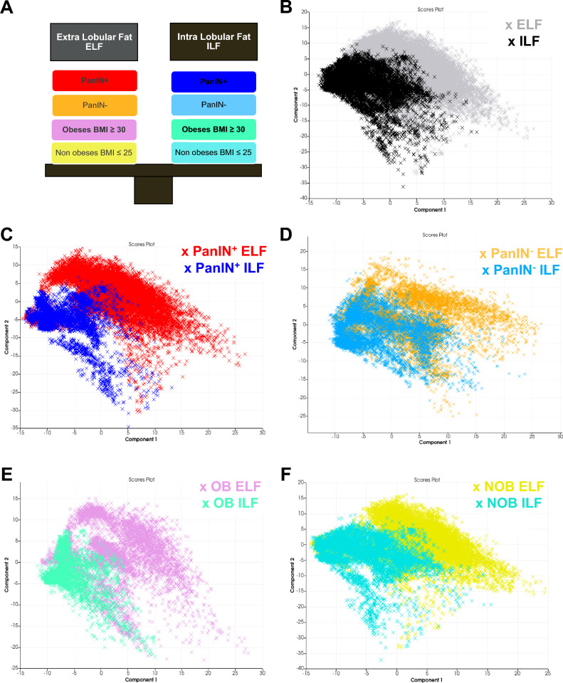
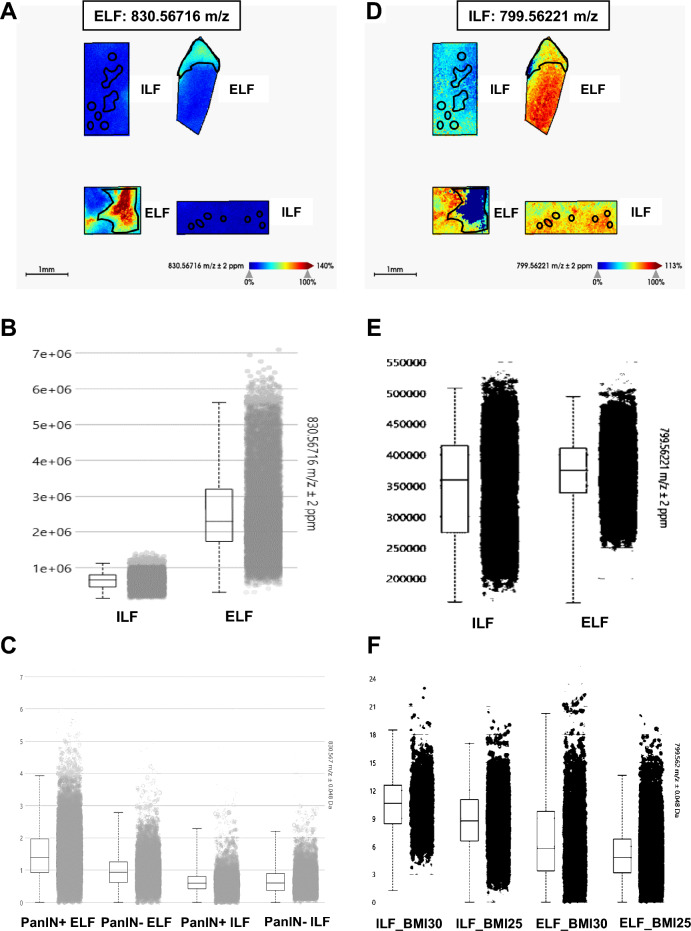
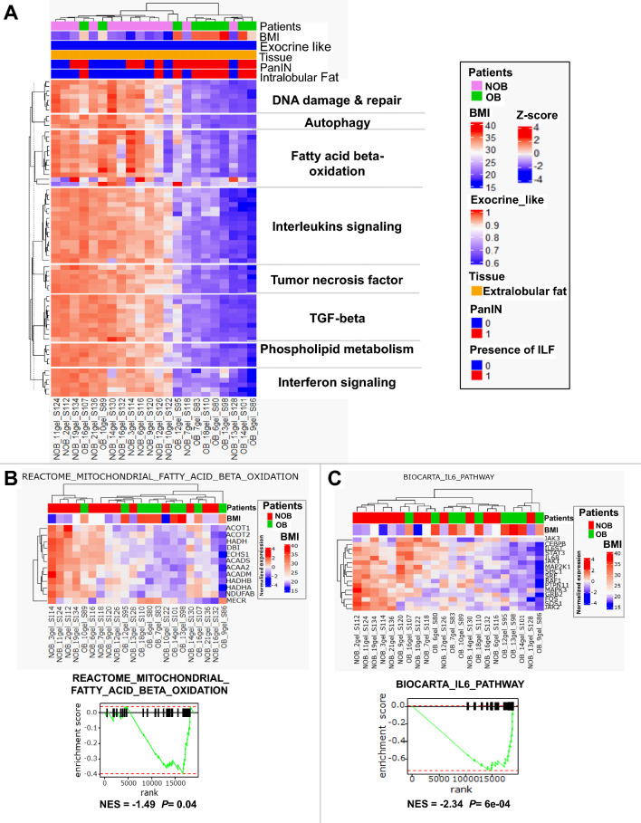
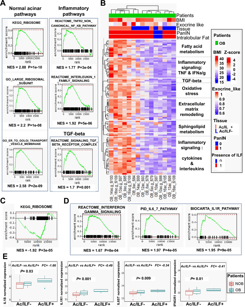
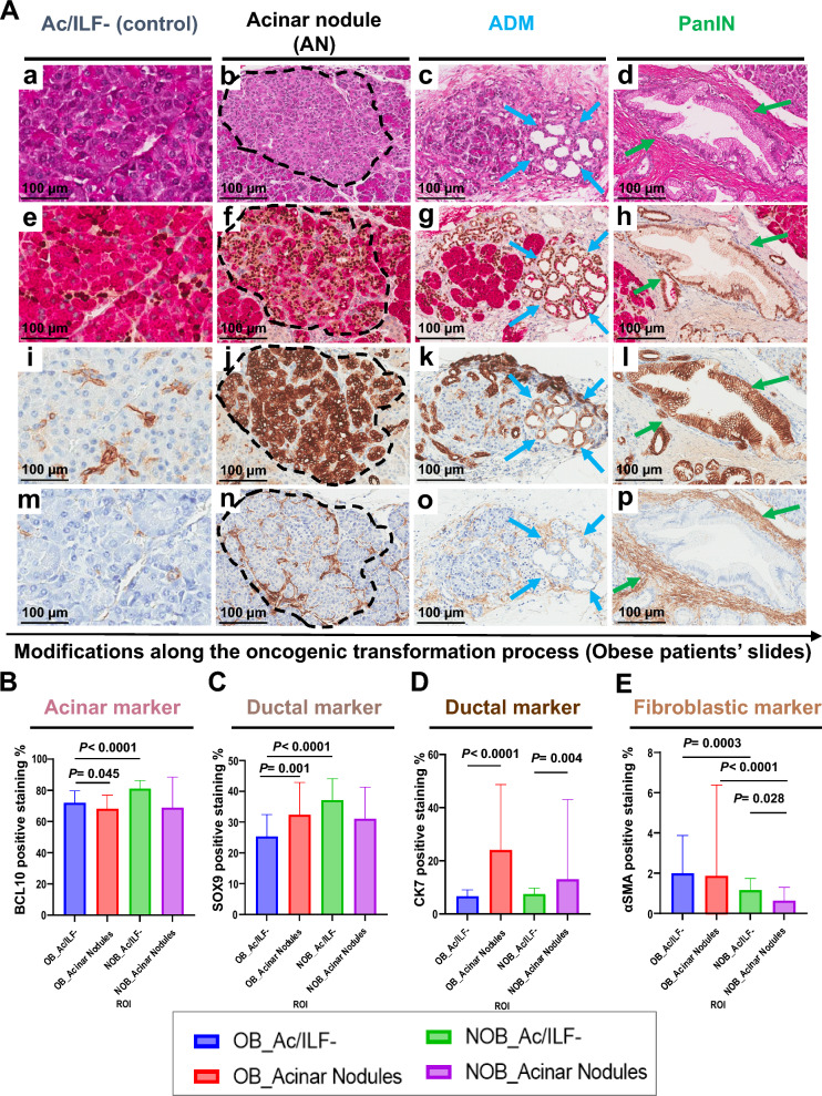
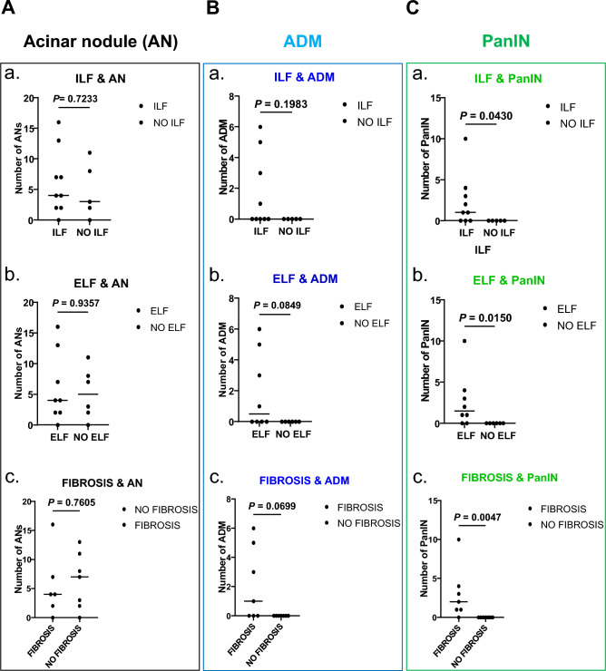
Although pancreatic precancerous lesions are known to be related to obesity and fatty pancreatic infiltration, the mechanisms remain unclear. We assessed the role of fatty infiltration in the process of pancreatic oncogenesis and obesity. A combined transcriptomic, lipidomic and pathological approach was used to explore neoplastic transformations. Intralobular (ILF) and extralobular (ELF) lipidomic profiles were analyzed to search for lipids associated with pancreatic intraepithelial neoplasia (PanINs) and obesity; the effect of ILF and ELF on acinar tissue and the histopathological aspects of pancreatic parenchyma changes in obese (OB) and non-obese patients. This study showed that the lipid composition of ILF was different from that of ELF. ILF was related to obesity and ELF-specific lipids were correlated to PanINs. Acinar cells were shown to have different phenotypes depending on the presence and proximity to ILF in OB patients. Several lipid metabolic pathways, oxidative stress and inflammatory pathways were upregulated in acinar tissue during ILF infiltration in OB patients. Early acinar transformations, called acinar nodules (AN) were linked to obesity but not ELF or ILF suggesting that they are the first reversible precancerous pancreatic lesions to occur in OB patients. On the other hand, the number of PanINs was higher in OB patients and was positively correlated to ILF and ELF scores as well as to fibrosis. Our study suggests that two types of fat infiltration must be distinguished, ELF and ILF. ILF plays a major role in acinar modifications and the development of precancerous lesions associated with obesity, while ELF may play a role in the progression of PDAC.

Keywords: Intra-lobular and extra-lobular fatty pancreatic infiltration; Lipidomic MALDI-TOF imaging mass spectrometry; Obesity; Pancreatic oncogenesis; Pancreatic precancerous lesions.


Conflict of interest statement

The authors declare no competing interests.

Figures

Figure 1
Figure 1
Principal component analysis (PCA) score plots of lipid peaks in OB and NOB patients. (A) Explanatory diagram of the different comparisons (the colors correspond to the representation of sample groups in each PCA). (B) PCA score plots of ILF (black dots) and ELF (gray dots) based on OB and NOB patient data indicating a segmentation between ILF and ELF in all patients. (C) PCA score plots of ILF (blue dots) and ELF (red dots) based on PanIN lesion (PanIN+) patient data allowing a segmentation between ILF and ELF. (D) PCA score plots of ILF (light blue dots) and ELF (orange dots) based on the data from patients without PanIN lesions (PanIN-) resulting in non-segmentation between ELF and ELF. (E and F) PCA score plots of ILF and ELF based on the BMI (OB in E and NOB in F) with a segmentation between ELF and ILF in OB patients. PCA score plots were generated using Scils Lab Pro® software. ILF, pancreatic intralobular fat; ELF, pancreatic extralobular fat; BMI, body mass index; OB, obese patient; NOB, non-obese patient; PanIN, pancreatic intraepithelial neoplasia.
Figure 2
Figure 2
Example of the specific peaks of interest in ILF or ELF analyzed by MALDI FTICR. FTICR image of the m/z 830.56716 ELF and PanINs—specific lipid peak (A) and box plot comparing peaks between ILF and ELF in FTICR analysis (B) or in MALDI TOF analysis according to the presence or absence of PanINs (C). FTICR image of the m/z 799.56221 OB patient ILF-specific lipid peaks (D) and box plot comparing ILF and ELF in FTICR analysis (E) or in MALDI TOF analysis according to BMI (F). ILF, pancreatic intralobular fat; ELF, pancreatic extralobular fat; PanIN, pancreatic intraepithelial neoplasia; BMI, body mass index; OB, obese patients; NOB, non-obese patients.
Figure 3
Figure 3
Gene set variation analysis of enriched pathways in ELF tissue. (A) Heat map of GSVA of pathways that were significantly enriched in the ELF tissue of NOB patients compared to OB patients. (B and C) Heat map of genes involved in beta-oxidation (B) and IL6 (C) enriched pathways and their respective leading edge. OB, obese; NOB, non-obese; BMI, body mass index; PanIN, pancreatic intraepithelial neoplasia; ILF, pancreatic intralobular fat; ELF, pancreatic extralobular fat. NES, normalized enrichment score.
Figure 4
Figure 4
Gene set variation analysis of enriched pathways in acinar tissue. (A) Leading edge of enriched pathways in the Ac/ILF- of OB patients compared to NOB patients. (B) Heat map of the most significantly enriched pathways in Ac/ILF + compared to Ac/ILF- in OB patients. (C) Leading edge of enriched pathways in Ac/ILF- compared to Ac/ILF + in OB patients. (D) Leading edge of enriched inflammatory cytokines in Ac/ILF + compared to Ac/ILF- in OB patients. (E) Boxplot showing the expression of IL1B, IL1R1, IL6ST and IFNGR1 in the Ac/ILF- of OB and NOB patients and in the Ac/ILF + of OB patients. OB, obese patients; NOB, non-obese patients; BMI, body mass index; PanIN, pancreatic intraepithelial neoplasia; NES, normalized enrichment score; ILF, pancreatic intralobular fat; Ac/ILF-, acinar tissue without ILF infiltration; Ac/ILF + , acinar tissue next to ILF infiltration.
Figure 5
Figure 5
Histopathological analyses (H&E and IHC stainings) of quantified ROIs. (A); (ad) H&E staining of Ac/ILF- ROIs as controls, acinar nodules (AN), ADM and PanIN lesions in OB patients. IHC staining of ROIs in and OB patient for BCL10/Sox9 (eh), CK7 (il) and αSMA (mp) of NA-control ROIs, ANs, ADM and PanIN lesions in an OB patient. (B to E) Quantification of IHC staining of BCL10 (B) SOX9 (C), CK7 (D) and αSMA (E) of Ac/ILF- and AN areas from pancreatic tissue of OB and NOB patients. OB, obese; NOB, non-obese; ILF, pancreatic intralobular fat; Ac/ILF-, acinar tissue without ILF infiltration; AN, acinar nodule (black contouring); ADM, acinar-to-ductal metaplasia (blue arrows); PanIN, pancreatic intraepithelial neoplasia (green arrows).
Figure 6
Figure 6
Metaplastic and neoplastic precancerous lesions with ILF/ELF and fibrosis in obese and non-obese patients. (A) Scatter plot analysis of the number of AN in relation to the presence/absence of ILF (a), ELF (b) and fibrosis (c). (B) Scatter plot analysis of the number of ADM lesions in relation to the presence/absence of ILF (a), ELF (b) and fibrosis (c). (C) Scatter plot analysis of the total number of PanINs in relation to the presence/absence of ILF (a), ELF (b) and fibrosis (c). AN, acinar nodule; ADM, acinar-to-ductal metaplasia; PanIN, pancreatic intraepithelial neoplasia; ILF, pancreatic intralobular fat; ELF, pancreatic extralobular fat. Mann–Whitney test was performed for all the analyses.

References

    1. Maisonneuve P. Epidemiology and burden of pancreatic cancer. La Presse Médicale. 2019;48:e113–e123. doi: 10.1016/j.lpm.2019.02.030. - DOI - PubMed
    1. Rawla P, Thandra KC, Sunkara T. Pancreatic cancer and obesity: Epidemiology, mechanism, and preventive strategies. Clin. J. Gastroenterol. 2019;12:285–291. doi: 10.1007/s12328-019-00953-3. - DOI - PubMed
    1. Huang J, Lok V, Ngai CH, et al. Worldwide burden of, risk factors for, and trends in pancreatic cancer. Gastroenterology. 2021;160:744–754. doi: 10.1053/j.gastro.2020.10.007. - DOI - PubMed
    1. Avgerinos KI, Spyrou N, Mantzoros CS, et al. Obesity and cancer risk: Emerging biological mechanisms and perspectives. Metabolism. 2019;92:121–135. doi: 10.1016/j.metabol.2018.11.001. - DOI - PubMed
    1. Renehan AG, Tyson M, Egger M, et al. Body-mass index and incidence of cancer: a systematic review and meta-analysis of prospective observational studies. Lancet. 2008;371:569–578. doi: 10.1016/S0140-6736(08)60269-X. - DOI - PubMed

MeSH terms