

Near-real-time Mueller polarimetric image processing for neurosurgical intervention
- PMID: 38503943
- PMCID: PMC11178587
- DOI: 10.1007/s11548-024-03090-6
Near-real-time Mueller polarimetric image processing for neurosurgical intervention
Abstract
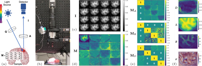
Purpose: Wide-field imaging Mueller polarimetry is a revolutionary, label-free, and non-invasive modality for computer-aided intervention; in neurosurgery, it aims to provide visual feedback of white matter fibre bundle orientation from derived parameters. Conventionally, robust polarimetric parameters are estimated after averaging multiple measurements of intensity for each pair of probing and detected polarised light. Long multi-shot averaging, however, is not compatible with real-time in vivo imaging, and the current performance of polarimetric data processing hinders the translation to clinical practice.
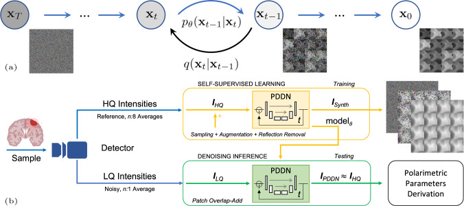
Methods: A learning-based denoising framework is tailored for fast, single-shot, noisy acquisitions of polarimetric intensities. Also, performance-optimised image processing tools are devised for the derivation of clinically relevant parameters. The combination recovers accurate polarimetric parameters from fast acquisitions with near-real-time performance, under the assumption of pseudo-Gaussian polarimetric acquisition noise.
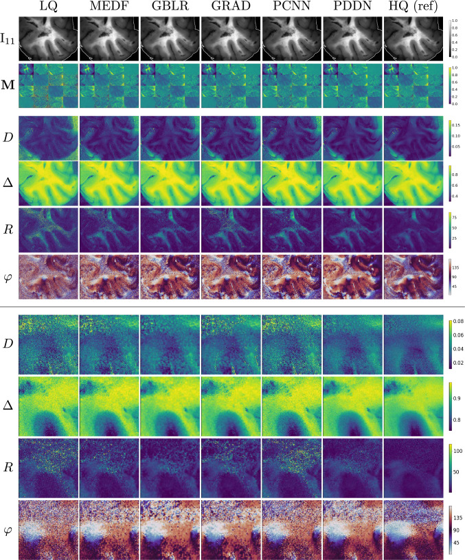
Results: The denoising framework is trained, validated, and tested on experimental data comprising tumour-free and diseased human brain samples in different conditions. Accuracy and image quality indices showed significant ( ) improvements on testing data for a fast single-pass denoising versus the state-of-the-art and high polarimetric image quality standards. The computational time is reported for the end-to-end processing.
Conclusion: The end-to-end image processing achieved real-time performance for a localised field of view ( ). The denoised polarimetric intensities produced visibly clear directional patterns of neuronal fibre tracts in line with reference polarimetric image quality standards; directional disruption was kept in case of neoplastic lesions. The presented advances pave the way towards feasible oncological neurosurgical translations of novel, label-free, interventional feedback.
Keywords: AI; Mueller polarimetric imaging; Neurosurgery; Real-time denoising.
© 2024. The Author(s).
Figures


References
-
- Qi J, Elson DS (2017) Mueller polarimetric imaging for surgical and diagnostic applications: a review. J Biophoton 10(8):950–982. 10.1002/jbio.201600152 - PubMed
-
- Li X, Han Y, Wang H, Liu T, Chen SC, Hu H (2022) Polarimetric imaging through scattering media: a review. Front Phys 10:815296. 10.3389/fphy.2022.815296
-
- Ramella-Roman JC, Saytashev I, Piccini M (2020) A review of polarization-based imaging technologies for clinical and preclinical applications. J Opt 22(12):123001. 10.1088/2040-8986/abbf8a
-
- Baba JS, Chung J-R, DeLaughter AH, Cameron BD, Cote GL (2002) Development and calibration of an automated Mueller matrix polarization imaging system. J Biom Opt 7(3):341–349. 10.1117/1.1486248 - PubMed
-
- Azzam RMA (2016) Stokes-vector and Mueller-matrix polarimetry. J Opt Soc Am A 33(7):1396–1408. 10.1364/JOSAA.33.001396 - PubMed
