. 2024 Jul;45(7):1349-1365.
doi: 10.1038/s41401-024-01245-4. Epub 2024 Mar 19.

Vagus nerve stimulation as a promising neuroprotection for ischemic stroke via α7nAchR-dependent inactivation of microglial NLRP3 inflammasome

Affiliations

Vagus nerve stimulation as a promising neuroprotection for ischemic stroke via α7nAchR-dependent inactivation of microglial NLRP3 inflammasome

Xiao-Mei Xia et al. Acta Pharmacol Sin. 2024 Jul.

Abstract

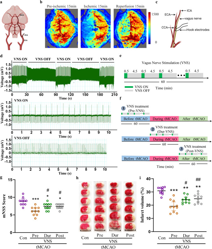
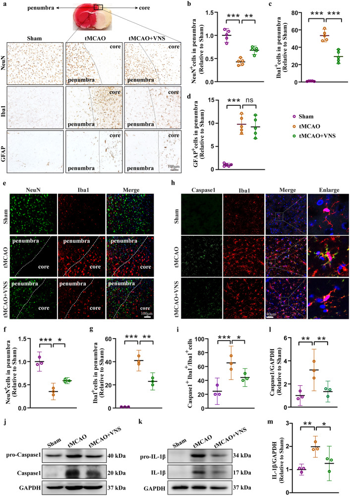
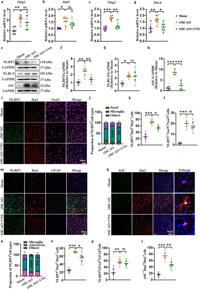
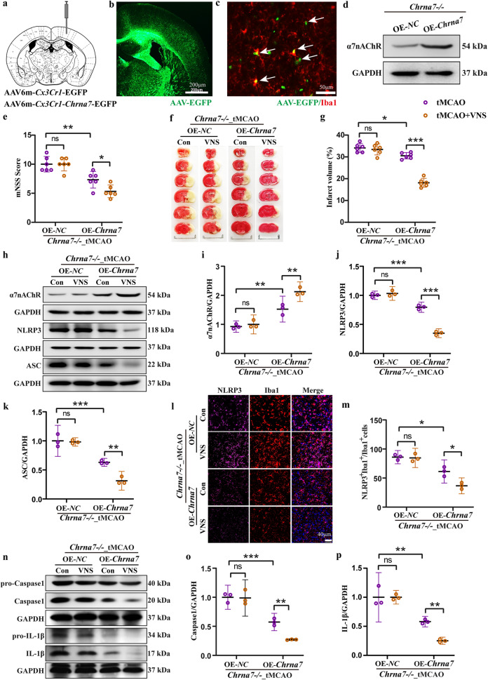
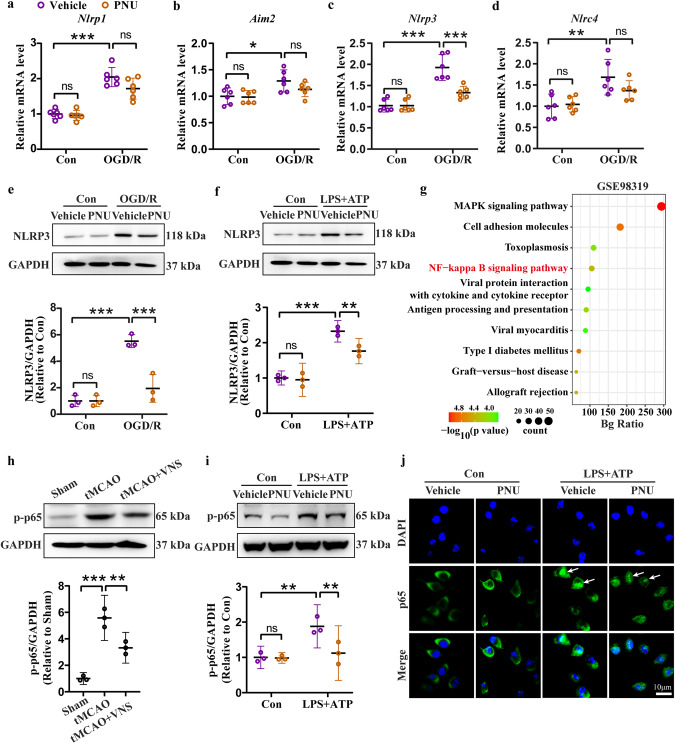
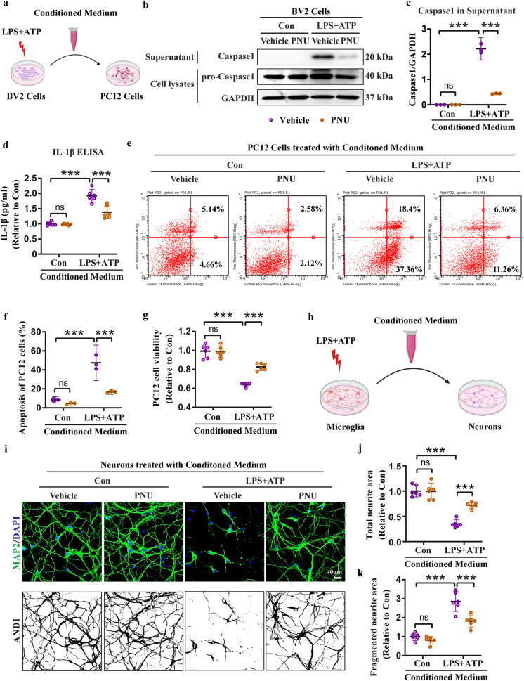
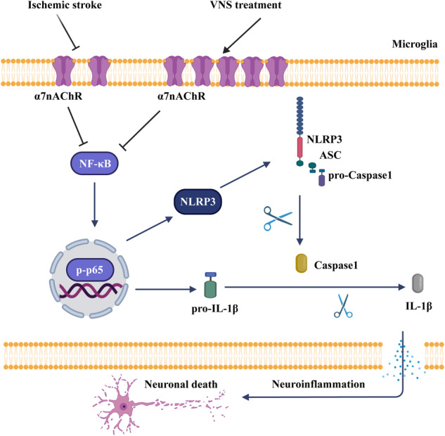
Ischemic stroke is a major cause of disability and death worldwide, and its management requires urgent attention. Previous studies have shown that vagus nerve stimulation (VNS) exerts neuroprotection in ischemic stroke by inhibiting neuroinflammation and apoptosis. In this study, we evaluated the timing for VNS intervention in ischemic stroke, and the underlying mechanisms of VNS-induced neuroprotection. Mice were subjected to transient middle cerebral artery occlusion (tMCAO) for 60 min. The left vagus nerve at cervical level was exposed and attached to an electrode connected to a low-frequency electrical stimulator. Vagus nerve stimulation (VNS) was given for 60 min before, during and after tMCAO (Pre-VNS, Dur-VNS, Post-VNS). Neurological function was assessed 24 h after reperfusion. We found that all the three VNS significantly protected against the tMCAO-induced injury evidenced by improved neurological function and reduced infarct volume. Moreover, the Pre-VNS was the most effective against the ischemic injury. We found that tMCAO activated microglia in the ischemic core and penumbra regions of the brain, followed by the NLRP3 inflammasome activation-induced neuroinflammation, which finally triggered neuronal death. VNS treatment preserved α7nAChR expression in the penumbra regions, inhibited NLRP3 inflammasome activation and ensuing neuroinflammation, rescuing cerebral neurons. The role of α7nAChR in microglial NLRP3 inflammasome activation in ischemic stroke was further validated using genetic manipulations, including Chrna7 knockout mice and microglial Chrna7 overexpression mice, as well as pharmacological interventions using the α7nAChR inhibitor methyllycaconitine and agonist PNU-282987. Collectively, this study demonstrates the potential of VNS as a safe and effective strategy to treat ischemic stroke, and presents a new approach targeting microglial NLRP3 inflammasome, which might be therapeutic for other inflammation-related diseases.

Keywords: NLRP3; ischemic stroke; microglia; neuroinflammation; vagus nerve stimulation; α7 nicotinic acetylcholine receptor.


Conflict of interest statement

The authors declare no competing interests.

Figures

Fig. 1
Fig. 1. VNS treatment improves neurological function and reduces infarct volume in the tMCAO mouse model.
a Illustration of transient middle cerebral artery occlusion. b Reduction in regional cerebral blood flow in the tMCAO model measured by the MoorFLPI full-field laser perfusion imager. c Illustration of vagus nerve stimulation. d Electrocardiograph showing a reduction in heart rate upon VNS treatment. e Illustration of VNS intervention in 60 min. f Illustration of VNS treatment timing in the tMCAO model. g mNSS score of tMCAO mice showing the improvement of VNS treatment in neurological function, n = 10. h, i TTC staining and quantitative analysis showing a reduction in infarct volume by VNS treatment. Scale bar: 1.0 cm, n = 10, *P < 0.05, **P < 0.01, ***P < 0.001 vs. Con group, #P < 0.05, ##P < 0.01 vs. Pre-VNS group.
Fig. 2
Fig. 2. VNS treatment rescues neuronal loss via inhibiting microglia activation-induced neuroinflammation.
ad Representative immunohistochemical images of NeuN, Iba1, and GFAP, and quantification of NeuN positive, Iba1 positive, and GFAP positive cells in the penumbra region showing the rescue of neuronal death and inhibition of microglia activation by VNS treatment, n = 5, scale bar: 100 μm. eg Representative immunofluorescence images of NeuN and Iba1, and quantification of NeuN positive and Iba1 positive cells in the penumbra region showing the rescue of neuronal death and inhibition of microglia activation by VNS treatment, n = 3, scale bar: 100 μm. h, i Representative immunofluorescence images and quantification of Caspase1 and Iba1 positive cells showing the inhibition of microglial inflammasome activation by VNS treatment, n = 3, scale bar: 40 μm. jm Representative blots and quantitative analyses showing the reduction in the expression of caspase1 and IL-1β by VNS treatment, n = 3. *P < 0.05, **P < 0.01, ***P < 0.001, ns no significance.
Fig. 3
Fig. 3. VNS treatment inhibits microglial NLRP3 inflammasome activation in the penumbra region of tMCAO mice.
ad Effects of VNS treatment on mRNA levels of Nlrp1, Aim2, Nlrp3, and Nlrc4 in the penumbra area of tMCAO mice examined by RT-PCR, n = 6. eh Representative blots quantitative analyses showing the effects of VNS treatment on the expression of NLRP3, NLRC4, and ASC in the penumbra area of tMCAO mice, n = 3–4. il Representative immunofluorescence images and quantification of NLRP3 positive cells in neurons and microglia, n = 3, *P < 0.05, **P < 0.01, ***P < 0.001 vs. Sham groups (NLRP3 positive microglia in j), scale bar: 40 μm. mp Representative immunofluorescence images and quantification of NLRP3 positive cells in microglia and astrocytes, n = 3, **P < 0.01, ***P < 0.001 vs. Sham groups (NLRP3 positive microglia in n), scale bar: 40 μm. q, r Representative immunofluorescence images and quantification of ASC positive microglia, n = 3, scale bar: 40 μm. *P < 0.05, **P < 0.01, ***P < 0.001, ns no significance.
Fig. 4
Fig. 4. VNS treatment preserves α7nAChR expression in the penumbra area of tMCAO mice.
a Heatmap showing the differentially expressed genes related to nicotinic acetylcholine receptors reanalyzed using GSE98319 dataset. bj Effects of VNS treatment on the mRNA levels of Chrns in the penumbra area, n = 6. k, l Representative blots and quantification of α7nAChR showing the upregulation of α7nAChR by VNS treatment in the penumbra area of tMCAO mice, n = 3. *P < 0.05, **P < 0.01, ***P < 0.001, ns no significance.
Fig. 5
Fig. 5. Pharmacological inhibition of α7nAChR abrogates the protective effect of VNS treatment on ischemic injury.
a mNSS score showing the blockade of VNS-improved neurological function in the MLA-treated mice subjected to tMCAO model, n = 6. b, c TTC staining and quantification showing the blockade of VNS-induced reduction in infarct volume in the MLA-treated mice subjected to tMCAO model. Scale bar: 1.0 cm, n = 6. df Representative blots and quantification of NLRP3 and ASC in the penumbra area, n = 3. g, h Representative immunofluorescence images and quantification of NLRP3 positive microglia, n = 3, scale bar: 40 μm. ik Representative blots and quantification of Caspase1 and IL-1β in the penumbra area, n = 3. *P < 0.05, **P < 0.01, ***P < 0.001, ns no significance. Mice were administered MLA at 2.5 mg/kg intraperitoneally 1 h before tMCAO model.
Fig. 6
Fig. 6. Genetic deletion of Chrna7 abolishes the therapeutic effect of VNS treatment on ischemic injury.
a Genotyping of wide type (WT) and Chrna7 knockout (KO) mice, n = 6. b, c Undetectable mRNA level of Chrna7 in the brain of Chrna7 KO mice by qPCR, n = 6. d mNSS score showing the blockade of VNS-improved neurological function in the Chrna7 KO mice subjected to tMCAO model, n = 6. e, f TTC staining and quantification showing the blockade of VNS-induced reduction in infarct volume in the Chrna7 KO mice subjected to tMCAO model. Scale bar: 1.0 cm, n = 6. gi Representative blots and quantification of NLRP3 and ASC in the penumbra area, n = 3. j, k Representative immunofluorescence images and quantification of NLRP3 positive microglia, n = 3, scale bar: 40 μm. ln Representative blots and quantification of Caspase1 and IL-1β in the penumbra area, n = 3. *P < 0.05, **P < 0.01, ***P < 0.001, ns no significance.
Fig. 7
Fig. 7. Overexpression of microglial Chrna7 reinstates the therapeutic efficacy of VNS intervention on ischemic injury.
a Illustration of the injection site of recombinant adeno-associated virus (AAVs). b Representative immunofluorescence image showing the infection area of AAVs, scale bar: 200 μm. c Representative immunofluorescence image showing the infected microglia by AAVs, scale bar: 50 μm. d Representative blots showing the overexpression of α7nAChR in the brain of Chrna7 KO mice. e mNSS score showing the recovery of VNS treatment on neurological function by overexpression of microglial Chrna7 in the Chrna7 KO mice subjected to tMCAO model, n = 6. f, g TTC staining and quantification showing the retrieval of the effect of VNS treatment on reduction in infarct volume by overexpression of microglial Chrna7 in the Chrna7 KO mice subjected to tMCAO model. Scale bar: 1.0 cm, n = 6. hk Representative blots and quantification of α7nAChR, NLRP3, and ASC in the penumbra area, n = 3. l, m Representative immunofluorescence images and quantification of NLRP3 positive microglia, n = 3, scale bar: 40 μm. np Representative blots and quantification of Caspase1 and IL-1β in the penumbra area, n = 3. *P < 0.05, **P < 0.01, ***P < 0.001, ns no significance.
Fig. 8
Fig. 8. Activation of α7nAChR by PNU treatment inhibits NLRP3 expression through NF-κB signaling.
ad Effects of PNU-282987 (PNU) on mRNA levels of Nlrp1, Aim2, Nlrp3, and Nlrc4 in the OGD/R model, n = 6. e, f Representative blots and quantification of NLRP3 showing the inhibitory effect of PNU-282987 on NLRP3 expression in the OGD/R and LPS + ATP models, n = 3. g The NF-κB signaling is involved in the ischemic stroke injury analyzed using GSE98319 dataset by KEGG. h, i Representative blots and quantification showing the effects of VNS treatment and PNU treatment on the phosphorylation of p65, n = 3. j Representative immunofluorescence images of p65 showing the inhibitory effect of PNU on p65 nucleus translocation in the LPS + ATP model, scale bar: 10 μm. *P < 0.05, **P < 0.01, ***P < 0.001, ns: no significance. BV2 cells were pretreated with PNU-282987 (PNU) at 0.1 μM followed by OGD/R model or LPS (100 ng/mL, 5.5 h) + ATP (5 mM, 0.5 h) model.
Fig. 9
Fig. 9. Activation of α7nAChR by PNU treatment inhibits microglial NLRP3 inflammasome-induced neuronal death.
a Workflow of conditioned medium collection from BV2 cells for PC12 cells culture. b, c Representative blots and quantification of Caspase1 in the conditioned medium collected from BV2 cells, n = 3. d ELISA assay showing the release of IL-1β in the conditioned medium collected from BV2 cells, n = 6. e, f Flow cytometry assay and quantification showing the protective effect of PNU-containing conditioned medium on apoptosis of PC12 cells, n = 3. g Increased viability of PC12 cells by PNU-containing conditioned medium treatment examined by CCK-8 assay, n = 6. h Workflow of conditioned medium collection from microglia for primary neurons culture. ik Morphology analyses of neurons treated with conditioned medium using MAP2 staining and ImageJ (ANDI, automated neurite degeneration index), n = 6, scale bar: 40 μm. ***P < 0.001, ns no significance. BV2 cells and microglia were pretreated with PNU-282987 (PNU) at 0.1 μM followed by LPS (100 ng/mL, 5.5 h) + ATP (5 mM, 0.5 h) model, and then conditioned medium was collected. PC12 cells and neurons were cultured in the conditioned medium for 24 h followed by further experiments.
Fig. 10
Fig. 10. Schematic illustration of α7nAChR-dependent inactivation of microglial NLRP3 inflammasome.
In stroke, activation of the NF-κB signaling upregulates the expression of NLRP3 and pro-IL-1β, followed by assembly and activation of the NLRP3 inflammasome, which promotes the maturation and release of IL-1β, resulting in neuroinflammation and neuronal death. However, VNS treatment activates microglial α7nAChR, which negatively regulates the NF-κB signaling, thereby inhibiting the NLRP3 inflammasome-induced neuroinflammation and neuronal death.

References

    1. Feigin VL, Brainin M, Norrving B, Martins S, Sacco RL, Hacke W, et al. World Stroke Organization (WSO): global stroke fact sheet 2022. Int J Stroke. 2022;17:18–29. doi: 10.1177/17474930211065917. - DOI - PubMed
    1. Paul S, Candelario-Jalil E. Emerging neuroprotective strategies for the treatment of ischemic stroke: an overview of clinical and preclinical studies. Exp Neurol. 2021;335:113518. doi: 10.1016/j.expneurol.2020.113518. - DOI - PMC - PubMed
    1. Jayaraj RL, Azimullah S, Beiram R, Jalal FY, Rosenberg GA. Neuroinflammation: friend and foe for ischemic stroke. J Neuroinflamm. 2019;16:142. doi: 10.1186/s12974-019-1516-2. - DOI - PMC - PubMed
    1. Liao S, Wu J, Liu R, Wang S, Luo J, Yang Y, et al. A novel compound DBZ ameliorates neuroinflammation in LPS-stimulated microglia and ischemic stroke rats: role of Akt(Ser473)/GSK3beta(Ser9)-mediated Nrf2 activation. Redox Biol. 2020;36:101644. doi: 10.1016/j.redox.2020.101644. - DOI - PMC - PubMed
    1. Jia H, Qi X, Fu L, Wu H, Shang J, Qu M, et al. NLRP3 inflammasome inhibitor ameliorates ischemic stroke by reprogramming the phenotype of microglia/macrophage in a murine model of distal middle cerebral artery occlusion. Neuropathology. 2022;42:181–9. doi: 10.1111/neup.12802. - DOI - PubMed

Substances

LinkOut - more resources