. 2024 Mar 19;43(1):86.
doi: 10.1186/s13046-024-02984-2.

Unveiling CXCR2 as a promising therapeutic target in renal cell carcinoma: exploring the immunotherapeutic paradigm shift through its inhibition by RCT001

Affiliations

Unveiling CXCR2 as a promising therapeutic target in renal cell carcinoma: exploring the immunotherapeutic paradigm shift through its inhibition by RCT001

Christopher Montemagno et al. J Exp Clin Cancer Res. .

Abstract

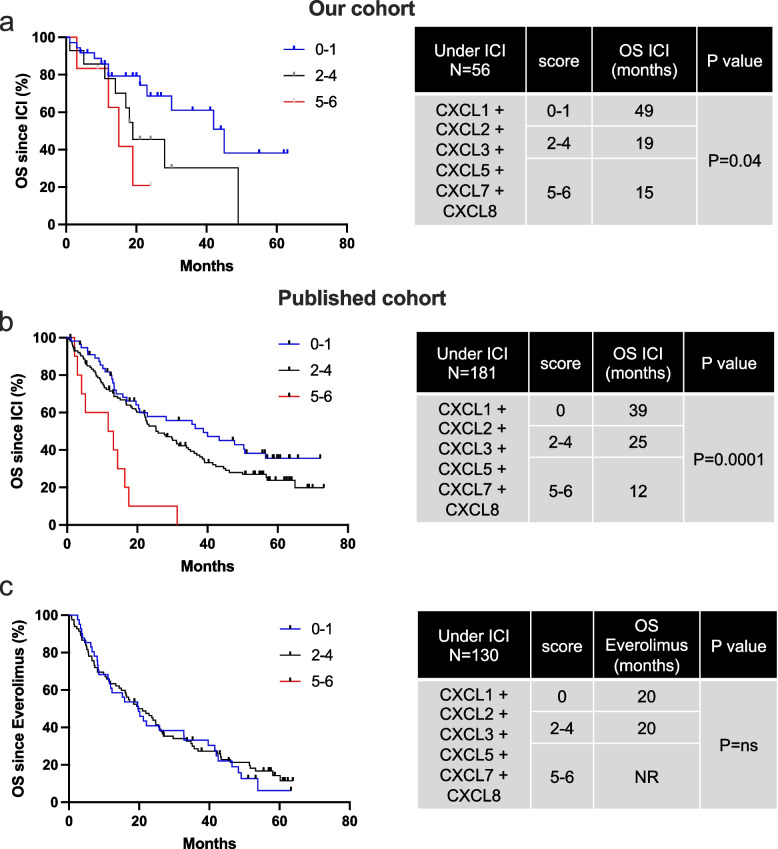
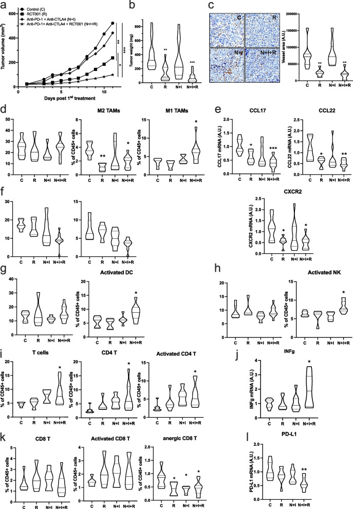
Background: In clear cell renal cell carcinoma (ccRCC), first-line treatment combines nivolumab (anti-PD-1) and ipilimumab (anti-CTLA4), yielding long-term remissions but with only a 40% success rate. Our study explored the potential of enhancing ccRCC treatment by concurrently using CXCR2 inhibitors alongside immunotherapies.

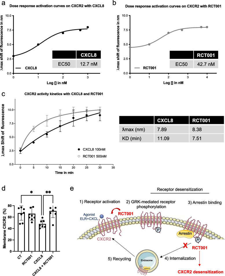
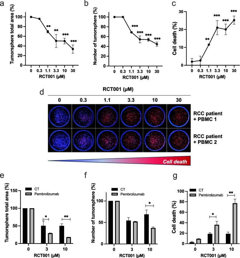
Methods: We analyzed ELR + CXCL levels and their correlation with patient survival during immunotherapy. RCT001, a unique CXCR2 inhibitor, was examined for its mechanism of action, particularly its effects on human primary macrophages. We tested the synergistic impact of RCT001 in combination with immunotherapies in both mouse models of ccRCC and human ccRCC in the presence of human PBMC.

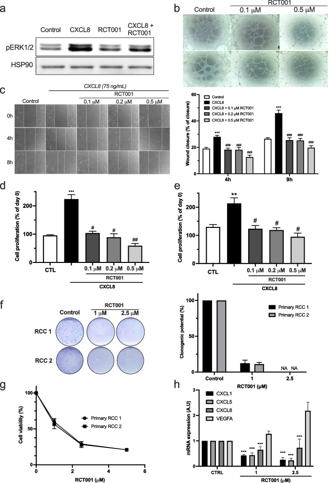
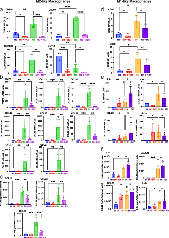
Resuts: Elevated ELR + CXCL cytokine levels were found to correlate with reduced overall survival during immunotherapy. RCT001, our optimized compound, acted as an inverse agonist, effectively inhibiting angiogenesis and reducing viability of primary ccRCC cells. It redirected M2-like macrophages without affecting M1-like macrophage polarization directed against the tumor. In mouse models, RCT001 enhanced the efficacy of anti-CTLA4 + anti-PD1 by inhibiting tumor-associated M2 macrophages and tumor-associated neutrophils. It also impacted the activation of CD4 T lymphocytes, reducing immune-tolerant lymphocytes while increasing activated natural killer and dendritic cells. Similar effectiveness was observed in human RCC tumors when RCT001 was combined with anti-PD-1 treatment.

Conclusions: RCT001, by inhibiting CXCR2 through its unique mechanism, effectively suppresses ccRCC cell proliferation, angiogenesis, and M2 macrophage polarization. This optimization potentiates the efficacy of immunotherapy and holds promise for significantly improving the survival prospects of metastatic ccRCC patients.

Keywords: CXCR2 inhibitors; Immunotherapies resistance; Ipilimumab; M2 tumor associated macrophages; Nivolumab; Renal Cell Carcinoma.


Conflict of interest statement

We disclose that RB, CR, GP, MD are the co-founders of the start-up Roca Therapeutics who develops RCT001.

Figures

Fig. 1
Fig. 1
High ELR+CXCL expression is associated with poor OS under ICI in RCC patients. Levels of ELR+CXCLs (CXCL1, CXCL2, CXCL3, CXCL5, CXCL8) and PD-L1 mRNA expression in metastatic RCC patients on ICIs or everolimus were measured by RT-qPCR and correlated with OS in patients from our cohort (ICI treatment, 56 patients, a) and in published cohort (ICI treatment, 181 patients, b or everolimus, 130 patients, c). The value of the third quartile of ELR+CXCL expression was chosen as the cut-off value. We created a score reflecting the overexpression of each ELR+CXCL (score 0 = no ELR+CXCL overexpressed, score 6 = all 6 ELR+CXCL overexpressed). The score was correlated with OS. The Kaplan–Meier method was used to generate survival curves and analyses of censored data were performed using Cox models. Statistical significance (p-values) is indicated
Fig. 2
Fig. 2
RCT001 is an inverse agonist of CXCR2. a-c The recombinant CXCR2 reconstituted in detergent buffer containing lipids that mimic the lipid composition of the receptor, is then labelled with a non-modifying probe that allow detection of the activation state and conformational change of the receptor upon addition of a ligand. The maximum wavelength of the probe emission spectrum (λmax) shifts according to the activation (TM6 opening)/inactivation (TM6 closing) of the receptor. The kinetics of receptor activation/inactivation can be followed by observing the λmax shift over time after addition of a ligand. a CXCR2 activation was measured by the λmax shift 20 min after stimulation by different doses of CXCL8. b CXCR2 activation was measured by the λmax shift 20 min after stimulation by a dose response of RCT001. c CXCR2 kinetic activation (0 to 30 min) was measured by the λmax shift after stimulation by CXCL8 (100 nM) or RCT001 (500 nM). d Starved endothelial cells were stimulated with 50 ng/ml CXCL8 for 1 h. Membrane-associated CXCR2 protein levels were quantified by flow cytometry. Results are presented as the mean of three independent experiments ± SD. Statistics were performed using the ANOVA test: *p < 0.05; **p < 0.01. e Summary schematic of the mechanism of action of RCT001 as an inverse CXCR2 agonist
Fig. 3
Fig. 3
RCT001 inhibits angiogenesis and viability of primary RCC cells in vitro. a Immunoblot analysis of pERK1/2 expression in serum-depleted endothelial cells determined 24 h after stimulation by CXCL8 (75 ng/mL) and/or treatment with RCT001 (1 μM). b Matrigel-based tube formation assay was performed with endothelial cells 4 h after treatment with RCT001 (0.1 or 0.5 μM). c. Wound scratch assay was performed with serum-depleted endothelial cells treated or not with CXCL8 (75 ng/mL) in the presence of RCT001 (0.1, 0.2 or 0.5 μM). Wound closure was determined 4 and 8 h after treatment. d-e Cell proliferation assay of serum-depleted endothelial cells treated or not with CXCL7 (75 ng/mL) (d) or CXCL8 (75 ng/mL) (e) in the presence of RCT001 (0.1, 0.2 or 0.5 μM) for 72 h. f Clonogenic assay with two primary RCC cells in the presence or not of RCT001 (1 or 2.5 μM). g Viability of two primary RCC cells treated with RCT001 for 48 h. h Evaluation of CXCL1/5/8 and VEGFA mRNA levels in primary RCC cells after 24 h of treatment with RCT001 treatment (1 or 2.5 μM) by qPCR. Results are presented as the mean of three independent experiments ± SD. Statistics were performed using the ANOVA test: **p < 0.01, *** p < 0.001 vs control conditions, #p < 0.01, ###p < 0.001 vs CXCL8 conditions
Fig. 4
Fig. 4
RCT001 reverses the polarization of M2-like macrophages polarization. Purified monocytes from healthy human donors (n = 5) were differentiated into M0-like macrophages (5 days with CSF1) and then polarized into M0-like macrophages (CSF1), or M2-like macrophages (IL4) or M1-like macrophages (LPS) for 48 h. The already polarized macrophages then were treated with RCT001 (2.5 µM) in the presence of the respective cytokines (CSF1 for M0, IL4 for M2 and LPS for M1 macrophages) for 48 h. MFI: Mean Fluorescence Intensity a-c M0 and M2-like macrophages were treated with RCT001 for 48 h. a Specific M2 macrophage membrane markers (CD206, CD209, CD200R, CD163) were examined by flow cytometry. b Specific M2 macrophage mRNA markers (TIMP3, CCL13, CCL14, CCL17, CCL18, CCL22, CCL23, CCL24) were evaluated by qPCR. c. Specific cytokines secreted by M2 macrophages (CCL13, CCL22, CCL24) were determined by ELISA. d-f M0 and M1-like macrophages were treated with RCT001 for 48 h. d Specific M1 macrophage membrane markers (CD80, CD86) were evaluated by flow cytometry. e Specific M1 macrophages mRNA markers (IL8, CXCL11, CCL20, IL-1a) were evaluated by qPCR. f Specific cytokines secreted by M1 macrophages (IL8, CXCL11, CCL20, IL-1a) were determined by ELISA. Results are presented as the mean of five independent experiments ± SD. Statistics were performed using the ANOVA test: *p < 0.05, **p < 0.01, *** p < 0.001
Fig. 5
Fig. 5
RCT001 efficiently inhibits RCC growth and synergizes with ICIs. RENCA cells were injected subcutaneously. Once the tumor reached 30 mm3, mice were divided into different groups: Control (n = 10), RCT001 alone (n = 10, 20 mg/kg), anti-PD-1 + anti-CTLA4 (n = 10, 100 μg each) and the combination of RCT001 + PD-1 + anti-CTLA4 (n = 10). a Tumor volume was measured each day. Results are expressed as mean ± sd. b Tumor weights at the end of the experiment. c Blood vessels were visualized and quantified by IHC for αSMA d Tumor-associated macrophages (TAMs, total population), M2 TAMs and M1 TAMs were evaluated by cytometry. e The specific mRNA markers of M2 TAMs (CCL17 and CCL22) and mRNA CXCR2 were determined by qPCR. f Tumor-associated neutrophils (TANs, total population) and mature TANs were evaluated by cytometry. g Intratumor dendritic cells (DCs, total population) and activated dendritic cells were evaluated by flow cytometry. h Intratumor natural killer (NKs, total population) and activated natural killer were evaluated by cytometry. i Intratumor T cells (total population) and CD4 + T, Activated CD4 + T were evaluated by flow cytometry. j Intratumor activated T cell specific mRNA marker (INFγ) mRNA was evaluated by qPCR. k Intratumor CD8 T, activated CD8 T and anergic CD8 T were evaluated by flow cytometry. l Intratumor anergic T cell specific mRNA marker (PD-L1) mRNA was evaluated by qPCR. Statistics were performed using the ANOVA test: *p < 0.01, **p < 0.01, *** p < 0.001
Fig. 6
Fig. 6
RCT001 inhibits proliferation of human RCC tumor spheres and synergize with anti PD-1. MEXF486 RCC tumors were grown as subcutaneous tumor xenografts in immunodeficient mice. After removal, the tumors are placed in layers of Versagel® from Cypre to achieve spheroid formation with human dermal fibroblasts (HDF). After the 3D tumors are established (7 days), they are treated with RCT001 (0.3 to 30 µM) for 7 days. a The total area of tumor spheroids was determined. b The number of tumor spheroids was determined. c—d Cell death was analyzed by DRAQ7 labelling. Quantification (C) and representative images (D) were shown. e–g Activated human PBMCs are first added to the cell culture medium (1 day before treatment); over time, they gradually infiltrate and attack the tumor spheres in the gel. Then tumor spheres + PBMCs were treated with RCT001 (3.3 and 10 µM) ± anti- PD -1 (pembrolizumab, 100 µg/ml). e The total area of tumorspheres was determined. f The number of tumorspheres was determined. g Cell death was analyzed by DRAQ7 labelling

References

    1. Sung H, Ferlay J, Siegel RL, Laversanne M, Soerjomataram I, Jemal A, et al. Global Cancer Statistics 2020: GLOBOCAN Estimates of Incidence and Mortality Worldwide for 36 Cancers in 185 Countries. CA Cancer J Clin. 2021;71(3):209–249. doi: 10.3322/caac.21660. - DOI - PubMed
    1. Siegel RL, Miller KD, Fuchs HE, Jemal A. Cancer Statistics, 2021. CA Cancer J Clin. 2021;71(1):7–33. doi: 10.3322/caac.21654. - DOI - PubMed
    1. Muglia VF, Prando A. Renal cell carcinoma: histological classification and correlation with imaging findings. Radiol Bras. 2015;48(3):166–174. doi: 10.1590/0100-3984.2013.1927. - DOI - PMC - PubMed
    1. Padala SA, Kallam A. Clear Cell Renal Carcinoma. StatPearls. Treasure Island (FL)2023. - PubMed
    1. Choueiri TK, Escudier B, Powles T, Tannir NM, Mainwaring PN, Rini BI, et al. Cabozantinib versus everolimus in advanced renal cell carcinoma (METEOR): final results from a randomised, open-label, phase 3 trial. Lancet Oncol. 2016;17(7):917–927. doi: 10.1016/S1470-2045(16)30107-3. - DOI - PubMed