. 2024 Mar 19;26(1):48.
doi: 10.1186/s13058-024-01802-z.

TMEM120B strengthens breast cancer cell stemness and accelerates chemotherapy resistance via β1-integrin/FAK-TAZ-mTOR signaling axis by binding to MYH9

Affiliations

TMEM120B strengthens breast cancer cell stemness and accelerates chemotherapy resistance via β1-integrin/FAK-TAZ-mTOR signaling axis by binding to MYH9

Ran Hu et al. Breast Cancer Res. .

Abstract

Background: Breast cancer stem cell (CSC) expansion results in tumor progression and chemoresistance; however, the modulation of CSC pluripotency remains unexplored. Transmembrane protein 120B (TMEM120B) is a newly discovered protein expressed in human tissues, especially in malignant tissues; however, its role in CSC expansion has not been studied. This study aimed to determine the role of TMEM120B in transcriptional coactivator with PDZ-binding motif (TAZ)-mediated CSC expansion and chemotherapy resistance.

Methods: Both bioinformatics analysis and immunohistochemistry assays were performed to examine expression patterns of TMEM120B in lung, breast, gastric, colon, and ovarian cancers. Clinicopathological factors and overall survival were also evaluated. Next, colony formation assay, MTT assay, EdU assay, transwell assay, wound healing assay, flow cytometric analysis, sphere formation assay, western blotting analysis, mouse xenograft model analysis, RNA-sequencing assay, immunofluorescence assay, and reverse transcriptase-polymerase chain reaction were performed to investigate the effect of TMEM120B interaction on proliferation, invasion, stemness, chemotherapy sensitivity, and integrin/FAK/TAZ/mTOR activation. Further, liquid chromatography-tandem mass spectrometry analysis, GST pull-down assay, and immunoprecipitation assays were performed to evaluate the interactions between TMEM120B, myosin heavy chain 9 (MYH9), and CUL9.

Results: TMEM120B expression was elevated in lung, breast, gastric, colon, and ovarian cancers. TMEM120B expression positively correlated with advanced TNM stage, lymph node metastasis, and poor prognosis. Overexpression of TMEM120B promoted breast cancer cell proliferation, invasion, and stemness by activating TAZ-mTOR signaling. TMEM120B directly bound to the coil-coil domain of MYH9, which accelerated the assembly of focal adhesions (FAs) and facilitated the translocation of TAZ. Furthermore, TMEM120B stabilized MYH9 by preventing its degradation by CUL9 in a ubiquitin-dependent manner. Overexpression of TMEM120B enhanced resistance to docetaxel and doxorubicin. Conversely, overexpression of TMEM120B-∆CCD delayed the formation of FAs, suppressed TAZ-mTOR signaling, and abrogated chemotherapy resistance. TMEM120B expression was elevated in breast cancer patients with poor treatment outcomes (Miller/Payne grades 1-2) than in those with better outcomes (Miller/Payne grades 3-5).

Conclusions: Our study reveals that TMEM120B bound to and stabilized MYH9 by preventing its degradation. This interaction activated the β1-integrin/FAK-TAZ-mTOR signaling axis, maintaining stemness and accelerating chemotherapy resistance.

Keywords: Breast cancer; Focal adhension kinase; MYH9; Stemness; TMEM120B.


Conflict of interest statement

The authors declare no competing interests.

Figures

Fig. 1
Fig. 1
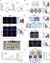
TMEM120B was highly expressed in breast cancer specimens and cell lines. (A) TCGA database was assessed to explore the mRNA expression of TMEM120B in pan-cancer and normal tissues, N for Normal, T for Tumor, Meta for metastasis. (B) Representative images of immunohistochemistry staining of TMEM120B in normal breast epithelial cells (a), normal intestinal epithelial cells (c), normal gastric epithelial cells (e), normal lung epithelial cells(g), normal ovarian epithelial cells (i) breast cancer epithelial cells (b), colon cancer epithelial cells (d), gastric carcinoma epithelial cells (f), lung cancer epithelial cells (h) and ovarian cancer epithelial cells (j), N for Normal, T for Tumor. (C-D) TMEM120B mRNA levels were identified between non-cancerous and cancerous tissues using the TCGA database (E) Representative images of immunohistochemistry staining of TMEM120B in (a) both normal and cancerous tissues in the same specimen, (b) adjacent normal tissue and breast cancer with diverse staining (c, weak, d, moderate, e, strong), N for Normal, T for Tumor. (F) Kaplan–Meier curves showed a correlation between mRNA expression of TMEM120B and overall survival in breast cancer patients. (G) Kaplan–Meier curves showing a correlation between TMEM120B protein expression and overall survival of patients with breast cancer. (H) TMEM120B protein level in 16 pairs of freshly isolated samples from patients with breast cancer was analyzed by western blotting (I) The protein expression of TMEM120B in breast cancer cell lines and normal breast cells. (J) Immunofluorescence assay was used to evaluate the subcellular localization of TMEM120B in breast cancer cells (scale bar = 20 μm). Quantification data are expressed as mean ± SD of three independent experiments (t-test, two-sided, *P < 0.05, **P < 0.01, ***P < 0.001)
Fig. 2
Fig. 2
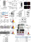
Overexpression of TMEM120B promoted breast cancer cell proliferation and invasion both in vitro and in vivo. The MTT assay (A), colony formation assay (B), and EdU assay (C, scale bar = 100 μm) were performed to examine the effects on the proliferation of after overexpressing or silencing TMEM120B in SK-BR-3 or MDA-231 cells. Transwell (D) and wound healing (E) assays were used to assess the effects of TMEM120B-myc, TMEM120B sgRNA, and the control on cell invasion and migration in SK-BR-3 and MDA-231 cells. Representative examples of explanted tumors (F) and lung metastases (G) in the negative SK-BR-3-control (NC) and SK-BR-3- overexpressing TMEM120B groups. Quantification data are expressed as mean ± SD of three independent experiments (t-test, two-sided, *P < 0.05, **P < 0.01, ***P < 0.001)
Fig. 3
Fig. 3
Overexpression of TMEM120B enhanced stemness of breast cancer cells. (A) GO analysis was performed to detect the biological process significantly correlated with the deletion of TMEM120B in MDA-453 cells. (B) Bioinformatics analysis for the mRNAsi Stemness score of TMEM120B (C) Immunoblotting of Myc-tag, ALDH1, OCT4, NANOG, SOX2, and GAPDH after overexpressing or deleting TMEM120B in SK-BR-3 and MDA-231 cells. Both the first (D, scale bar = 250 μm) and second round of sphere formation assays (E) were performed to examine the effects on stemness of cells after overexpressing or knocking out TMEM120B in SK-BR-3 or MDA-231 cells. (F) Immunoblotting of Myc-tag, OCT4, NANOG, ALDH1, SOX2, and GAPDH in MDA-231 cells in both non-sphere and sphere groups. (G) Flow cytometry assay detected the ratio of ALDH1+ cells upon overexpression or deletion of TMEM120B in SK-BR-3 or MDA-231 cells (H) Diluted injection xenograft assays to explore the effects on stemness of breast cancer cells upon depleting TMEM120B in MDA-231 cells in vivo. Quantification data are expressed as mean ± SD of three independent experiments (t-test, two-sided, *P < 0.05, **P < 0.01, ***P < 0.001)
Fig. 4
Fig. 4
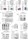
Overexpressing TMEM120 accelerated breast cancer stemness by activating TAZ-mTOR signaling axis. (A) KEGG analysis was conducted to detect the signaling pathway significantly correlated with deletion of TMEM120B in MDA-453 cells. (B) Phosphorylation antibodies array kit was used to explore the key signaling pathway involved in TMEM120B overexpression in SK-BR-3 cells. (C) Immunoblotting of Myc-tag, TMEM120B, AKT, p-AKT, mTOR, p-mTOR, YAP, TAZ, and GAPDH after overexpressing or silencing TMEM120B in SK-BR-3 or MDA-231 cells. (D) qPCR assay was used to investigate the alteration of the target genes of YAP/TAZ within ectopic or deleted TMEM120B in SK-BR-3 or MDA-231 cells. (E) Immunoblotting of Myc-tag, mTOR, p-mTOR, TAZ, and GAPDH after overexpressing TMEM120 with or without mTOR signaling pathway inhibitor rapamycin in SK-BR-3 cells. (F) Immunoblotting of Myc-tag, mTOR, p-mTOR, TAZ, and GAPDH after overexpressing TMEM120 with or without knocking down TAZ by siRNA in SK-BR-3 cells. Subcellular localization of TAZ was evaluated by immunofluorescence assay (G) or western blotting assay (H) within ectopic TMEM120B in SK-BR-3 cells. Scale bar = 20 μm. (I) After being treated with CHX at indicated time points, the expression of TAZ was evaluated by western blotting after overexpressing or silencing TMEM120B in SK-BR-3 or MDA-231 cells. Quantification data are expressed as mean ± SD of three independent experiments (t-test, two-sided, ***, P < 0.001)
Fig. 5
Fig. 5
TMEM120B promoted breast cancer cell stemness by binding with MYH9 via their coil-coil domains. (A) Mass spectrometry (MS) analysis was performed to identify candidates for interaction with TMEM120B after overexpressing TMEM120B in SK-BR-3 cells. Endogenous (B) and exogenous co-IP assay (C) were assessed to detect the interaction between MYH9 and TMEM120B in MDA-231 and SK-BR-3 cells. (D) GST pull-down assay was used to confirm the direct binding between MYH9 and TMEM120B in SK-BR-3 cells. (E) Immunofluorescence assay was used to show the co-localization of TMEM120B and MYH9 in SK-BR-3 cells, subcellular location coefficient of TMEM120B–MYH9 interaction was quantified by Fiji software (scale bar = 50 μm) (F) Divergent TMEM120B and MYH9 splicing mutant plasmids were designed to examine the domain responsible for the interaction between TMEM120B and MYH9.(I) GST pull-down assay was performed to confirm the direct interaction between MYH9 and TMEM120B after overexpressing TMEM120B-WT or TMEM120B-∆CCD plasmids in SK-BR-3 cells. Colony formation assay (J), Transwell assay (K), and sphere formation assay (L) were performed to detect the effects on the proliferation, invasion, and stemness of breast cancer cells after transfecting TMEM120B, TMEM120B-∆CCD, and control plasmid in SK-BR-3 cells. (M) Immunoblotting of Myc-tag, mTOR, p-mTOR, TAZ, and GAPDH after overexpressing TMEM120B or TMEM120B-∆CCD in SK-BR-3 cells. (N) Immunoblotting was used to evaluate the expression of cytosolic or nuclear TAZ after overexpressing TMEM120B or TMEM120B-∆CCD in SK-BR-3 cells. The effects on proliferation, metastasis, and stemness were verified in nude mice by subcutaneous tumorigenesis (O), tail vein injection (P), and diluted injection xenografts assays (Q) by overexpressing TMEM120B and TMEM120B-∆CCD in SK-BR-3 cells. Quantification data are expressed as mean ± SD of three independent experiments (t-test, two-sided, **, P < 0.01, ***, P < 0.001)
Fig. 6
Fig. 6
TMEM120B stabilized MYH9 by preventing its ubiquitin-mediated degradation from CUL9. (A and B) Western blotting and qPCR assays were performed to determine the protein and mRNA expression of MYH9, respectively, after overexpressing or deleting TMEM120B in SK-BR-3 or MDA-231 cells. (C-D) Western blotting and qPCR assays were performed to assess the protein and mRNA expression of TMEM120B, respectively, after overexpressing or deleting MYH9 in SK-BR-3 or MDA-231 cells. (E) After being treated with CHX at indicated time points, the expression of MYH9 was evaluated by western blotting after overexpressing or silencing TMEM120B in SK-BR-3 or MDA-231 cells. (F) The ubiquitination level of MYH9 was detected using western blotting after being transfected with TMEM120B, TMEM120B-∆CCD, and control plasmids in SK-BR-3 cells. (G) Mass spectrometry (MS) analysis was performed to identify candidates for interaction with ectopic TMEM120B or TMEM120B-∆CCD in SK-BR-3 cells, respectively. (H) Endogenous co-IP assay was performed to detect the interaction between MYH9, CUL9, and TMEM120B in MDA-231 cells. (I and J) Protein levels of MYH9 and the ubiquitination level were assessed using western blotting after transfecting TMEM120B alone or co-transfecting both TMEM120B and CUL9 in SK-BR-3 cells. (K) Co-IP assay was used to evaluate the interaction among TMEM120B, MYH9, and CUL9 after overexpressing TMEM120B and CUL9 in different doses in SK-BR-3 cells. Quantification data are expressed as mean ± SD of three independent experiments (t-test, two-sided)
Fig. 7
Fig. 7
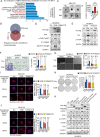
TMEM120B–MYH9 interaction activated the TAZ-mTOR axis by accelerating FAK assembly. (A) GO analysis for TMEM120B interaction candidates from MS analysis after overexpressing TMEM120B in SK-BR-3 cells. (B) Venn analysis for the overlap between RNA-seq and MS analysis. (C) 3D collagen gel invasion assay was performed after overexpressing TMEM120B or TMEM120B-∆CCD and control in MDA-231 cells. (D) Immunoblotting assay was performed to evaluate the expression of Myc-tag, FAK, p-FAK (Tyr397), β1-integrin, active-β1-integrin, and LaminB1 after transfecting TMEM120B-myc, TMEM120B-∆CCD-myc, and control plasmids in SK-BR-3 cells. (E) Immunoblotting of Myc-tag, p-mTOR, TAZ, and GAPDH after overexpressing TMEM120B with or without FAK signaling pathway inhibitor PF562271 in SK-BR-3 cells. Transwell assay (F), sphere formation assay (G), and colony formation assay (H) were performed to detect the effect on the invasion, stemness, and proliferation of breast cancer cells upon ectopic TMEM120B or ∆TMEM120B-CCD in SK-BR-3 cells. Representative immunofluorescence images of p-FAK (I) and β1-integrin (J) after treatment with nocodazole (NZ), followed by washout for 0, 30, and 60 min. Scale bar = 10 μm. (K) Immunoblotting of Myc-tag, Flag-tag, FAK, p-FAK(Tyr397), mTOR, p-mTOR, TAZ, ALDH1, and GAPDH after transfected with TMEM120B-myc, TMEM120B-∆CCD-myc, MYH9-flag, MYH9-delCCD-flag alone, or TMEM120B-myc + MYH9-flag, TMEM120B-∆CCD-myc + MYH9-flag, or TMEM120B-myc + MYH9-delCCD-flag in SK-BR-3 cells, respectively. Quantification data are expressed as mean ± SD of three independent experiments (t-test, two-sided, *P < 0.05, **P < 0.01, ***P < 0.001)
Fig. 8
Fig. 8
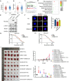
Overexpression of TMEM120B promoted chemotherapy resistance both in vitro and in vivo. The TCGA database was used to examine the relationship between TMEM120B expression and the effect of chemotherapy with docetaxel (A) and doxorubicin (B). GSEA from TCGA database (C) and RNA-seq by deleting TMEM120B in MDA-453 cells (D). (E) The expression of Myc-tag, RAD51, γ-H2AX, and GAPDH was evaluated by western blotting after overexpressing TMEM120B or TMEM120B-∆CCD and control in SK-BR-3 cells. (F) Representative immunofluorescence images of the foci number of γ-H2AX after overexpressing TMEM120B or TMEM120B-∆CCD and control in SK-BR-3 cells (scale = 10 μm). IC50 values in SK-BR-3 cells overexpressing TMEM120B after treatment with docetaxel (G) or docetaxel (H). (I) Xenografts assays were assessed after overexpressing TMEM120B or TMEM120B-∆CCD in SK-BR-3 cells with docetaxel or docetaxel. Quantification data are expressed as mean ± SD of three independent experiments (t-test, two-sided, *P < 0.05, **P < 0.01, ***P < 0.001)
Fig. 9
Fig. 9
TMEM120 expression positively correlated with TAZ, p-mTOR, SOX2, and chemotherapy resistance in breast cancer specimens. (A) Representative images of immunohistochemistry staining of TMEM120B, phosphorylated mTOR, and TAZ in human breast cancer specimens. (B) Representative images of immunohistochemistry staining of TMEM120B and SOX2 human breast cancer specimens. (C) Representative images of immunohistochemistry staining of TMEM120B in specimens from breast cancer patients with varying therapeutic effects evaluated using Miller/Payne Grades after neoadjuvant chemotherapy. (D) Pathway diagram for TMEM120B functional activity interaction with MYH9 in breast cancer cells. Quantification data are expressed as mean ± SD of three independent experiments (t-test, two-sided, ***, P < 0.001)

References

    1. Sung H, Ferlay J, Siegel RL, Laversanne M, Soerjomataram I, Jemal A, et al. Global Cancer statistics 2020: GLOBOCAN estimates of incidence and Mortality Worldwide for 36 cancers in 185 countries. CA Cancer J Clin. 2021;71(3):209–49. doi: 10.3322/caac.21660. - DOI - PubMed
    1. Drăgănescu M, Carmocan C. Hormone therapy in breast Cancer. Chirurgia (Bucur) 2017;112(4):413–7. doi: 10.21614/chirurgia.112.4.413. - DOI - PubMed
    1. Shien T, Iwata H. Adjuvant and neoadjuvant therapy for breast cancer. Jpn J Clin Oncol. 2020;50(3):225–9. doi: 10.1093/jjco/hyz213. - DOI - PubMed
    1. Jacobs AT, Martinez Castaneda-Cruz D, Rose MM, Connelly L. Targeted therapy for breast cancer: an overview of drug classes and outcomes. Biochem Pharmacol. 2022;204:115209. doi: 10.1016/j.bcp.2022.115209. - DOI - PubMed
    1. Bai X, Ni J, Beretov J, Graham P, Li Y. Cancer stem cell in breast cancer therapeutic resistance. Cancer Treat Rev. 2018;69:152–63. doi: 10.1016/j.ctrv.2018.07.004. - DOI - PubMed

Publication types