Review
. 2024;22(11):1826-1845.
doi: 10.2174/1570159X22666240319123151.

Histaminergic System and Vestibular Function in Normal and Pathological Conditions

Affiliations
Review

Histaminergic System and Vestibular Function in Normal and Pathological Conditions

Brahim Tighilet et al. Curr Neuropharmacol. 2024.

Abstract

Most neurotransmitter systems are represented in the central and peripheral vestibular system and are thereby involved both in normal vestibular signal processing and the pathophysiology of vestibular disorders. However, there is a special relationship between the vestibular system and the histaminergic system. The purpose of this review is to document how the histaminergic system interferes with normal and pathological vestibular function. In particular, we will discuss neurobiological mechanisms such as neuroinflammation that involve histamine to modulate and allow restoration of balance function in the situation of a vestibular insult. These adaptive mechanisms represent targets of histaminergic pharmacological compounds capable of restoring vestibular function in pathological situations. The clinical use of drugs targeting the histaminergic system in various vestibular disorders is critically discussed.

Keywords: Vestibular system; histaminergic drugs; histaminergic system; neuroinflammation; vertigo.; vestibular disorders.


Conflict of interest statement

The authors declare no conflict of interest, financial or otherwise.

Figures

Fig. (1)
Fig. (1)
Anatomic and functional organization of the vestibular system. The vestibular nerve is in contact with the sensory hair cells located in the peripheral vestibular system. This nerve projects ipsilaterally to the four vestibular nuclei (VNs): superior, lateral, medial and inferior, located in the brainstem. From these nuclei, several output pathways exist. The vestibulo-oculomotor pathway originates from the lateral nuclei that project to the oculomotor nuclei via the medial longitudinal fasciculus (MLF), allowing the stabilization of gaze during head movements. The vestibulospinal pathway includes a lateral vestibulospinal fasciculus (LVSF) that connects the ipsilateral lateral VN to the spinal cord and a medial vestibulospinal fasciculus (MVSF) that connects the contralateral medial, inferior, and lateral VNs to the musculature of the neck and upper body axis. This organization establishes postural control and muscle tone. The vestibulo-vegetative pathway consists of the superior and medial VNs that activate the vagus nerve, responsible for vital functions. VNs are linked to several neurovegetative nuclei, such as the dorsal nucleus of the vagus nerve (DNV), solitary nucleus (NTS) and area postrema. Various vestibulo-cortical pathways originate from all VNs and project bilaterally to the cortex. The diversity of output from the VNs underlines the broad role of the vestibular system in posturo-locomotor, oculomotor and higher cognitive functions. The balanced resting activity between the bilateral VNs is crucial for these functions. Created with bioRender.
Fig. (2)
Fig. (2)
Unbalanced resting activity within the vestibular nuclei complex and vestibular syndrome expression after unilateral vestibular neurectomy. Electrophysiological imbalance after unilateral vestibular neurectomy (UVN) between the bilateral vestibular nuclei (VNs) is responsible for the acute vestibular syndrome. After UVN, the ipsilateral VNs are deafferented and show reduced excitability in contrast to the VNs contralateral to the lesion. This effect is explained by the absence of vestibular peripheral inputs from the lesion side. The syndrome generated by this unbalanced resting activity in the VNs is composed of oculomotor, posturo-locomotor, vegetative and perceptive-cognitive signs and symptoms. Over time, the syndrome disappears as neuroplasticity mechanisms result in a rebalanced activity between the bilateral VNs - a mechanism called vestibular compensation. Created with bioRender.
Fig. (3)
Fig. (3)
Synthesis, catabolism, and distribution of histamine in the adult mammalian brain. (a) Neuronal histamine is synthetized from the amino acid L-histidine by the L-histidine decarboxylase enzyme. Histamine is then degraded by two enzymes, histamine N-methyltransferase and monoamine oxidase, resulting in tele-methylimidazoleacetic. (b) Histaminergic neurons are restricted to the tuberomammillary nucleus of the posterior hypothalamus, from where they project widely into the brain, including in the vestibular nuclei complexes. Created with bioRender.
Fig. (4)
Fig. (4)
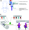
Localization of histaminergic receptors in the central (a) and peripheral (b) vestibular system. (a) The vestibular nuclei complexes (VNCs) contain three types of histaminergic receptors (HR). The secondary vestibular neurons express histaminergic type 1 (H1R) and histaminergic type 2 (H2R) receptor while the afferents from the tuberomammillary histaminergic neurons (in purple) express the histaminergic type 3 (H3R) autoreceptor. It should be noted that other afferents releasing various neurotransmitter in the VNCs can express a histaminergic type 3 (H3R) heteroreceptor. (b) The peripheral vestibular system contains all types of histaminergic receptors. Every type of HR is expressed in the Scarpa’s ganglion while only H1R and H3R are expressed in the endolymphatic sac. H1R, H3R and H4R are expressed in both type II and type I vestibular hair cells, while H2R is only found in type I vestibular hair cells. These schematic representations were created based on information found in [36, 58, 69] for the upper part and [71-73] for the lower part. Created with biorender.
Fig. (5)
Fig. (5)
Involvement of histamine in vestibular compensation after unilateral vestibular neurectomy. The unilateral vestibular neurectomy (UVN) leads to an electrophysiological imbalance between the vestibular nuclei, which is conveyed to the posterior hypothalamus through direct vestibulo-hypothalamic loops. In consequence, the synthesis and release of histamine (HA) from the tuberomammillary nucleus (TMN) to the deafferented vestibular nuclei (VN) increases. In VN, HA will bind to the three types of histamine receptors. By the histamine H3 receptor (H3R), more HA will be synthesized and released because of the abolition of the negative feedback. By the histamine H1 (H1R) and H2 (H2R) receptors, HA will restore the electrophysiological imbalance underlying the vestibular compensation. Created with bioRender.
Fig. (6)
Fig. (6)
Dual role of histamine depending on the context of the cellular environment. Histamine has a rather pro-inflammatory or anti-inflammatory role via the four histamine receptors expressed by microglia, depending on the micro-environment. Histamine has a pro-inflammatory role via the histamine H1 (H1R) and H4 (H4R) in a physiological context. Their activation leads to increased secretion of pro-inflammatory factors such as tumor necrosis factor-alpha (TNF-α), interleukin 6 (IL-6), interleukin 1 beta (IL-1β) and prostaglandin E2 (PGE2), but also to increased phagocytosis and reactive oxygen species (ROS) production. Conversely, histamine has a rather anti-inflammatory role in an inflammatory context via the histamine H2 (H2R) and H3 (H3R) receptors. Their activation leads to the inhibition of the inflammatory process of lipopolysaccharide (LPS), to the increase of secretion of anti-inflammatory factors such as interleukin 10 (IL-10) and interleukin 4 (IL-4), to the inhibition of pro-inflammatory factors (TNF-α and IL-1β), cluster of differentiation 14 (CD14) and interferon-gamma (IFN-γ). Finally, activation of these receptors also decreases phagocytosis and ROS production. Created with bioRender.
Fig. (7)
Fig. (7)
Anti-inflammatory and neuroprotective effect of betahistine after vestibular lesion. The vestibular lesion induces an inflammatory context in the deafferented vestibular nuclei (VN). Betahistine inhibits the histamine H3 receptor (H3R), which leads to the activation of a cAMP/PKA/CREB signaling pathway that induces glial-mediated inhibition of inflammation. These elements lead to a neuroprotective environment and would explain the decrease of neuronal differentiation in deafferented VN under this treatment. Created with bioRender.
Fig. (8)
Fig. (8)
Pharmacological mechanisms of betahistine underlying the rebalanced activity between the deafferented and intact vestibular nuclei after a vestibular lesion. Betahistine increases histamine (HA) synthesis and secretion by blocking histamine H3 (H3R) auto-receptors. Several events participate in the restoration of vestibular functions. Histaminergic activation of histamine H1 receptor (H1R) by betahistine and histamine and of histamine H2 receptor (H2R) by histamine in excitatory neurons of the deafferented vestibular nuclei (VN) induces depolarization. Betahistine and histamine bind to H1R on ipsilesional commissural GABAergic neurons, inhibiting the VN contralateral to the lesion. Peripherally, betahistine could increase the cochlear and vestibular blood flow, improving the microcirculation of the inner ear via histamine H3 heteroreceptors, while activation of H1R by betahistine and histamine induces a reduction in the activity of intact vestibular inputs. Created with bioRender.

References

    1. Kingma H., van de Berg R. Anatomy, physiology, and physics of the peripheral vestibular system. Handb. Clin. Neurol. 2016;137:1–16. doi: 10.1016/B978-0-444-63437-5.00001-7. - DOI - PubMed
    1. Angelaki D.E., Cullen K.E. Vestibular system: The many facets of a multimodal sense. Annu. Rev. Neurosci. 2008;31(1):125–150. doi: 10.1146/annurev.neuro.31.060407.125555. - DOI - PubMed
    1. Azzena G.B., Mameli O., Tolu E. Distribution of visual input to the vestibular nuclei. Arch. Ital. Biol. 1980;118(2):196–204. - PubMed
    1. Cullen K.E. The vestibular system: Multimodal integration and encoding of self-motion for motor control. Trends Neurosci. 2012;35(3):185–196. doi: 10.1016/j.tins.2011.12.001. - DOI - PMC - PubMed
    1. Gdowski G.T., McCrea R.A. Neck proprioceptive inputs to primate vestibular nucleus neurons. Exp. Brain Res. 2000;135(4):511–526. doi: 10.1007/s002210000542. - DOI - PubMed

MeSH terms

LinkOut - more resources