Review
. 2024 Mar 5:18:1336886.
doi: 10.3389/fncel.2024.1336886. eCollection 2024.

Out of the core: the impact of focal ischemia in regions beyond the penumbra

Affiliations
Review

Out of the core: the impact of focal ischemia in regions beyond the penumbra

Ludmila Koukalova et al. Front Cell Neurosci. .

Abstract

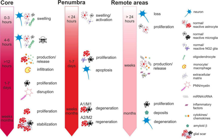
The changes in the necrotic core and the penumbra following induction of focal ischemia have been the focus of attention for some time. However, evidence shows, that ischemic injury is not confined to the primarily affected structures and may influence the remote areas as well. Yet many studies fail to probe into the structures beyond the penumbra, and possibly do not even find any significant results due to their short-term design, as secondary damage occurs later. This slower reaction can be perceived as a therapeutic opportunity, in contrast to the ischemic core defined as irreversibly damaged tissue, where the window for salvation is comparatively short. The pathologies in remote structures occur relatively frequently and are clearly linked to the post-stroke neurological outcome. In order to develop efficient therapies, a deeper understanding of what exactly happens in the exo-focal regions is necessary. The mechanisms of glia contribution to the ischemic damage in core/penumbra are relatively well described and include impaired ion homeostasis, excessive cell swelling, glutamate excitotoxic mechanism, release of pro-inflammatory cytokines and phagocytosis or damage propagation via astrocytic syncytia. However, little is known about glia involvement in post-ischemic processes in remote areas. In this literature review, we discuss the definitions of the terms "ischemic core", "penumbra" and "remote areas." Furthermore, we present evidence showing the array of structural and functional changes in the more remote regions from the primary site of focal ischemia, with a special focus on glia and the extracellular matrix. The collected information is compared with the processes commonly occurring in the ischemic core or in the penumbra. Moreover, the possible causes of this phenomenon and the approaches for investigation are described, and finally, we evaluate the efficacy of therapies, which have been studied for their anti-ischemic effect in remote areas in recent years.

Keywords: NG2-glia; astrocyte; future outlooks; microglia; oligodendrocytes; remote areas; stroke; therapy.


Conflict of interest statement

The authors declare that the research was conducted in the absence of any commercial or financial relationships that could be construed as a potential conflict of interest.

Figures

FIGURE 1
FIGURE 1
Time-dependent ischemia-induced changes in the ischemic core (left), penumbra (middle) and remote areas (right). Core: Profound cell swelling occurs several minutes after the onset of ischemia. Within 6 hours, neurons undergo necrosis and general loss of cells can be observed. Around 12 hours after ischemia, the surviving astrocytes begin to release chemokines attracting migration and infiltration of monocytes/macrophages. One to seven days after ischemia, the number of cells increases mostly due to the infiltration of microglia and macrophages and the massive proliferation of microglia. The numbers of neurons are severely reduced, and perineuronal nets and myelin sheaths are disrupted. One month or more after ischemia, the number of astrocytes increases, due to their proliferation and migration from the penumbra. The lesion contracts and the glial scar is stabilized by the extracellular matrix produced by reactive astrocytes. Penumbra: Swelling and activation of astrocytes, NG2 glia and microglia are delayed in comparison with the core but occur within 24 hours after ischemic insult. One to seven days after ischemia, neurons are still visible but their numbers have declined due to apoptosis. Astrocytes, microglia, NG2-glia and oligodendrocytes intensively proliferate. Proliferating astrocytes and microglia create heterogenic groups, where distinct subtypes differ in gene expression and membrane properties. Astrocytes and microglia with pro-inflammatory phenotypes in the vicinity of the core contribute to the formation of glial scar. Those with anti-inflammatory phenotypes in the outer parts of the penumbra with less severe hypoperfusion begin to produce growth factors and cytokines contributing to tissue regeneration. One month or more after ischemia, the numbers of glial cells are moderated, and depending on the duration and severity of the hypoperfusion, neurodegenerative or regenerative processes are activated. Remote areas: The first changes in the remote areas can be observed not earlier than 24 hours after ischemic insult but typically several days, weeks or even months following it. Depending on the brain region, changes in the cell structure and numbers are subtle or there are none. A slight decline in the numbers of neurons and a moderate increase in the numbers of microglia or astrocytes can be observed. However, morphological changes typical for their reactive states are mostly missing, even though the cells express markers of activation. More distinct alterations can be observed in the cellular functions, gene and protein expression profiles or production of cytokines. Induced delayed neuroinflammation may evoke neurodegenerative processes and amyloid deposits. For more details see the text.

Similar articles

Cited by

References

    1. Acosta-Martinez M. (2020). Shaping microglial phenotypes through estrogen receptors: Relevance to sex-specific neuroinflammatory responses to brain injury and disease. J. Pharmacol. Exp. Ther. 375 223–236. 10.1124/jpet.119.264598 - DOI - PubMed
    1. Agrawal K. L., Mittal B. R., Bhattacharya A., Khandelwal N., Prabhakar S. (2011). Crossed cerebellar diaschisis on F-18 FDG PET/CT. Indian J. Nucl. Med. 26 102–103. 10.4103/0972-3919.90263 - DOI - PMC - PubMed
    1. Akabori H., Moeinpour F., Bland K. I., Chaudry I. H. (2010). Mechanism of the anti-inflammatory effect of 17beta-estradiol on brain following trauma-hemorrhage. Shock 33 43–48. 10.1097/SHK.0b013e3181b0ebcb - DOI - PubMed
    1. Aleithe S., Blietz A., Mages B., Hobusch C., Hartig W., Michalski D. (2019). Transcriptional response and morphological features of the neurovascular unit and associated extracellular matrix after experimental stroke in mice. Mol. Neurobiol. 56 7631–7650. 10.1007/s12035-019-1604-4 - DOI - PMC - PubMed
    1. Allen N. J., Lyons D. A. (2018). Glia as architects of central nervous system formation and function. Science 362 181–185. 10.1126/science.aat0473 - DOI - PMC - PubMed

LinkOut - more resources