Review
. 2023 Apr 21;4(2):021301.
doi: 10.1063/5.0127122. eCollection 2023 Jun.

Sonomechanobiology: Vibrational stimulation of cells and its therapeutic implications

Affiliations
Review

Sonomechanobiology: Vibrational stimulation of cells and its therapeutic implications

Lizebona August Ambattu et al. Biophys Rev (Melville). .

Abstract

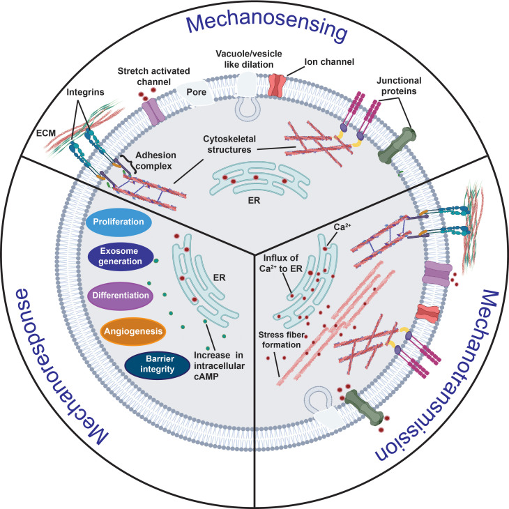
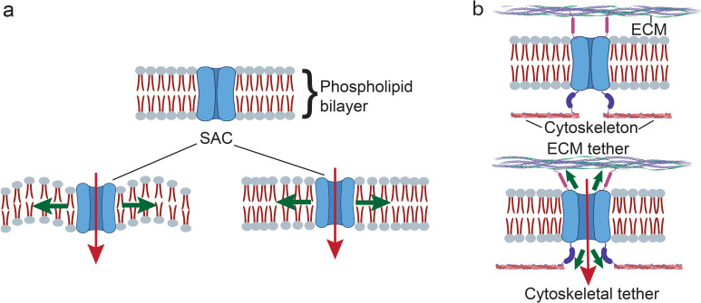
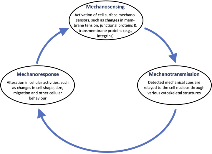
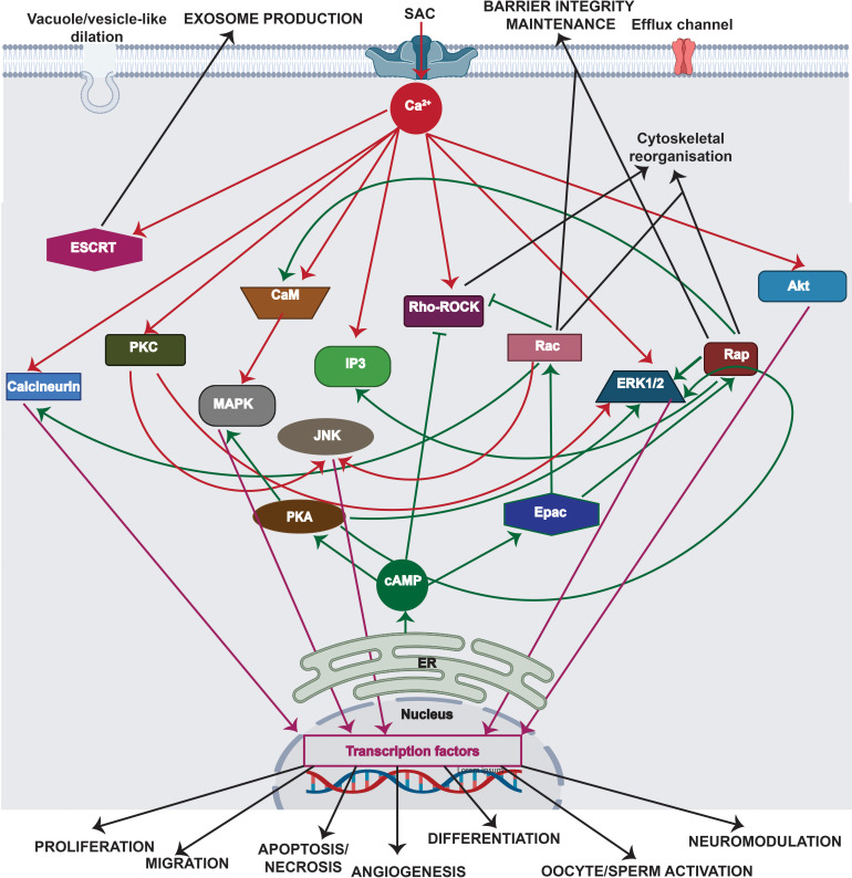
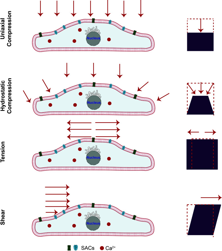
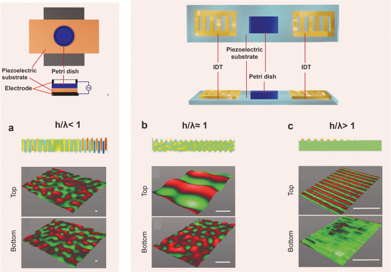
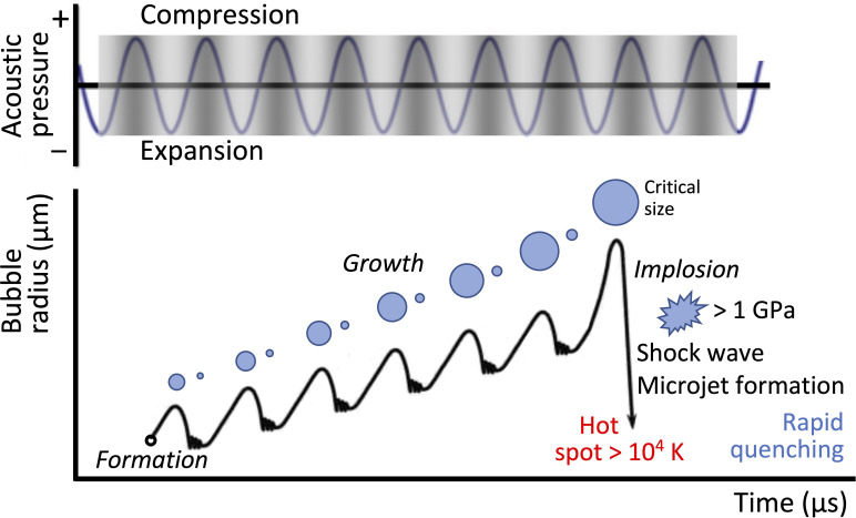
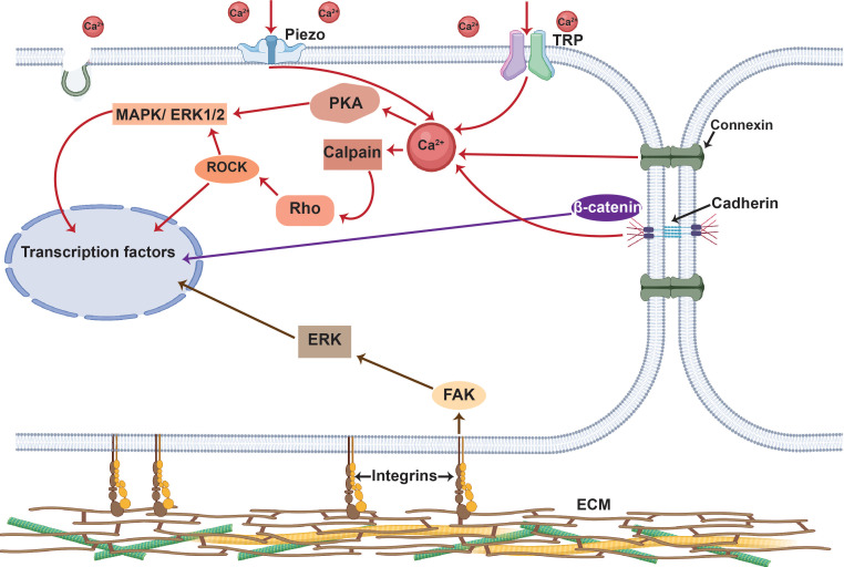
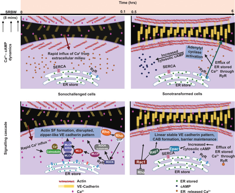
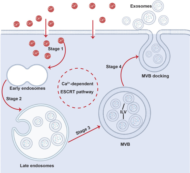
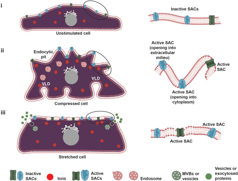
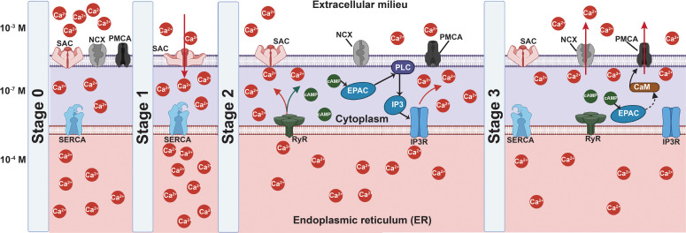
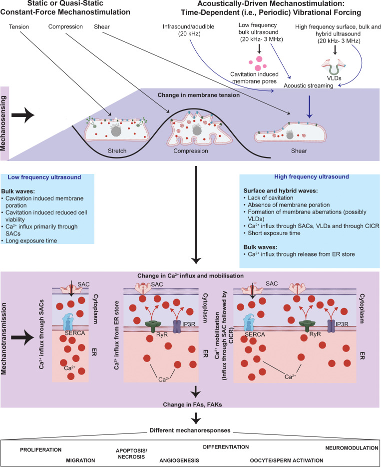
All cells possess an innate ability to respond to a range of mechanical stimuli through their complex internal machinery. This comprises various mechanosensory elements that detect these mechanical cues and diverse cytoskeletal structures that transmit the force to different parts of the cell, where they are transcribed into complex transcriptomic and signaling events that determine their response and fate. In contrast to static (or steady) mechanostimuli primarily involving constant-force loading such as compression, tension, and shear (or forces applied at very low oscillatory frequencies (1 Hz) that essentially render their effects quasi-static), dynamic mechanostimuli comprising more complex vibrational forms (e.g., time-dependent, i.e., periodic, forcing) at higher frequencies are less well understood in comparison. We review the mechanotransductive processes associated with such acoustic forcing, typically at ultrasonic frequencies (>20 kHz), and discuss the various applications that arise from the cellular responses that are generated, particularly for regenerative therapeutics, such as exosome biogenesis, stem cell differentiation, and endothelial barrier modulation. Finally, we offer perspectives on the possible existence of a universal mechanism that is common across all forms of acoustically driven mechanostimuli that underscores the central role of the cell membrane as the key effector, and calcium as the dominant second messenger, in the mechanotransduction process.


Conflict of interest statement

The authors have no conflicts to disclose.

Figures

FIG. 1.
FIG. 1.
Overview of the mechanotransduction process in which a cell recognizes and responds to mechanical cues through three distinct stages. In the mechanosensing stage, the cell detects mechanical cues from the extracellular milieu primarily through local changes or perturbations along its plasma membrane, such as vacuole/vesicle-like dilations (VLDs) or pore formation, that arise as a consequence of the external force on the cell; alternatively, or additionally, other key mechanosensors such as transmembrane proteins (e.g., integrins) or junctional proteins [e.g., adherens junctions (AJs) such as cadherins, or, gap junctions (GJs) such as connexins] can also be involved in the process. In the mechanotransduction stage, the aforementioned conformational changes in the cytoskeletal structures associated with the mechanosensors as a result of the mechanical cues are then relayed to intracellular sensing structures; phosphorylation, ion transport (primarily Ca2+), and the activation of other second messengers, such as cyclic adenosine monophosphate (cAMP), also play a crucial role in the process. These transmitted cues subsequently trigger multiple downstream signaling cascades in the mechanoresponse stage, resulting in the activation of different cellular activities, such as cell migration, proliferation and differentiation, angiogenesis, exosome biogenesis, and barrier integrity maintenance.
FIG. 2.
FIG. 2.
Two accepted models for SAC gating. (a) In the force-from-lipid (FFL) model, the reorganization of the lipid bilayer structure of the cell membrane in response to the applied mechanical loading exerts a force on the SAC to regulate its gating. (b) In the force-from-filament (FFF) model, the SACs are tethered to the extracellular matrix (ECM) or the cytoskeleton, through which the force is exerted.
FIG. 3.
FIG. 3.
The closed-feedback-loop nature of the mechanotransduction process. Changes in mechanosensors are relayed to the nucleus, which alter various cellular activities and behavior. These include changes to cell shape and size, as well as the propensity of the cell to migrate, which, in turn, can act as a trigger for further sensing to maintain cellular homeostasis.
FIG. 4.
FIG. 4.
Major signaling cascades associated with the mechanotransduction process (for simplicity, only the key pathways involved in mechanotransduction are shown). These can be broadly categorized into Ca2+-facilitated pathways (red lines) and cAMP-assisted pathways (green lines) that either result in transient (e.g., cytoskeletal reorganization, barrier integrity maintenance, or exosome production) or permanent (e.g., cell migration, proliferation, apoptosis, angiogenesis and differentiation, or cellular activation or neuromodulation) changes in cell behavior. Transient changes can be initiated by phosphorylation of substrates associated with the Rho–ROCK proteins and through ESCRT pathways. Permanent changes, on the other hand, can be initiated by signaling cascades, such as the MAPK, JNK, ERK1/2, or Akt pathways, which activate transcription factors, such as c-Fos, c-Jun or CREB, that are triggered by both Ca2+ and cAMP. The normal arrows depict the activation of signaling cascades, whereas the inhibitory arrows ⊣ denote inhibitory effects. (Rho: Ras homologous protein; ROCK: Rho-associated protein kinase; MAPK: mitogen-activated protein kinase; ESCRT: endosomal sorting complexes required for transport; Rap: Ras-related protein; PKC: protein kinase C; CaM: calmodulin; IP3: inositol 1,4,5-trisphosphate; Rac: Ras-related C3 botulinum toxin substrate; ERK: extracellular signal-regulated kinase; Akt: protein kinase B; cAMP: cyclic adenosine monophosphate; Epac: exchange factor directly activated by cAMP; PKA: cAMP-dependent protein kinase; c-Fos: AP-1 transcription factor subunit; c-Jun: transcription factor AP-1; CREB: cAMP responsive element binding protein; JNK: c-Jun N-terminal protein kinases).
FIG. 5.
FIG. 5.
Static forms of mechanostimulation. Schematic depiction of different extrinsic mechanical load, which can induce compression (uniaxial compression or hydrostatic compression), tension or shear stress, on the cells. Adapted from Ref. .
FIG. 6.
FIG. 6.
The different sound wave modes and the way in which they are generated on piezoelectric substrates: (a) bulk acoustic waves (BAWs), (b) hybrid surface and bulk acoustic waves [known as surface reflected bulk waves (SRBWs)131], and (c) surface acoustic waves (SAWs). The top row comprises top and side view sketches illustrating how these different wave forms are generated on piezoelectric (often, lithium niobate is used) substrates: (a) BAWs are generated by applying an AC electric field through a conducting layer or strip on the top and bottom surfaces of the piezoelectric substrate, whereas (b) SRBWs and (c) SAWs are typically generated using the same setup in which interdigitated transducer (IDT) electrodes are patterned on the piezoelectric substrate to which the AC electric field is applied; the thickness of the substrate h relative to the wavelength λ and hence resonant frequency f=c/λ is what determines which wave form is generated: (a) BAWs usually arise when h/λ<1, (b) SRBWs when h/λ1, and (c) SAWs when h/λ>1. The bottom row shows actual laser Doppler vibrometry scans measuring the displacement velocity on the top and bottom surfaces of the piezoelectric substrate for each case. Reproduced in part with permission from Rezk et al., Adv. Mater. 28, 1970 (2016). Copyright 2016 Wiley-VCH GmbH & Co. KGaA.
FIG. 7.
FIG. 7.
Acoustic cavitation. Schematic illustration depicting the formation, growth, and implosion of cavitation bubbles under low frequency ultrasonic excitation. Rezk et al., Adv. Sci. 8, 2001983 (2021); licensed under a Creative Commons Attribution (CC BY) license.
FIG. 8.
FIG. 8.
Schematic representation of the typical osteogenic pathways triggered by acoustically driven mechanostimulation of stem cells. The stimulation activates SACs, such as piezo and TRP channels, in addition to cell junctional proteins, such as connexins and cadherins, that facilitate influx of Ca2+. The change in the intracellular Ca2+ profile is responsible for initiating various Ca2+-signaling cascades (red arrows), such as Rho–ROCK (possibly through calpain) and PKA (cAMP-dependent protein kinase) signaling. While the former is responsible for regulating nuclear translocation of osteogenic factors such as TAZ and RUNX2, it can also initiate ERK/MAPK signaling such as PKA to instigate transcription factors, such as c-Fos, to induce osteogenesis. Concurrently, mechanostimulated integrins initiate the kinase pathway (brown arrows) through FAKs. The dissociation of β-catenin from cadherin, on the other hand, activates the Wnt canonical signaling pathway (purple arrows). ERK: extracellular signal-related kinase; FAK: focal adhesion kinase; MAPK: mitogen-activated protein kinase, Rho: Ras homologous protein; ROCK: Rho-associated protein kinase; TRP: transient receptor potential channel.
FIG. 9.
FIG. 9.
Calcium–cAMP signaling cascades induced by SRBW mechanostimulation. The immediate response of the SRBW-challenged cells is the sudden influx in intracellular Ca2+ as a consequence of membrane aberrations and piezo channel activation, which reduces Ca2+ levels in the immediate vicinity of the cells and drives transient invagination of VE-cadherin, while inducing Rho–ROCK signaling that causes actin stress fibers to be produced. Consequently, immature zipper-like VE-cadherin conformation ensues in the initial sonochallenge phase immediately following the 8 min SRBW excitation. As the cell subsequently relaxes, the increased intracellular Ca2+ level is ameliorated by their storage in the ER through SERCA, after which Ca2+ is slowly released back into the extracellular milieu through RyR activation. cAMP, formed by RyR activation, then initiates the Epac1–Rap1 pathway which further triggers the formation of circumferential actin bundles and mature VE-cadherin conformation in this subsequent, though simultaneous, sonotransformation phase in which the endothelial barrier integrity is enhanced. Reproduced with permission from Ambattu et al., Biomaterials 292, 121866 (2023). Copyright 2023 Elsevier Ltd.
FIG. 10.
FIG. 10.
Proposed mechanism for SRBW-facilitated exosome production. The Ca2+ influx through membrane aberrations or piezo channel activation (stage 1) promotes the recruitment of ALIX—an accessory protein that makes up part of the ESCRT machinery—to the site along the membrane where the perturbation is introduced. The resultant ESCRT activation leads to the formation of early endosomes (stage 2) and their maturation into late endosomes (stage 3). These then transform into MVBs that subsequently dock onto the cell membrane (stage 4) to release the intraluminal vesicles (ILVs) within them as exosomes, all the while during which the aberrations in the cell membrane are being healed.
FIG. 11.
FIG. 11.
Response of the cell membrane, originally at rest under homeostatic conditions in which the SACs are (i) inactive, to subsequent mechanical stimuli involving (ii) compression or (iii) stretch; the illustrations in the right column are a magnification of the lipid bilayer structure along a representative portion of the cell membrane as indicated by the circle in the illustrations in the left column. Under (ii) compression, the cell membrane folds to form VLDs and endocytic pits to counteract the decrease in membrane tension, leading to the activation of SACs through the FFL gating mechanism illustrated in Fig. 2. Depending on the location of the SAC with respect to the deformation region, it may either open into the ECM to allow ions to bind to the SAC, or into the cytoplasm to release ions into the cell. In contrast, the membrane aberrations and pores that form in cells under (iiii) stretch or tension activate the SACs directly due to the increase in membrane tension, again through the FFL mechanism, leading to an increased propensity for the fusion of multivesicular bodies (MVB) to ease the membrane tension, and resulting in exocytosis and exosome release.
FIG. 12.
FIG. 12.
Calcium mobilization stages and the various signaling cascades they induce. Stage 0: The cytoplasmic Ca2+ level is maintained at relatively low levels ( 107 M) in resting cells by influx and efflux through plasma membrane Ca2+ ATPase (PMCA) pumps and Na+/Ca2+ exchangers (NCX) along the cell membrane and via smooth endoplasmic reticular Ca2+ ATPase (SERCA) transporters along the ER membrane. Stage 1: With a change in membrane tension, SACs are activated, leading to an influx of Ca2+ into the cytoplasm that increases the cytoplasmic Ca2+ levels. This is rapidly restored by activation of the SERCA pumps that transport the Ca2+ ions into internal stores, mainly the ER. Stage 2: The increase in intracellular Ca2+ is followed by slow release of Ca2+ back into cytoplasm through calcium-induced calcium release (CICR) primarily from the ER via Ca2+-sensitive ryanodine receptors (RyR), accompanied by an increase in intracellular cAMP that activates the Epac signaling cascade. This, in turn, triggers the formation of the phospholipase C (PLC) pathway to activate the IP3 receptor (IP3R) that stimulates further release of Ca2+ from the ER store. Stage 3: The Ca2+ that is released from the ER is slowly effluxed back into the ECM through NCX. Meanwhile, Epac activates Ca2+/calmodulin (CaM) to trigger Ca2+ efflux via PMCA back into the ECM.
FIG. 13.
FIG. 13.
Proposed universal mechanism that underpins a cell's response to different forms of mechanostimuli, wherein the cell membrane plays a central role as a universal transducer/effector, and the second messenger Ca2+ as a key mechanotransduction mechanism. Static or quasi-static constant force mechanostimuli in the form of compression, stretch, or shear directly alter membrane fluidity to drive influx of Ca2+ into the cell that, in turn, trigger different downstream signaling pathways to elicit a variety of mechanoresponses. More complex dynamical forms of mechanostimuli, such as those driven acoustically, invoke changes to the membrane tension indirectly by either inducing cavitation, perturbations or aberrations to the membrane, or the generation of acoustic streaming in the cell media. They nevertheless lead to similar mechanoresponses given that these effectively result in compression, stretch, or shear to the cell, or more complex combinations thereof. Where mechanoresponses differ, however, is a consequence of how Ca2+ is regulated and hence mobilized within the cell in response to these mechanotransductive processes.

Similar articles

Cited by

References

    1. Ambrosi D., Ben Amar M., Cyron C. J., DeSimone A., Goriely A., Humphrey J. D., and Kuhl E., “ Growth and remodelling of living tissues: Perspectives, challenges and opportunities,” J. R. Soc. Interface 16, 20190233 (2019).10.1098/rsif.2019.0233 - DOI - PMC - PubMed
    1. Ingber D. E., “ Mechanobiology and diseases of mechanotransduction,” Ann. Med 35, 564–577 (2003).10.1080/07853890310016333 - DOI - PubMed
    1. Shah N., Morsi Y., and Manasseh R., “ From mechanical stimulation to biological pathways in the regulation of stem cell fate,” Cell Biochem. Funct 32, 309–325 (2014).10.1002/cbf.3027 - DOI - PubMed
    1. Bukoreshtliev N. V., Haase K., and Pelling A. E., “ Mechanical cues in cellular signalling and communication,” Cell Tissue Res 352, 77–94 (2013).10.1007/s00441-012-1531-4 - DOI - PubMed
    1. Wang J., Lu D., Mao D., and Long M., “ Mechanomics: An emerging field between biology and biomechanics,” Protein Cell 5, 518–531 (2014).10.1007/s13238-014-0057-9 - DOI - PMC - PubMed

LinkOut - more resources