. 2024 Jan 10;8(2):ziad008.
doi: 10.1093/jbmrpl/ziad008. eCollection 2024 Feb.

Loss of the auxiliary α2δ1 voltage-sensitive calcium channel subunit impairs bone formation and anabolic responses to mechanical loading

Affiliations

Loss of the auxiliary α2δ1 voltage-sensitive calcium channel subunit impairs bone formation and anabolic responses to mechanical loading

Madison M Kelly et al. JBMR Plus. .

Abstract

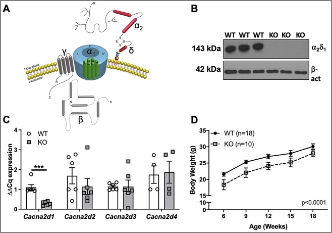
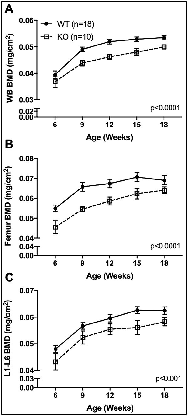
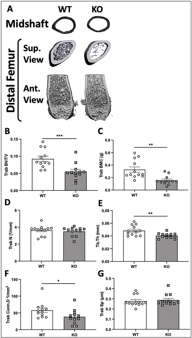
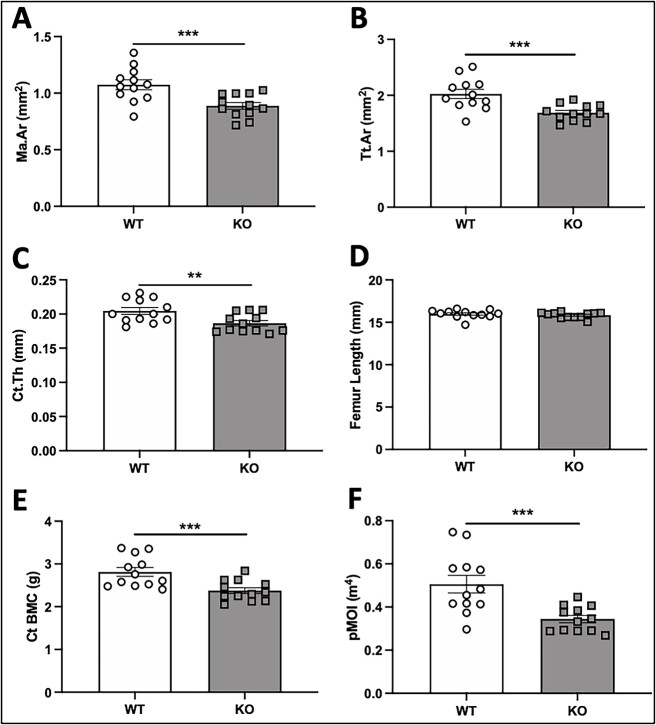
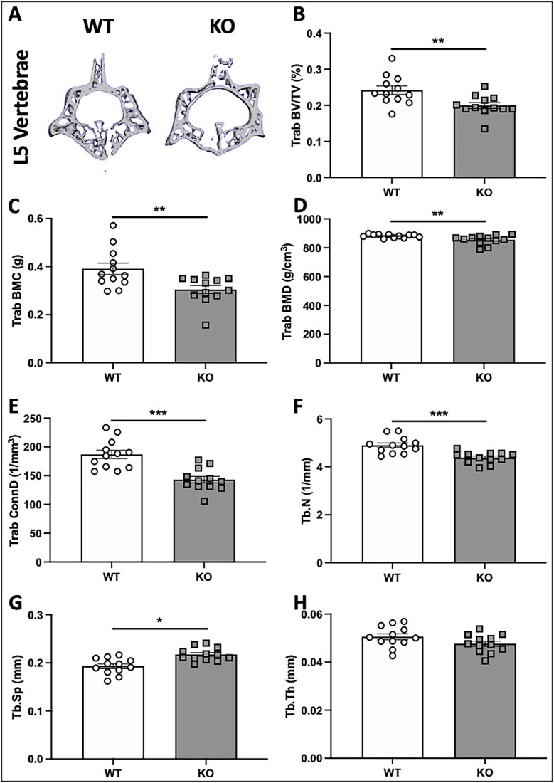
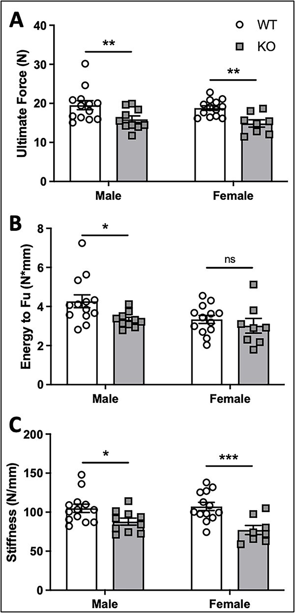
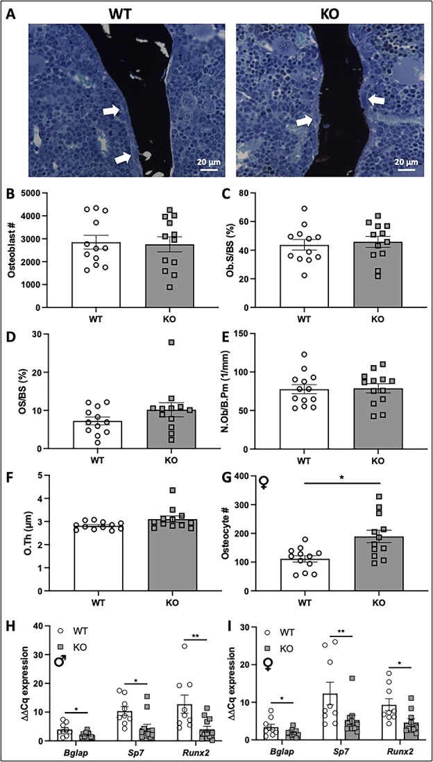
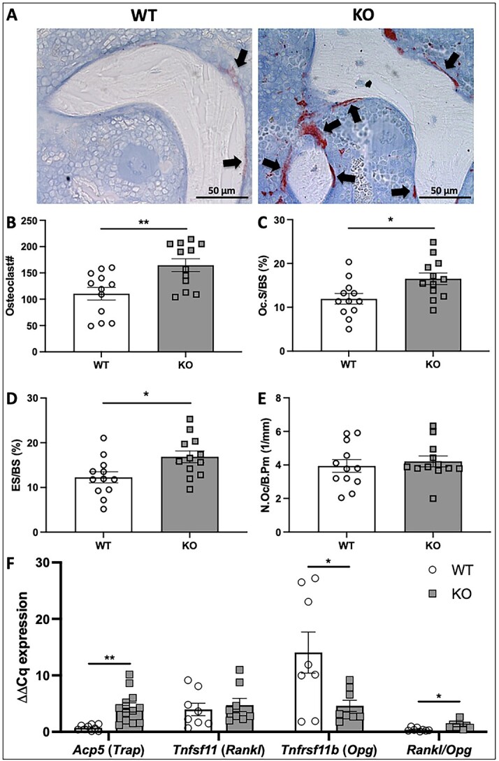
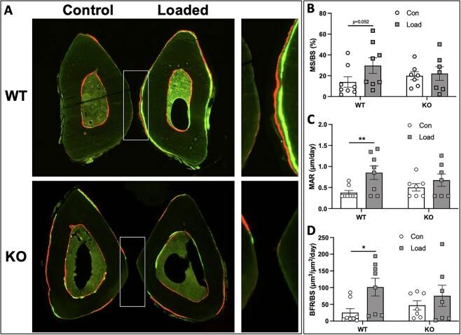
Voltage-sensitive calcium channels (VSCCs) influence bone structure and function, including anabolic responses to mechanical loading. While the pore-forming (α1) subunit of VSCCs allows Ca2+ influx, auxiliary subunits regulate the biophysical properties of the pore. The α2δ1 subunit influences gating kinetics of the α1 pore and enables mechanically induced signaling in osteocytes; however, the skeletal function of α2δ1 in vivo remains unknown. In this work, we examined the skeletal consequences of deleting Cacna2d1, the gene encoding α2δ1. Dual-energy X-ray absorptiometry and microcomputed tomography imaging demonstrated that deletion of α2δ1 diminished bone mineral content and density in both male and female C57BL/6 mice. Structural differences manifested in both trabecular and cortical bone for males, while the absence of α2δ1 affected only cortical bone in female mice. Deletion of α2δ1 impaired skeletal mechanical properties in both sexes, as measured by three-point bending to failure. While no changes in osteoblast number or activity were found for either sex, male mice displayed a significant increase in osteoclast number, accompanied by increased eroded bone surface and upregulation of genes that regulate osteoclast differentiation. Deletion of α2δ1 also rendered the skeleton insensitive to exogenous mechanical loading in males. While previous work demonstrates that VSCCs are essential for anabolic responses to mechanical loading, the mechanism by which these channels sense and respond to force remained unclear. Our data demonstrate that the α2δ1 auxiliary VSCC subunit functions to maintain baseline bone mass and strength through regulation of osteoclast activity and also provides skeletal mechanotransduction in male mice. These data reveal a molecular player in our understanding of the mechanisms by which VSCCs influence skeletal adaptation.

Keywords: bone formation; loading; mechanotransduction; voltage-sensitive calcium channel; α2δ1 subunit.


Conflict of interest statement

None declared.

Figures

Figure 1
Figure 1
Deletion of α2δ1 does not alter expression of other α2δ isoforms. (A) Schematic depicting VSCC subunit structure and spatial relationship. Schematic modified from previously published image. (B) Protein expression of α2δ1 assessed by Western blot using whole protein lysates from tibias of male WT and α2δ1 KO mice. Each lane is a lysate (20 μg) prepared from individual mice. Membranes were blotted for α2δ1 or β-actin as a loading control. (C) Expression of α2δ isoforms from mRNA isolated from tibias of male WT and α2δ1 KO mice. Each gene was normalized to Gapdh (n = 6 for Cacna2d1-3; n = 4 for Cacna2d4). (D) Body mass of male WT (n = 18) and KO (n = 10) mice was measured every 3 wk from 6 to 18 wk of age. Significance of qPCR data was tested by unpaired Student’s t-tests. Longitudinal body mass was determined using repeated measures ANOVA. ***P < .001.
Figure 2
Figure 2
Longitudinal in vivo DXA scans of male WT and α2δ1 KO mice. WT (n = 18, solid circle) and α2δ1 KO (n = 10, dotted square) mice were scanned every 3 wk from 6 to 18 wk of age and analyzed for BMD at the (A) whole body (WB), (B) femur, and (C) spine (L1-L6 vertebrae). Longitudinal data were tested for significance between genotype by two-way repeated measures ANOVA.
Figure 3
Figure 3
μCT-derived measures of trabecular bone in the distal femur of 22-wk-old male WT and α2δ1 KO mice. (A) Representative 3D reconstructions of midshaft femur (top row), distal metaphysis (middle row, proximal view), and caudal region of the distal femur (bottom row). Quantitative differences in (B) trabecular bone volume fraction (BV/TV) of the femur; (C) trabecular BMC; (D) trabecular number (Trab N); (E) trabecular thickness (Trab Th); (F) trabecular connectivity (Trab conn D); (G) trabecular spacing (Trab Sp). Significance between genotypes was tested using two-way ANOVA followed by Fisher’s LSD post hoc test, *P < .05, **P < .01, ***P < .001. n = 12 per group.
Figure 4
Figure 4
μCT-derived measures of cortical bone at the midshaft femur of 22-wk-old male WT and α2δ1 KO mice. Measures derived from cortical bone at the mid-diaphysis of the femur including (A) marrow area; (B) total area; (C) cortical thickness (cortical Th); (D) femur length; (E) cortical BMC; (F) polar moment of inertia (pMOI). Significance between genotypes tested using two-way ANOVA followed by Fisher’s LSD post hoc test, **P < .01, ***P < .001. n = 12 per group.
Figure 5
Figure 5
μCT-derived measures of L5 vertebrae of 22-wk-old male WT and α2δ1 KO mice. (A) Representative images of 3D reconstructions of L5 vertebrae from WT and α2δ1 KO mice. Quantification of μCT measures of L5 vertebrae included (B) trabecular bone volume fraction (BV/TV); (C) trabecular BMC; (D) trabecular BMD; (E) trabecular connectivity (Trab Conn D); (F) trabecular number (Trab N); (G) trabecular spacing (Trab Sp); (H) trabecular thickness (Trab Th). Significance between genotypes tested using two-way ANOVA followed by Fisher’s LSD post hoc test, *P < .05, **P < .01, ***P < .001. n = 12 per group.
Figure 6
Figure 6
Mechanical testing via monotonic three-point bending to failure on whole femora from 22-wk-old WT and α2δ1 KO mice. Quantification of (A) ultimate force, (B) energy to ultimate force, and (C) stiffness reveal significant reductions in the mechanical properties of bone following deletion of α2δ1 in male mice. Reductions in (A) ultimate force and (C) stiffness were observed in female mice, with no significant differences in energy to ultimate force in females. Data were tested for significance using two-way ANOVA followed by Fisher’s LSD post hoc test, *P < .05, **P < .01, ***P < .001. WT: n = 13, KO: n = 10 for males; WT: n = 13, KO: n = 8 for females.
Figure 7
Figure 7
Histological analyses of von Kossa/MacNeal–stained sections of 22-wk-old femora. (A) Representative images of distal femur sections from male WT and α2δ1 KO mice stained with von Kossa/MacNeal. White arrows indicate osteoblasts on the bone surface. Quantification of osteoblast parameters from male mice including (B) osteoblast number, (C) osteoblast surface normalized to bone surface (Ob.S/BS), (D) osteoid surface normalized to bone surface (OS/BS), (E) osteoblast number normalized to bone perimeter (N.Ob/B.Pm), and (F) osteoid thickness (O.Th). (G) Osteocyte number was quantified from distal femurs of female WT and KO mice. Expression of osteoblast regulatory genes from male (H) and female (I) mice, including osteocalcin (Bglap), osterix (Sp7), and Runx2 were all normalized to Gapdh. Data were tested for significance using two-way ANOVA followed by Fisher’s LSD post hoc test, *P < .05, **P < .01. n = 12 per group for histomorphometry analyses. For qPCR, WT: n = 9, KO: n = 12 for males; WT: n = 9, KO: n = 10 for females.
Figure 8
Figure 8
Histomorphometric analyses of TRAP-stained sections of 22-wk-old male femora. (A) Representative images of distal femur sections from WT and α2δ1 KO mice stained for TRAP. Multinucleated osteoclasts are stained crimson/red (black arrows). Histomorphometric quantification of osteoclast parameters include (B) osteoclast number, (C) osteoclast surface normalized to total bone surface (Oc.S/BS), (D) eroded surface normalized to total bone surface (ES/BS), (E) number of osteoclasts as a function of total bone perimeter (N.Oc/B.Pm). (F) Expression of osteoclast regulatory genes including Acp5, Tnfsf11, Tnfrsf11b, and the ratio of Rankl/Opg were all normalized to Gapdh. Data were tested for significance using two-way ANOVA followed by Fisher’s LSD post hoc test, *P < .05, **P < .01. n = 12 per group for histomorphometry analyses. For qPCR, WT: n = 9, KO: n = 8 to 12.
Figure 9
Figure 9
Axial ulnar loading in 18-wk-old male WT and α2δ1 KO mice. Strain-matched peak forces were applied to ulnae of male mice. (A) Representative images of control (nonloaded) and loaded ulnae from WT and KO mice. (B) Mineralizing surface (MS/BS), (C) mineral apposition rate (MAR), and (D) bone formation rate (BFR/BS) were increased in WT mice but had no significant changes in α2δ1 KO mice. Data were tested for significance using two-way ANOVA followed by Fisher’s LSD post hoc test, *P < .05, **P < .01. WT: n = 8, KO: n = 7.

Similar articles

References

    1. Warden SJ, Thompson WR. Become one with the force: optimising mechanotherapy through an understanding of mechanobiology. Br J Sports Med. 2017;51(13):989–990. Epub 2017/06/25. 10.1136/bjsports-2017-097634. - DOI - PMC - PubMed
    1. Thompson WR, Scott A, Loghmani MT, Ward SR, Warden SJ. Understanding mechanobiology: physical therapists as a force in mechanotherapy and musculoskeletal regenerative rehabilitation. Phys Ther. 2016;96(4):560–569. 10.2522/ptj.20150224. - DOI - PMC - PubMed
    1. Bonewald LF. The amazing osteocyte. J Bone Miner Res. 2011;26(2):229–238. 10.1002/jbmr.320. - DOI - PMC - PubMed
    1. Thompson WR, Rubin CT, Rubin J. Mechanical regulation of signaling pathways in bone. Gene. 2012;503(2):179–193. 10.1016/j.gene.2012.04.076. - DOI - PMC - PubMed
    1. Thompson WR, Modla S, Grindel BJ, et al. Perlecan/Hspg2 deficiency alters the pericellular space of the lacunocanalicular system surrounding osteocytic processes in cortical bone. J Bone Miner Res. 2011;26(3):618–629. 10.1002/jbmr.236. - DOI - PMC - PubMed