Case Reports
. 2024 Feb;86(1):142-148.
doi: 10.18999/nagjms.86.1.142.

Potential arrhythmic substrate of atrial fibrillation at the left atrial diverticulum

Affiliations
Case Reports

Potential arrhythmic substrate of atrial fibrillation at the left atrial diverticulum

Takehiko Takayanagi. Nagoya J Med Sci. 2024 Feb.

Abstract

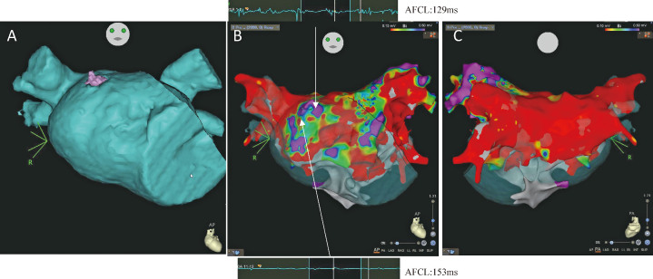
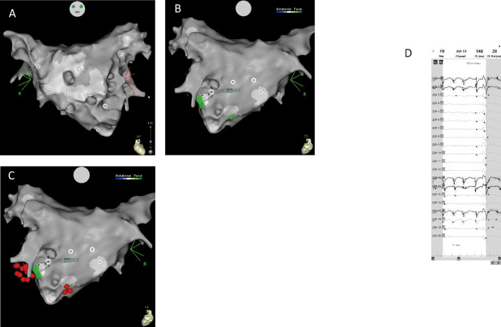
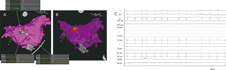
Catheter ablation therapy for persistent atrial fibrillation (PeAF) is both difficult and has limited outcomes. The mechanisms underlying the development and persistence of atrial fibrillation (AF) are not fully understood; therefore, ablation strategies are diverse. A 45-year-old man was referred to our hospital for persistent atrial fibrillation to undergo radiofrequency catheter insertion (RFCA). In the first session we conducted pulmonary vein isolation and additional linear ablation, including that of the roof line and posterior inferior line (posterior box lesion) as the stepwise ablation. However, AF was recurred in six months, therefore he was readmitted for second session ablation preoperative 3D computed tomography (CT) scan for drug-refractory PeAF was performed. The additional isolation of the left superior pulmonary vein and potential drivers of AF by mapping wavefront propagation using multipolar catheters by CARTOFINDER (Biosense Webster, Inc, Diamond Bar, CA, USA) was conducted. However, AF did not terminate. Tomography revealed that the left atrial (LA) diverticulum (LAD) was found uniquely. Electrophysiological findings showed focal firing of the myocardial sleeve and LA diverticulum by an approach for defragmented potentials by re-visiting in interval confidence level (ICL) mode included in the electroanatomical mapping system (CARTO 3, Biosense Webster, Inc, Diamond Bar, CA, USA) and the ablation by encircling this site finally made AF terminate. The AF has not recurred for more than 12 months without the use of antiarrhythmic drugs. This case report suggests that additional ablation around substrates in LAD may be effective for treating refractory AF.

Keywords: CARTOFINDER; CFAE; persistent atrial fibrillation; radiofrequency ablation.


Conflict of interest statement

The author declares no conflict of interest for this article.

Figures

Fig. 1
Fig. 1
High density left atrial voltage maps Fig. 1A: Preoperative 3D-CT that detects a diverticulum on the posteroseptal wall of the LA. High density left atrial voltage maps created during AF ablation are shown using CARTO (Biosense Webster, Inc, Diamond Bar, CA, USA) recorded by a PentaRay catheter (Biosense Webster, Inc, Diamond Bar, CA, USA). Purple color represents areas greater than the threshold of 0.5 mV. Fig. 1B, 1C:Anterior-posterior (AP) view (B) and posterior-anterior (PA) view (C). They show the voltage map after the first session ablation procedure with isolation of PVI and post wall isolation. PV reconnection was also identified in the left anterior PV and the roof with atrial fibrillation recurrence after undergoing the pulmonary vein isolation (PVI) procedure. Figure 1B illustrates representative electrograms and cycle lengths (CL) for left atrial diverticulum (LAD) and the surrounding area. AFCL: atrial fibrillation cycle length
Fig. 2
Fig. 2
CARTOFINDER (Biosense Webster, Inc, Diamond Bar, CA, USA) module maps and electrograms Fig. 2A, 2B: Anterior-posterior (AP) view (A) and posterior-anterior (PA) view (B). They show the CARTOFINDER module map around the diverticulum after pulmonary vein isolation and box isolation. Drivers with a focal repetitive activation pattern (labeled in green) were identified at the posteroinferior and posterolateral sites (B). Fig. 2C: Posterior-anterior (PA) view after ablation. Red tags represent ablation lesions. Fig. 2D: The unipolar electrogram on the PentaRay catheter (Biosense Webster, Inc, Diamond Bar, CA, USA). This figure illustrates the unipolar electrogram on the PentaRay catheter (Biosense Webster, Inc, Diamond Bar, CA, USA) recorded posteroinferior and posterolateral sites showing a QS pattern (20A1, 2, 13, 14, and17) (cycle length 146 ms).
Fig. 3
Fig. 3
The distribution of CFAE potential (CFAE map), the degree of fragmented potentials (CFAE potential) by module of CARTO system, and electrogram under ablation of AF Fig. 3A: Complex fractionated atrial electrograms (CFAE) map by interval confidence level (ICL) mode of the CARTO system during AF before CFAE ablation (anterior-posterior [AP] view) and representative electrograms for LAD and the surrounding area. The areas with the highest number of ICLs are shown in red, while those with a low number of ICLs are displayed in purple. The color bar is configured to display 60% of the maximum ICL value. Fig. 3B: CFAE map after atrial fibrillation (AF) termination. Red tags represent the ablation lesions for CFAE. Fig. 3C: Intracardiac electrograms. These electrograms are shown to be wider and have more fragmented deflections in the left atrial diverticulum (LAD) than in the right atrium (RA) and coronary sinus (CS). Atrial fibrillation terminated into sinus rhythm during ablation encircling a diverticulum source at the posteroseptal left atrium. SVC dis: distal superior vena cava SVC pro: proximal superior vena cava RA dis: distal right atrium RA pro: proximal right atrium CS dis: distal coronary sinus CS pro: sproximal coronary sinus RV dis: distal right ventricular RV pro: proximal right ventricular Abl dis: distal bipole of the ablation catheter Abl pro: proximal bipole of the ablation catheter

Similar articles

Cited by

References

    1. Calkins H, Hindricks G, Cappato R, et al. 2017 HRS/EHRA/ECAS/APHRS/SOLAECE expert consensus statement on catheter and surgical ablation of atrial fibrillation. Heart Rhythm. 2017;14(10):e275-e444. doi:10.1016/j.hrthm.2017.05.012. - DOI - PMC - PubMed
    1. Ohe M, Haraguchi G, Kumanomido J, et al. New tailored approach using a revised assessment of fragmented potentials for persistent atrial fibrillation: Early area defragmentation by modified CFAE module. J Cardiovasc Electrophysiol. 2019;30(6):844–853. doi:10.1111/jce.13888. - DOI - PubMed
    1. Igawa O, Miake J, Adachi M. The small diverticulum in the right anterior wall of the left atrium. Europace. 2008;10(1):120. doi:10.1093/europace/eum275. - DOI - PubMed
    1. Peng LQ, Yu JQ, Yang ZG, et al. Left atrial diverticula in patients referred for radiofrequency ablation of atrial fibrillation: assessment of prevalence and morphologic characteristics by dual-source computed tomography. Circ Arrhythm Electrophysiol. 2012;5(2):345–350. doi:10.1161/CIRCEP.111.965665. - DOI - PubMed
    1. Killeen RP, O’Connor SA, Keane D, Dodd JD. Ectopic focus in an accessory left atrial appendage: radiofrequency ablation of refractory atrial fibrillation. Circulation. 2009;120(8):e60-e62. doi:10.1161/CIRCULATIONAHA.109.855569. - DOI - PubMed

Publication types

LinkOut - more resources