. 2024 Jul 2;150(1):49-61.
doi: 10.1161/CIRCULATIONAHA.123.066433. Epub 2024 Mar 20.

Virus-Induced Acute Respiratory Distress Syndrome Causes Cardiomyopathy Through Eliciting Inflammatory Responses in the Heart

Affiliations

Virus-Induced Acute Respiratory Distress Syndrome Causes Cardiomyopathy Through Eliciting Inflammatory Responses in the Heart

Jana Grune et al. Circulation. .

Abstract

Background: Viral infections can cause acute respiratory distress syndrome (ARDS), systemic inflammation, and secondary cardiovascular complications. Lung macrophage subsets change during ARDS, but the role of heart macrophages in cardiac injury during viral ARDS remains unknown. Here we investigate how immune signals typical for viral ARDS affect cardiac macrophage subsets, cardiovascular health, and systemic inflammation.

Methods: We assessed cardiac macrophage subsets using immunofluorescence histology of autopsy specimens from 21 patients with COVID-19 with SARS-CoV-2-associated ARDS and 33 patients who died from other causes. In mice, we compared cardiac immune cell dynamics after SARS-CoV-2 infection with ARDS induced by intratracheal instillation of Toll-like receptor ligands and an ACE2 (angiotensin-converting enzyme 2) inhibitor.

Results: In humans, SARS-CoV-2 increased total cardiac macrophage counts and led to a higher proportion of CCR2+ (C-C chemokine receptor type 2 positive) macrophages. In mice, SARS-CoV-2 and virus-free lung injury triggered profound remodeling of cardiac resident macrophages, recapitulating the clinical expansion of CCR2+ macrophages. Treating mice exposed to virus-like ARDS with a tumor necrosis factor α-neutralizing antibody reduced cardiac monocytes and inflammatory MHCIIlo CCR2+ macrophages while also preserving cardiac function. Virus-like ARDS elevated mortality in mice with pre-existing heart failure.

Conclusions: Our data suggest that viral ARDS promotes cardiac inflammation by expanding the CCR2+ macrophage subset, and the associated cardiac phenotypes in mice can be elicited by activating the host immune system even without viral presence in the heart.

Keywords: CCR2; SARS-CoV-2; acute respiratory distress syndrome; macrophage.


Conflict of interest statement

Disclosures M.N. has received funds or material research support from Alnylam, Biotronik, CSL Behring, GlycoMimetics, GSK, Medtronic, Novartis, and Pfizer, as well as consulting fees from Lilly, Biogen, Gimv, IFM Therapeutics, Molecular Imaging, Sigilon, and Verseau Therapeutics. The other authors report no conflicts.

Figures

Figure 1.
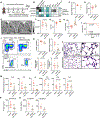
Figure 1.. A mouse model of VLARDS.
(A) Experimental outline. All assays were done on day 5 after the first instillation. (B) Body weight in (g) (n=6 mice per group). (C) Body temperature in (°C). (D) Kaplan-Meier curve indicating mortality of VLARDS (n=11) and vehicle-treated control (n=8 mice, log rank Mantel-Cox test. (E) X-ray computed tomography images and (F) lung opacity quantification reported in Hounsfield Units (HU). (G) sO2 and PaO2 in arterial blood. (H) Lung H&E staining indicating increased cellularity in lungs of mice with ARDS. Scale bar 250 μm (left) and 50 μm (right). (I) Representative flow plots for lung tissue and (J) corresponding quantification of leukocyte, neutrophil and macrophage counts per lung lobe. (K) qRT-PCR analysis of total lung tissue. (L) ELISA for IL6 in serum. (M) ELISA for IL6 and IL-1ß in bronchoalveolar lavage fluid (BALF). *p < 0.05; **p < 0.01; ***p < 0.001; ****p < 0.0001 vs. naive control group. One-way ANOVA followed by Bonferroni’s multiple comparisons test or Kruskal-Wallis test followed by Dunn’s multiple comparison’s test, as appropriate. Data are mean ± SEM.
Figure 2.
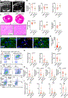
Figure 2.. Phenotypic changes of cardiac macrophage subsets in VLARDS.
(A) Experimental outline. (B) B-mode images of the left ventricle in a parasternal long axis view and (C) quantitative analyses. (D) Troponin levels measured in whole blood samples. (E) H&E-stained cardiac cross-sections and 20x magnifications, demonstrating cellular infiltration. Scale bar 2.5 mm (left) or 100 μm (right). (F) Experimental outline. Flow cytometry was performed on day 3, 5 and 14 after the first instillation. (G) Exemplary flow plots of cardiac tissue. (H) Quantification of leukocyte populations and (I) macrophage subsets in hearts from naive and VLARDS mice. One-way ANOVA followed by Bonferroni’s multiple comparisons test or Kruskal-Wallis test followed by Dunn’s multiple comparison’s test, as appropriate. (J) qRT-PCR analyses of bulk heart tissue on day 5 after first instillation. Unpaired parametric two-tailed t-test or Mann-Whitney test, as appropriate. *p < 0.05, **p < 0.01; ***p < 0.001, ****p < 0.0001 vs. naive control group. Data are mean ± SEM.
Figure 3.
Figure 3.. Human SARS-CoV-2-associated ARDS raises cardiac macrophage recruitment.
(A) Immunofluorescence staining of myocardium from control and COVID-19 patients. Tissue was stained with DAPI (blue), CD68-AF88 (green) and CCR2-DyLight 594 (red). Scale bar is 50 μm or 20 μm, respectively. (B) Quantification of CD68+ macrophages per field of view (FOV) and (C) CCR2+ CD68+ macrophages in control and COVID-19 patients. (D) CD68+ cells and (E) CCR2+ CD68+ cells per FOV stratified according to cause of death. **p < 0.01; ***p < 0.001; ****p < 0.0001 vs. control group. Mann-Whitney test or Kurskal-Wallis test followed by Dunn’s multiple comparisons test, as appropriate. Data are mean ± SEM.
Figure 4.
Figure 4.. Anti-TNFα therapy protects against VLARDS.
(A) Experimental outline of anti-inflammatory drug screen. All readouts were taken on day 5 after the first instillation. (B) Heat map indicating readouts of drug screen expressed as percent change compared to the average of untreated VLARDS mice in (%) (n=3 mice per group), leukocytes were enumerated in the heart. (C) Body weight loss in (%). (D) Body temperature in (°C). (E) Computed tomography images and (F) quantification of lung opacity in Hounsfield Units (HU). (G) sO2 and (H) PaO2 in arterial blood. (I) Kaplan-Meier curve indicating mortality of VLARDS (n=11) and VLARDS in mice treated with anti-TNFα antibody (n=10 mice, Log rank Mantel-Cox test). (J) Representative flow plots of lung tissue and (K) quantification of leukocytes, neutrophils and macrophages per lung lobe. (L) H&E staining of lung tissue. Scale bar 200 μm (left) or 50 μm (right). (M) Gene expression analysis in lung tissue. (N) ELISA for IL6 in serum. (O) ELISA for IL6 and IL-1ß in bronchoalveolar lavage fluid (BALF). *p < 0.05; **p < 0.01; ***p < 0.001 vs. VLARDS group. Unpaired parametric two-tailed t-test or Mann-Whitney test, as appropriate. Data are mean ± SEM.
Figure 5.
Figure 5.. Anti-TNFα therapy prevents ARDS-induced changes in macrophage subsets and improves cardiac function.
(A) B-mode images in a parasternal long axis view and (B) analyses of left ventricular systolic function. (C) H&E-stained cardiac cross-sections and 20x magnifications, demonstrating cellular infiltrations. Scale bar 2.5 mm (up) or 100 μm (low). (D) Representative images from cardiac cross-sections stained with DAPI (blue), troponin (green) and TUNEL (red), scale = 25 μm (E) Quantification of Troponin+ TUNEL+ cells. Nested T-test. (F) Representative flow plots of cardiac tissue and (G) corresponding quantification of leukocytes and (H) macrophage subsets, based on CCR2 and MHCII. (I) qRT-PCR analysis of heart tissue. *p < 0.05; **p < 0.01; ***p < 0.001; ****p < 0.0001 vs. VLARDS group. Unpaired parametric two-tailed t-test or Mann-Whitney test, as appropriate. Data are mean ± SEM.
Figure 6.
Figure 6.. Pre-existing HFpEF increases VLARDS mortality.
(A) Experimental outline of inducing heart failure with preserved ejection fraction (HFpEF) in mice via L-NAME (drinking water) and 60% high-fat diet, both ad libitum over 12 weeks. (B) Echocardiographic assessment of ejection fraction (EF) and (C) diastolic function, indicated by the late diastolic trans-mitral flow velocity (E/A); the ratio between mitral inflow velocity and mitral valve annular early diastolic velocity (E/e’) and the global longitudinal peak strain (GLS). (D) Blood leukocyte numbers assessed by flow cytometry in naive controls and mice with HFpEF. (E) Experimental outline of VLARDS induction in naive mice (Ctrl) and mice with pre-existing HFpEF. (F) Body temperature in (°C) measured on day 4 after first intratracheal instillation. (G) Kaplan-Meier curve indicating mortality of VLARDS (n=11) and HFpEF + VLARDS (n=8) mice. (H) Experimental outline indicating treatment of HFpEF+ARDS mice with anti-TNFα therapy. (I) Body temperature in (°C) measured on day 4 after first intratracheal instillation. (J) Kaplan-Meier curve indicating mortality of HFpEF + VLARDS (n=8) and HFpEF + VLARDS + anti-TNFα treatment (n=8, Log rank Mantel-Cox test).*p < 0.05; ***p < 0.001 and ****p < 0.0001 vs. naive group (C-D), vs. VLARDS group (F) or vs. HFpEF + VLARDS (I). Unpaired parametric two-tailed t-test or Mann-Whitney test, as appropriate. Data are mean ± SEM.

References

    1. Bos LDJ, Sjoding M, Sinha P, Bhavani SV, Lyons PG, Bewley AF, Botta M, Tsonas AM, Serpa Neto A, Schultz MJ, Dickson RP, Paulus F, PRoVENT-COVID CG. Longitudinal respiratory subphenotypes in patients with COVID-19-related acute respiratory distress syndrome: results from three observational cohorts. Lancet Respir Med. 2021;9:1377–1386. - PMC - PubMed
    1. Yang L, Xie X, Tu Z, Fu J, Xu D, Zhou Y. The signal pathways and treatment of cytokine storm in COVID-19. Signal Transduct Target Ther. 2021;6:255. - PMC - PubMed
    1. Metkus TS, Sokoll LJ, Barth AS, Czarny MJ, Hays AG, Lowenstein CJ, Michos ED, Nolley EP, Post WS, Resar JR, Thiemann DR, Trost JC, Hasan RK. Myocardial Injury in Severe COVID-19 Compared With Non-COVID-19 Acute Respiratory Distress Syndrome. Circulation. 2021;143:553–565. - PMC - PubMed
    1. Xie Y, Xu E, Bowe B, Al-Aly Z. Long-term cardiovascular outcomes of COVID-19. Nat Med. 2022;28:583–590. - PMC - PubMed
    1. Nicholls JM, Poon LL, Lee KC, Ng WF, Lai ST, Leung CY, Chu CM, Hui PK, Mak KL, Lim W, Yan KW, Chan KH, Tsang NC, Guan Y, Yuen KY, Peiris JS. Lung pathology of fatal severe acute respiratory syndrome. Lancet. 2003;361:1773–1778. - PMC - PubMed

MeSH terms