. 2024 Apr 12;134(8):1006-1022.
doi: 10.1161/CIRCRESAHA.123.323655. Epub 2024 Mar 20.

AKAP12 Upregulation Associates With PDE8A to Accelerate Cardiac Dysfunction

Affiliations

AKAP12 Upregulation Associates With PDE8A to Accelerate Cardiac Dysfunction

Hanan Qasim et al. Circ Res. .

Abstract

Background: In heart failure, signaling downstream the β2-adrenergic receptor is critical. Sympathetic stimulation of β2-adrenergic receptor alters cAMP (cyclic adenosine 3',5'-monophosphate) and triggers PKA (protein kinase A)-dependent phosphorylation of proteins that regulate cardiac function. cAMP levels are regulated in part by PDEs (phosphodiesterases). Several AKAPs (A kinase anchoring proteins) regulate cardiac function and are proposed as targets for precise pharmacology. AKAP12 is expressed in the heart and has been reported to directly bind β2-adrenergic receptor, PKA, and PDE4D. However, its roles in cardiac function are unclear.

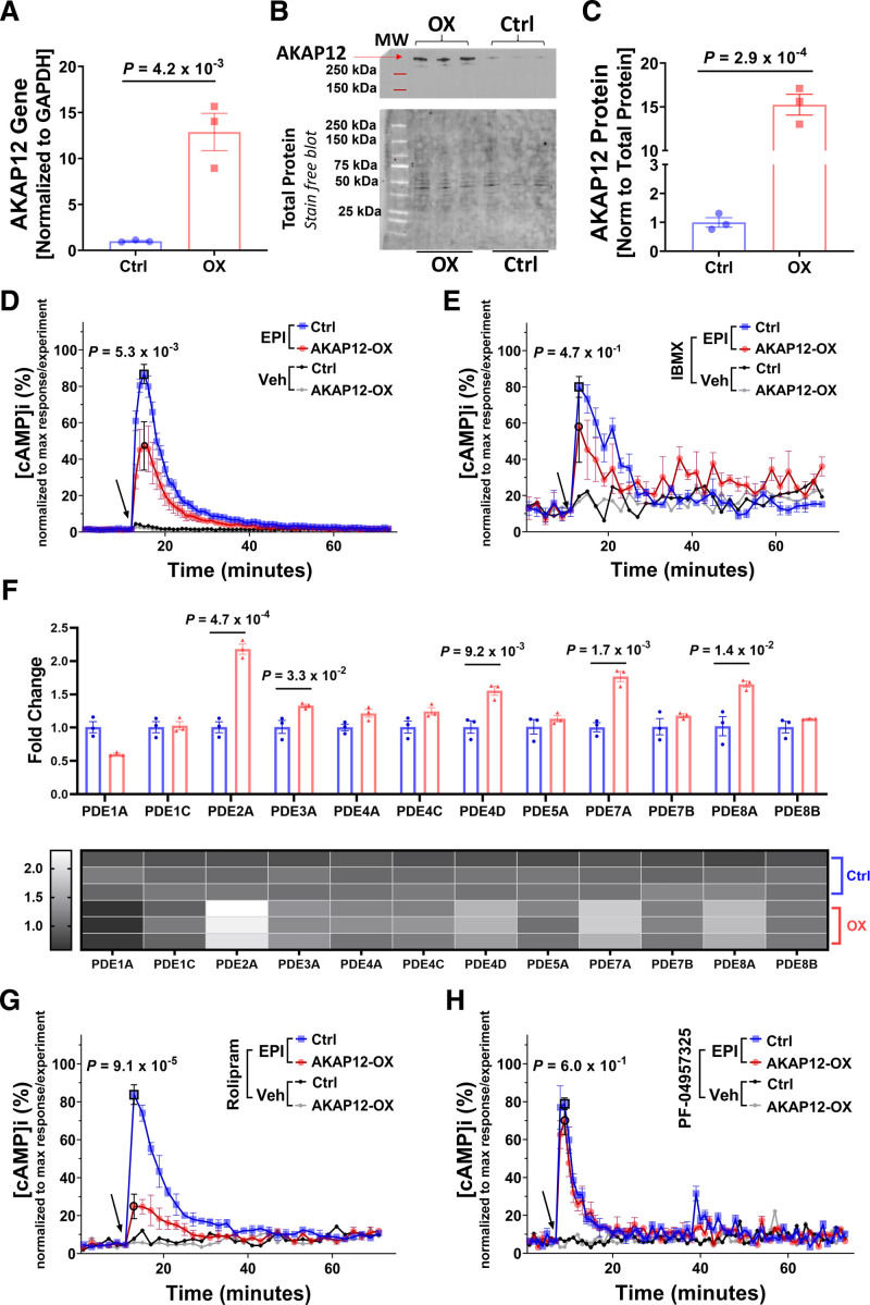
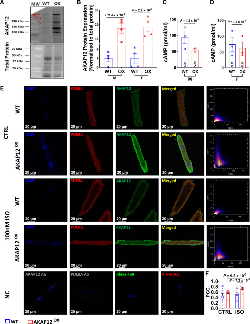
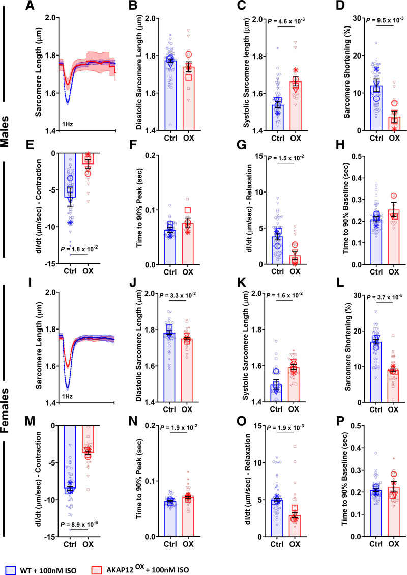
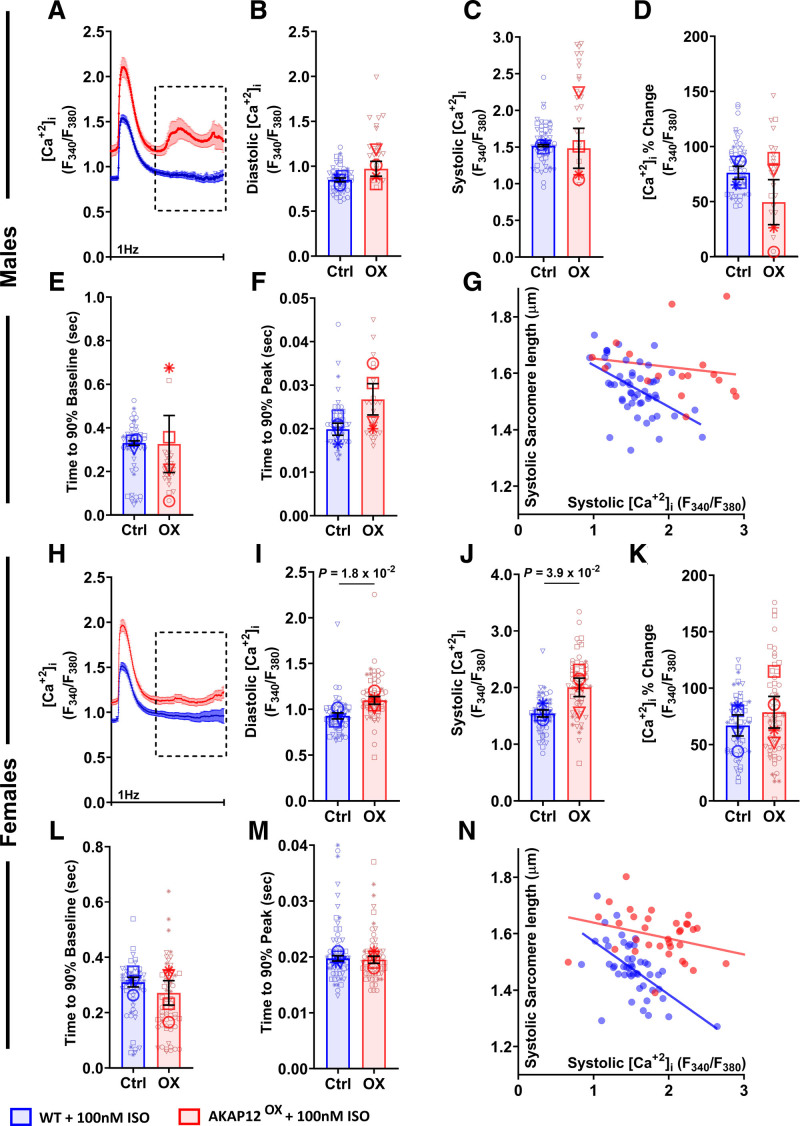
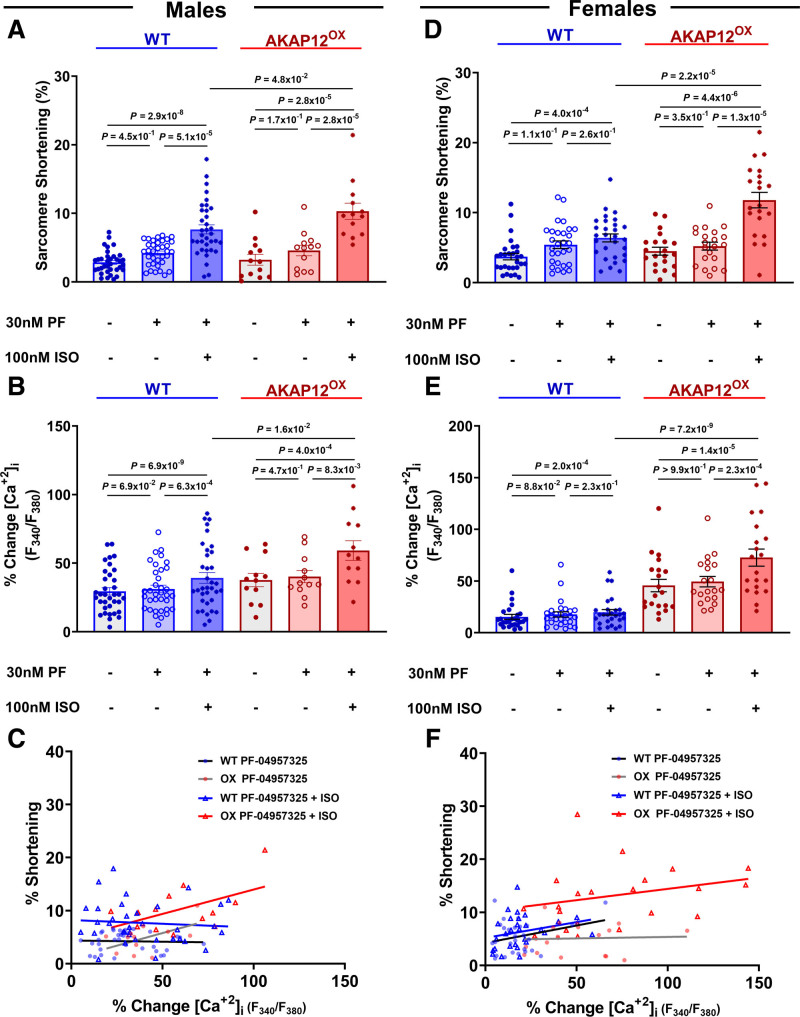
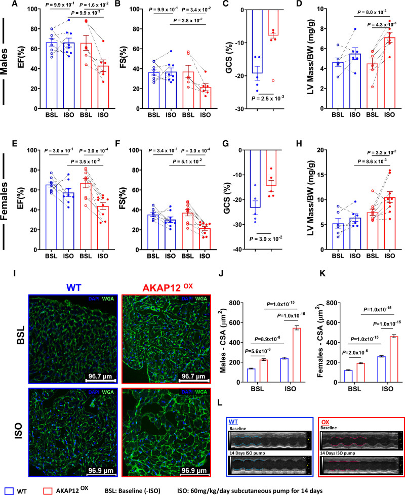
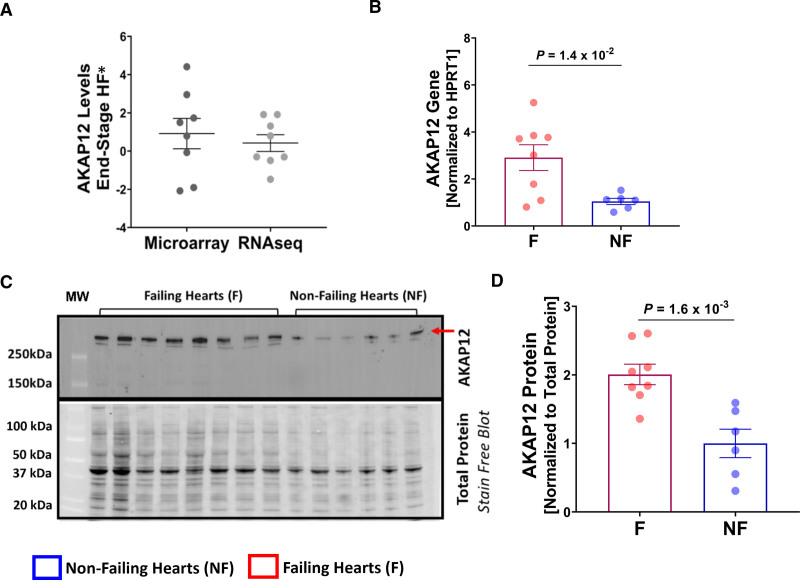
Methods: cAMP accumulation in real time downstream of the β2-adrenergic receptor was detected for 60 minutes in live cells using the luciferase-based biosensor (GloSensor) in AC16 human-derived cardiomyocyte cell lines overexpressing AKAP12 versus controls. Cardiomyocyte intracellular calcium and contractility were studied in adult primary cardiomyocytes from male and female mice overexpressing cardiac AKAP12 (AKAP12OX) and wild-type littermates post acute treatment with 100-nM isoproterenol (ISO). Systolic cardiac function was assessed in mice after 14 days of subcutaneous ISO administration (60 mg/kg per day). AKAP12 gene and protein expression levels were evaluated in left ventricular samples from patients with end-stage heart failure.

Results: AKAP12 upregulation significantly reduced total intracellular cAMP levels in AC16 cells through PDE8. Adult primary cardiomyocytes from AKAP12OX mice had significantly reduced contractility and impaired calcium handling in response to ISO, which was reversed in the presence of the selective PDE8 inhibitor (PF-04957325). AKAP12OX mice had deteriorated systolic cardiac function and enlarged left ventricles. Patients with end-stage heart failure had upregulated gene and protein levels of AKAP12.

Conclusions: AKAP12 upregulation in cardiac tissue is associated with accelerated cardiac dysfunction through the AKAP12-PDE8 axis.

Keywords: A kinase anchor proteins; calcium; echocardiography; heart; myocytes, cardiac; receptors, adrenergic, beta.


Conflict of interest statement

Disclosures H. Qasim was employed by IonOptix LLC following the initial article submission. H. Qasim, M. Rajaei, Y. Xu, and B.K. McConnell have a Provisional Patent Application (no. 63/621 310; Modulation of AKAP12 Signalosome; UH Technology Disclosure ID no. 2024-023). The other authors report no conflicts.

Figures

Figure 1.
Figure 1.
AKAP12 (A kinase anchoring protein 12) upregulation in vitro reduces intracellular cAMP (cyclic adenosine 3′,5′-monophosphate) levels. A, Quantification of RTqPCR for AKAP12 gene expression in AC16 cells stably transfected with human AKAP12 plasmid (AKAP12-OX) and nontransfected AC16 cells (Ctrl); n=3 in each group. B, Representative Western blot comparing AKAP12 expression in AC16 cells stably transfected with human AKAP12 plasmid (AKAP12-OX) and nontransfected AC16 cells (Ctrl). C, Quantification of AKAP12 protein expression, n=3 in each group. Intracellular cAMP levels in AC16 cells were detected using Glosensor Luciferase assay under different pretreatments followed by 10-µM Epinephrine (EPI); (D) without pre-treatment (E) pretreatment for 30 minutes with 0.1-mM IBMX (G) pretreatment for 30-minute 10-µM Rolipram, or (H) pretreatment for 30-minute 200-nM PF-04957325. F, Quantification of RTqPCR for PDEs (phosphodiesterases) in the AC16 cells. Data represented as % intracellular cAMP (normalized data; data was normalized for each experiment separately using the following equation: x new = ((x-x min)/(x max-x min))*100). The arrow indicates the start of EPI treatment or vehicle (Optimem) addition. All data represented as average mean±SEM; D and E, n=6, (Veh=3 for panel E) G and H; n=3. All experiments were performed as technical duplicates. Data in panels D, E, G and H are independent experiments and normalization of data was performed for each treatment separately. Data were determined to have a parametric distribution by the Shapiro-Wilk test; α=0.05 G. Data in panels A, C and F were analyzed using unpaired 2-tailed student t-test. Data in panels D, E, G and H were analyzed using two-way ANOVA at point of max response followed by Sidak multiple comparisons post hoc test. The point of max response has black borders. Veh indicates vehicle.
Figure 2.
Figure 2.
PDE8A (phosphodiestrase 8A) is in the vicinity of the AKAP12 (A kinase anchoring protein 12) signalosome in adult primary cardiomyocytes. A, Representative Western blot of left ventricular (LV) extracts from mice overexpressing AKAP12 (AKAP12OX) compared with wild-type (WT) littermates. B, Quantification of AKAP12 protein levels, N=4 in each group. C and D, ELISA assay comparing baseline cAMP (cyclic adenosine 3′,5′-monophosphate) levels in primary cardiomyocytes extracted from LVs of AKAP12OX males and female mice, respectively compared with WTs, N=3 in each group. E, Representative immunocytochemistry images of primary adult cardiomyocytes and their scatter plot for colocalization using Pearson’s correlation test. F, Quantification of PDE8A colocalization with AKAP12 in the absence of ISO; n=24 WT, n=7 AKAP12OX and in the presence of ISO; n=16 WT and n=8 AKAP12,OX pooled data. All data represented as average mean±SEM. Data were determined to have a parametric distribution by the Shapiro-Wilk test; α=0.05 and were analyzed using unpaired 2-tailed Student t test for all panels except for the AKAP12OX group in F which had nonparametric distribution, and data were compared within AKAP12OX group using Mann-Whitney U test. NC indicates negative controls.
Figure 3.
Figure 3.
Overexpressing AKAP12 (A kinase anchoring protein 12) in adult primary cardiomyocytes reduces their contractility post acute isoproterenol (ISO) treatment. A and I, Average tracings of sarcomere length from all primary adult cardiomyocytes isolated from male and female mice and treated acutely with ISO. B through H, Quantification of contractility parameters among the AKAP12OX and wild-type (WT) males, n=49 in WT and n=14-15 in AKAP12OX. J through P, Quantification of contractility parameters among the AKAP12OX and WT females, n=40-41 in WT and n=26 in AKAP12OX. Comparison of paced cells parameters measured includes (B and J) diastolic sarcomere length (μm), (C and K) systolic sarcomere length (μm), (D and L) sarcomere shortening (%), (E and M) dl/dt- contraction (μm/s), (F and N) time to 90% peak (seconds), (G and O) dl/dt- relaxation (μm/s), and (H and P) time to 90% baseline (second). All data represented as average mean±SEM of median values for each animal except A and I, which represents the mean±SEM of all cells. N=4 in each group. Data were determined to have a parametric distribution by the Shapiro-Wilk test; α=0.005 and were analyzed using an unpaired 2-tailed Student t test. ‡‡ indicates that the isolation buffer contained Blebbistatin instead of BDM.
Figure 4.
Figure 4.
Overexpressing AKAP12 (A kinase anchoring protein 12) in adult primary cardiomyocytes significantly increases intracellular calcium post acute isoproterenol (ISO) treatment. A and H, Average tracings of [Ca2+]i represented by Fura-2 fluorescence ratio (340 nm/380 nm) from all primary adult cardiomyocytes isolated from male and female mice treated acutely with ISO. B through F, Quantification of [Ca2+]i and calcium kinetics among the AKAP12OX and wild-type (WT) males; n=52 in WT and n=24 in AKAP12OX. I through M, Quantification of [Ca2+]i and calcium kinetics among the AKAP12OX and WT females; n=59 in WT and n=64 in AKAP12OX. Comparison of paced cells parameters measured includes B and I, diastolic [Ca2+]i (F340/F380); C and J, systolic [Ca2+]i (F340/F380); D and K, [Ca2+]i change (%; E and L) time to 90% baseline (seconds); F and M, time to 90% peak (seconds). G and N, Scatter plot of systolic [Ca2+]i (F340/F380) and systolic sarcomere length (µm). N=4 in each group. All data represented as average mean±SEM of median values for each animal except A and H, which represents the mean±SEM. of all cells. Data were determined to have a parametric distribution by the Shapiro-Wilk test; α=0.005 and were analyzed using an unpaired 2-tailed Student t test. ‡‡ indicates that the isolation buffer contained Blebbistatin instead of BDM.
Figure 5.
Figure 5.
PDE8 (phosphodiesterase 8) inhibitor (PF-04957325) reverses AKAP12OX effect on [Ca2+]i and contractility in primary adult mice cardiomyocytes. A through C, Quantification of PDE8 inhibition effects on cardiomyocytes calcium and contractility and their scatter plot among the AKAP12OX and wild-type (WT) males; n=35 in WT and n=12-13 in AKAP12OX. D through F, Quantification of PDE8 inhibition effects on cardiomyocytes calcium and contractility and their scatter plot among the AKAP12OX and WT females; n=27-29 in WT and n=20-21 in AKAP12OX. N=3 to 4 in each group. When data were determined to have a parametric distribution by the Shapiro-Wilk test; α=0.05, AKAP12OX groups in D and B, 1-way ANOVA was used to assess data within each group. Other data with nonparametric distribution were compared within each group using the Friedman test. Mann-Whitney U test was used to compare PF-04957325+ISO effects between groups in all except A; unpaired 2-tailed Student t test. ISO indicates isoproterenol; and PF; PF-04957325.
Figure 6.
Figure 6.
Cardiomyocytes AKAP12OX upregulates maladaptive genes in the left ventricle (LV). Heat map of maladaptive and adaptive gene expression (FPKM [fragments per kilobase of exon per million mapped fragments]), assessed in LV extracts from AKAP12OX and wild-type (WT) male and female mice (8–12 weeks old) in the absence of isoproterenol (ISO) treatment (Sham) and after 14 days post-isoproterenol treatment (ISO); N=3 in each group.
Figure 7.
Figure 7.
Cardiac AKAP12OX worsens systolic function and promotes left ventricular hypertrophy. A through C, Echocardiographic measurements of systolic cardiac function before and after 14 days of isoproterenol (ISO) treatment in AKAP12OX and wild-type (WT) males; N=9 in WT and N=6 in AKAP12OX. Comparison of parameters measured includes A, ejection fraction (%), B, fractional shortening (%), and C, global circumferential strain (%); N=5 in each group. D, Left ventricular (LV) hypertrophy measurement represented by corrected LV mass/body weight ratio before and after ISO treatment; N=6 in each group. E through G, Echocardiographic measurements of systolic cardiac function before and after 14 days of ISO treatment in AKAP12OX and WT females; N=8 in WT and N=9 in AKAP12OX. Comparison of parameters measured include E, ejection fraction (%), F, fractional shortening (%), and G, global circumferential strain (%); N=5 in each group. H, LV hypertrophy measurement represented by corrected LV mass/body weight ratio before and after ISO treatment; N=6 in WT and N=9 in AKAP12OX. I, Representative immunocytochemistry images of WGA staining in cardiac slices before and after ISO treatment to assess cardiac hypertrophy through the cross-sectional area. J and K, Quantification of the cross-sectional area of cells in males and females cardiac slices respectively; n=90, N=3 in each group. L, Representative echocardiogram images. All data represented as average mean±SEM. Data were determined to have a parametric distribution by the Shapiro-Wilk test except panels J&K which had non parametric distribution. Two-way ANOVA was used for data comparison in all except C and G, which were evaluated using unpaired 2-tailed Student t test. Sidak post hoc multiple comparison test was used for panels A, B, D, E, F and H while Tukey post hoc multiple comparison test was used for panels J and K. C, A red circle without filling is an excluded outlier, detected using the ROUT (robust regression and outlier removal) method; Q=2% and was not used in the statistical analysis.
Figure 8.
Figure 8.
AKAP12 gene and protein expression are upregulated in human failing hearts. A, Summary plot of AKAP12 gene expression data that was generated from a previous report of 16 meta-studies, each point represents average expression from 1 study represented as t-value resulting from the individual differential expression analysis. B, Quantification of AKAP12 gene expression in left ventricular (LV) samples collected from patients with failing hearts; n=8 and nonfailing hearts; n=6. C, Western blot of AKAP12 including all LV samples collected from patient failing and nonfailing hearts. D, Quantification of the Western blot failing hearts; n=8 and nonfailing hearts; n=6. All data represented as average mean±SEM. Data were determined to have a parametric distribution by the Shapiro-Wilk test; α=0.05 and were analyzed using unpaired 2-tailed Student t test for all panels.

Comment in

References

    1. Madamanchi A. Beta-adrenergic receptor signaling in cardiac function and heart failure. Mcgill J Med. 2007;10:99–104. doi: 10.26443/mjm.v10i2.458 - PMC - PubMed
    1. Tilley DG. G protein-dependent and g protein-independent signaling pathways and their impact on cardiac function. Circ Res. 2011;109:217–230. doi: 10.1161/CIRCRESAHA.110.231225 - PMC - PubMed
    1. Wang J, Gareri C, Rockman HA. G-protein–coupled receptors in heart disease. Circ Res. 2018;123:716–735. doi: 10.1161/CIRCRESAHA.118.311403 - PMC - PubMed
    1. Sadek MS, Cachorro E, El-Armouche A, Kämmerer S. Therapeutic implications for pde2 and cgmp/camp mediated crosstalk in cardiovascular diseases. Int J Mol Sci. 2020;21:7462. doi: 10.3390/ijms21207462 - PMC - PubMed
    1. Colombe A-S, Pidoux G. Cardiac camp-pka signaling compartmentalization in myocardial infarction. Cells. 2021;10:922. doi: 10.3390/cells10040922 - PMC - PubMed

MeSH terms