Restoration of virulence in the attenuated Candid#1 vaccine virus requires reversion at both positions 168 and 427 in the envelope glycoprotein GPC
- PMID: 38506509
- PMCID: PMC11019782
- DOI: 10.1128/jvi.00112-24
Restoration of virulence in the attenuated Candid#1 vaccine virus requires reversion at both positions 168 and 427 in the envelope glycoprotein GPC
Abstract
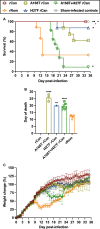
Live-attenuated virus vaccines provide long-lived protection against viral disease but carry inherent risks of residual pathogenicity and genetic reversion. The live-attenuated Candid#1 vaccine was developed to protect Argentines against lethal infection by the Argentine hemorrhagic fever arenavirus, Junín virus. Despite its safety and efficacy in Phase III clinical study, the vaccine is not licensed in the US, in part due to concerns regarding the genetic stability of attenuation. Previous studies had identified a single F427I mutation in the transmembrane domain of the Candid#1 envelope glycoprotein GPC as the key determinant of attenuation, as well as the propensity of this mutation to revert upon passage in cell culture and neonatal mice. To ascertain the consequences of this reversion event, we introduced the I427F mutation into recombinant Candid#1 (I427F rCan) and investigated the effects in two validated small-animal models: in mice expressing the essential virus receptor (human transferrin receptor 1; huTfR1) and in the conventional guinea pig model. We report that I427F rCan displays only modest virulence in huTfR1 mice and appears attenuated in guinea pigs. Reversion at another attenuating locus in Candid#1 GPC (T168A) was also examined, and a similar pattern was observed. By contrast, virus bearing both revertant mutations (A168T+I427F rCan) approached the lethal virulence of the pathogenic Romero strain in huTfR1 mice. Virulence was less extreme in guinea pigs. Our findings suggest that genetic stabilization at both positions is required to minimize the likelihood of reversion to virulence in a second-generation Candid#1 vaccine.IMPORTANCELive-attenuated virus vaccines, such as measles/mumps/rubella and oral poliovirus, provide robust protection against disease but carry with them the risk of genetic reversion to the virulent form. Here, we analyze the genetics of reversion in the live-attenuated Candid#1 vaccine that is used to protect against Argentine hemorrhagic fever, an often-lethal disease caused by the Junín arenavirus. In two validated small-animal models, we find that restoration of virulence in recombinant Candid#1 viruses requires back-mutation at two positions specific to the Candid#1 envelope glycoprotein GPC, at positions 168 and 427. Viruses bearing only a single change showed only modest virulence. We discuss strategies to genetically harden Candid#1 GPC against these two reversion events in order to develop a safer second-generation Candid#1 vaccine virus.
Keywords: Candid#1; Junín virus; arenavirus; envelope glycoprotein; live-attenuated; molecular genetics; reversion; vaccine; virulence determinants.
Conflict of interest statement
The authors declare no conflict of interest.
Figures


Similar articles
-
Epistastic Interactions within the Junín Virus Envelope Glycoprotein Complex Provide an Evolutionary Barrier to Reversion in the Live-Attenuated Candid#1 Vaccine.J Virol. 2017 Dec 14;92(1):e01682-17. doi: 10.1128/JVI.01682-17. Print 2018 Jan 1. J Virol. 2017. PMID: 29070682 Free PMC article.
-
Second-Generation Live-Attenuated Candid#1 Vaccine Virus Resists Reversion and Protects against Lethal Junín Virus Infection in Guinea Pigs.J Virol. 2021 Jun 24;95(14):e0039721. doi: 10.1128/JVI.00397-21. Epub 2021 Jun 24. J Virol. 2021. PMID: 33952638 Free PMC article.
-
The glycoprotein precursor gene of Junin virus determines the virulence of the Romero strain and the attenuation of the Candid #1 strain in a representative animal model of Argentine hemorrhagic fever.J Virol. 2015 Jun;89(11):5949-56. doi: 10.1128/JVI.00104-15. Epub 2015 Mar 25. J Virol. 2015. PMID: 25810546 Free PMC article.
-
Toward a vaccine against Argentine hemorrhagic fever.Bull Pan Am Health Organ. 1991;25(2):118-26. Bull Pan Am Health Organ. 1991. PMID: 1654168 Review.
-
Argentine hemorrhagic fever vaccines.Hum Vaccin. 2011 Jun;7(6):694-700. doi: 10.4161/hv.7.6.15198. Epub 2011 Jun 1. Hum Vaccin. 2011. PMID: 21451263 Review.
Cited by
-
Safety Toxicology Study of Reassortant Mopeia-Lassa Vaccine in Guinea Pigs.Future Pharmacol. 2025 Jun;5(2):26. doi: 10.3390/futurepharmacol5020026. Epub 2025 May 31. Future Pharmacol. 2025. PMID: 40667416 Free PMC article.
References
-
- Dan-Nwafor CC, Ipadeola O, Smout E, Ilori E, Adeyemo A, Umeokonkwo C, Nwidi D, Nwachukwu W, Ukponu W, Omabe E, et al. . 2019. A cluster of nosocomial Lassa fever cases in a tertiary health facility in Nigeria: description and lessons learned, 2018. Int J Infect Dis 83:88–94. doi:10.1016/j.ijid.2019.03.030 - DOI - PubMed
-
- NIAID . 2018. NIAID emerging infectious diseases/ pathogens. Available from: https://www.niaid.nih.gov/research/emerging-infectious-diseases-pathogens
MeSH terms
Substances
Supplementary concepts
Grants and funding
LinkOut - more resources
Full Text Sources

