Review
. 2024 Oct;61(10):1-13.
doi: 10.1007/s12035-024-04117-4. Epub 2024 Mar 20.

Research Advances in Neuroblast Migration in Traumatic Brain Injury

Affiliations
Review

Research Advances in Neuroblast Migration in Traumatic Brain Injury

Na Wu et al. Mol Neurobiol. 2024 Oct.

Abstract

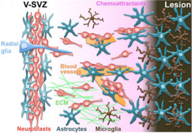
Neuroblasts were first derived from the adult mammalian brains in the 1990s by Reynolds et al. Since then, persistent neurogenesis in the subgranular zone (SGZ) of the hippocampus and subventricular zone (SVZ) has gradually been recognized. To date, reviews on neuroblast migration have largely investigated glial cells and molecular signaling mechanisms, while the relationship between vasculature and cell migration remains a mystery. Thus, this paper underlines the partial biological features of neuroblast migration and unravels the significance and mechanisms of the vasculature in the process to further clarify theoretically the neural repair mechanism after brain injury. Neuroblast migration presents three modes according to the characteristics of cells that act as scaffolds during the migration process: gliophilic migration, neurophilic migration, and vasophilic migration. Many signaling molecules, including brain-derived neurotrophic factor (BDNF), stromal cell-derived factor 1 (SDF-1), vascular endothelial growth factor (VEGF), and angiopoietin-1 (Ang-1), affect vasophilic migration, synergistically regulating the migration of neuroblasts to target areas along blood vessels. However, the precise role of blood vessels in the migration of neuroblasts needs to be further explored. The in-depth study of neuroblast migration will most probably provide theoretical basis and breakthrough for the clinical treatment of brain injury diseases.

Keywords: Neuroblast migration; Neurogenesis; Neuronal migration; Traumatic brain injury; Vascular migration.


Conflict of interest statement

The authors declare no competing interests.

Figures

Fig. 1
Fig. 1
Simplified overview of pathophysiology and therapy of TBI [21]. TBI, traumatic brain injury; ICP, intracranial pressure; BBB, blood–brain barrier; EPO, erythropoietin; NGF, nerve growth factor; VPA, valproic acid; IL-1RA, interleutin-1receptor antagonist; miR-21, microRNA-21; CsA, cyclosporine A; NNZ-2566, synthetic analogue of the endogenous N-terminus tripeptide glycine-proline-glutamate; Tβ4, thymosin beta 4; tPA, tissue plasminogen activator
Fig. 2
Fig. 2
Glutamate, reactive oxygen species, and apoptotic cell death pathways [22]. Fas-L, Fas ligand; ROS, reactive oxygen species
Fig. 3
Fig. 3
Cellular composition of the ventricular–subventricular zone (V-SVZ) [24]. Coronal section of adult mouse brain is shown in the upper right. The V-SVZ region indicated by the black arrow is shown enlarged in the lower left. Type B1 cells (blue; GFAP-positive) are the astrocytes that serve as the V-SVZ stem cell. These can divide and produce type C cells (green; Nestin-positive), which are rapidly dividing, transit amplifying cells. Type C cells give rise to type A cells (red; DCX-positive), the migratory neuroblasts. A blood vessel (BV, brown) is shown at the right. The apical surface of type B1 cells has a primary cilium and makes contact with the ventricle, which is at the left. These apical surfaces are found at the center of a “pinwheel” composed of multiciliated ependymal cells (yellow). The V-SVZ can be subdivided into three domains based on the structure and spatial arrangement of type B1 cells: Domain I (apical) contains the type B1 cells apical process and the body of ependymal cells; domain II (intermediate) contains the cell body of most type B1 cells, which are in contact with the type C and A cells; and domain III (basal) contains the B1 cell’s basal process with end-feet on blood vessels
Fig. 4
Fig. 4
Steps in neuronal migration and molecules involved [19]. a, b Polarized extension of the leading process. a PI3K signaling at the front of the cell regulates the balance of activation of the Rho GTPases Cdc42, Rac1, and RhoA. Inhibition of RhoA enhances leading-process outgrowth, whereas inhibition of Rac1 and Cdc42 impairs neurite outgrowth. Microtubule plus ends are recruited to the cortical actin meshwork. b In the intermediate segment of the leading process, microtubules (MT, green) are loosely organized, probably owing to the destabilizing activity of stathmin. γ-Tubulin and the microtubule-related protein ninein show a wide distribution in migrating neurons. c Forward movement of the centrosome. Cdc42 is found mainly in the perinuclear region. Forward movement of the centrosome (red rods) involves PARD6α and its associated kinase PKCξ; reorientation of the centrosome requires the activity of GSK3β, PKCξ, and the actin cytoskeleton. Focal-adhesion kinase (FAK) also contributes to centrosomal dynamics. Both centrioles split during the advance of the soma. d, e Movement of the nucleus (blue oval) toward the centrosome (nucleokinesis).d Nucleokinesis requires a microtubule motor complex based on dynein; proteins interacting within this include dynactin, LIS1, NDEL1, DISC1, and DCX. DCX molecules are found attached to microtubules that extend from the centrosome to the perinuclear “cage.” Ca2+ signaling might also operate at this stage. e Various components of the KASH family of proteins anchor the nucleus to the centrosome and cell membrane. Neurofilaments might contribute to connecting the nucleus to the cell cortex. f Trailing-process retraction. PTEN signaling at the back regulates RhoA. Actomyosin contraction has a role in driving the nucleus toward the centrosome
Fig. 5
Fig. 5
Glial-guided neuroblast migration [42]. Phase one involves radial movement of pyramidal neurons (dark green) from the site of origin at the ventricular surface to the subventricular zone (SVZ). In phase two, cells become multipolar and pause their migration in the lower intermediate zone (IZ) and subventricular zone (SVZ). Some neurons undergo phase three, which is characterized by retrograde motion toward the ventricle. Phase four is the final radial migration to the cortical plate (CP), guided by radial glial fibers. Radial glia (light green) remain mitotic, undergo interkinetic nuclear migration, and generate additional daughter cells (grey). MZ, marginal zone; R, radial glial cell; VZ, ventricular zone
Fig. 6
Fig. 6
Migration of neuroblasts from V-SVZ to the olfactory bulb(OB) [17]. Illustration showing the migration of V-SVZ neuroblasts to the olfactory bulb. Neuroblasts (red) are generated in the V-SVZ by neural stem cells (blue) through intermediate progenitors called transit-amplifying cells (green). The neuroblasts form elongated, chain-like aggregates that migrate tangentially through the rostral migratory stream (RMS) toward the olfactory bulb. After reaching the olfactory bulb (OB) and detaching from the chain, individual neuroblasts migrate radially to the outer layer where they differentiate into olfactory interneurons, granule cells (pink), or periglomerular cells (orange) and are integrated into the olfactory neuronal circuitry
Fig. 7
Fig. 7
Mechanisms supporting neuroblast migration toward an injury site in the brain [17]. After a brain insult, neuroblasts in the V-SVZ are redirected to the lesion by several diffusible attractive factors secreted by injury-activated astrocytes (blue-green), microglia (brown), and vascular endothelial cells (orange). Migrating neuroblasts use blood vessels, astrocytic processes, radial glial processes (light blue), and extracellular matrices (ECM, yellow-green) as scaffolds

Similar articles

Cited by

References

    1. Wu N, Sun XC, Zhou C, Yan J, Cheng JC (2023) Neuroblasts migration under control of reactive astrocyte-derived BDNF: a promising therapy in late neurogenesis after traumatic brain injury. Stem Cell Res Ther 14:1–14. 10.1186/s13287-022-03232-0 - PMC - PubMed
    1. Ghaith HS, Nawar AA, Gabra MD, Abdelrahman ME, Nafady MH, Bahbah EI, Ebada MA, Ashraf GM et al (2022) A literature review of traumatic brain injury biomarkers. Mol Neurobiol 59:4141–4158. 10.1007/s12035-022-02822-6 - PMC - PubMed
    1. Bressan C, Saghatelyan A (2020) Intrinsic mechanisms regulating neuronal migration in the postnatal brain. Front Cell Neurosci 14:620379. 10.3389/fncel.2020.620379 - PMC - PubMed
    1. Reynolds BA, Weiss S (1992) Generation of neurons and astrocytes from isolated cells of the adult mammalian central nervous system. Science 255:1707–1710 - PubMed
    1. Guerrero-Cazares H, Lavell E, Chen L, Schiapparelli P, Lara-Velazquez M, Capilla-Gonzalez V, Clements AC, Drummond G et al (2017) Brief Report: Robo1 regulates the migration of human subventricular zone neural progenitor cells during development. Stem Cells 35:1860–1865. 10.1002/stem.2628 - PMC - PubMed

LinkOut - more resources