. 2024 May;476(5):861-869.
doi: 10.1007/s00424-024-02941-0. Epub 2024 Mar 20.

XPR1: a regulator of cellular phosphate homeostasis rather than a Pi exporter

Affiliations

XPR1: a regulator of cellular phosphate homeostasis rather than a Pi exporter

David Burns et al. Pflugers Arch. 2024 May.

Abstract

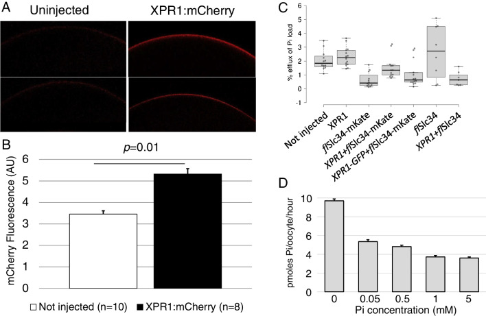
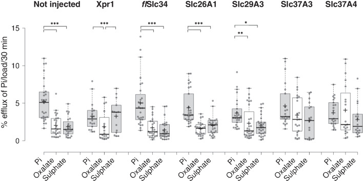
Phosphate (Pi) is an essential nutrient, and its plasma levels are under tight hormonal control. Uphill transport of Pi into cells is mediated by the two Na-dependent Pi transporter families SLC34 and SLC20. The molecular identity of a potential Pi export pathway is controversial, though XPR1 has recently been suggested by Giovannini and coworkers to mediate Pi export. We expressed XPR1 in Xenopus oocytes to determine its functional characteristics. Xenopus isoforms of proteins were used to avoid species incompatibility. Protein tagging confirmed the localization of XPR1 at the plasma membrane. Efflux experiments, however, failed to detect translocation of Pi attributable to XPR1. We tested various counter ions and export medium compositions (pH, plasma) as well as potential protein co-factors that could stimulate the activity of XPR1, though without success. Expression of truncated XPR1 constructs and individual domains of XPR1 (SPX, transmembrane core, C-terminus) demonstrated downregulation of the uptake of Pi mediated by the C-terminal domain of XPR1. Tethering the C-terminus to the transmembrane core changed the kinetics of the inhibition and the presence of the SPX domain blunted the inhibitory effect. Our observations suggest a regulatory role of XPR1 in cellular Pi handling rather than a function as Pi exporter. Accordingly, XPR1 senses intracellular Pi levels via its SPX domain and downregulates cellular Pi uptake via the C-terminal domain. The molecular identity of a potential Pi export protein remains therefore elusive.

Keywords: Xenopus oocytes; Cellular phosphate balance; Phosphate exporter; XPR1.


Conflict of interest statement

The authors declare no competing interests.

Figures

Fig. 1
Fig. 1
XPR1 is expressed at the membrane of Xenopus oocytes. Various constructs including the flurescent tags mCherry, mKate and GFP were tested with qualitatively comparable results for both localization and efflux of Pi. A mCherry-tagged XPR1 displays enhanced fluorescence (right panel) as compared to non-injected oocytes (left panel). B Quantification of fluorescence in arbitrary units. C Xenopus XPR1 wild type and tagged with fluorescent proteins did not affect Pi efflux from oocytes. D Native oocytes were exposed to varying concentrations of Pi for 24 h followed by measurement of Pi uptake. The reduction of Pi transport to even small increases in Pi in the medium indicates that oocytes have an endogenous system for sensing and responding to environmental Pi
Fig. 2
Fig. 2
Pi efflux from oocytes injected with XPR1, flSlc34 and potential cofactors under various experimental conditions. A Oocytes were injected with XPR1 and flSlc34 + Xpr1 and efflux was assayed at pH 6 and 7.4, respectively. B Oocytes were injected with RNAs encoding XPR1, flSlc34 and total kidney RNA from Xenopus. In general, oocytes with higher net uptake of Pi (i.e. expressing flSlc34) tend to show lower efflux, as observed in many experiments. No significant effect of XPR1 on Pi export is detectable (compare the two groups on the right). The apparent change in efflux between 3 and 5 day incubation in groups 1 and 2 on the left reflects intrinsic changes in oocytes rather than the effects of injected material. A representative experiment is shown. C Oocytes were injected with flSlc34 and a mix of pooled IP6K1 and IP6K2 cRNAs. Co-expression of the kinases did not affect Pi efflux. Oocytes from three different experiments are included
Fig. 3
Fig. 3
Pi uptake into oocytes expressing XPR1 constructs that lack the SPX domain. Constructs that lacked either part or the entire SPX domain were generated and expressed in oocytes. Oocytes were incubated for 5 days (left group) and 7 days (right group). Non-injected oocytes and the truncated constructs alone did not stimulate Pi uptake. flSlc34 and flSlc34 + XPR1-177 showed significant Pi uptake that increased and became more variable between days 5 and 7 (light and dark blue bars). flSlc34 + XPR1-205 showed a downregulation of Pi uptake to basal level between day 5 and day 7 (orange bars). Right panel, alpha fold-based model of human XPR1. The individual protein domains are indicated: SPX, SPX-domain; TM, transmembrane domain or ‘core’ and C-Term, C-terminal domain
Fig. 4
Fig. 4
Pi uptake into oocytes expressing XPR1 protein domains. A Schematic representation of XPR1 with the domains SPX, core and C-terminus. B Cells were injected with constructs of XPR1 domains; SPX domain, core domain, the C- terminus and XPR1 without C-terminus (SPX-core) and assayed after 5 days. C The effect is specific for Pi transport as expression of XPR1 fragments has no effect on.3H arginine uptake into oocytes (representative experiment of 3 repetitions is shown). One-way ANOVA with Tukey’s post hoc test, *p < 0.05
Fig. 5
Fig. 5
Efflux measurements with oocytes expressing potential Pi exporters. Anion exchangers were identified as potential Pi exporters based on substrate specificity and expression pattern. The transporters (Slc26A1, Slc29A3, Slc37A3 and Slc37A4) were expressed in oocytes, and efflux was assessed in the presence of 2 mM Pi, 50 µM oxalate or 2 mM suphate. Whereas the exogenous exchangers did not show enhanced export activity, though intrinsic export shows preference of Pi over oxalate and sulphate. One-way ANOVA with Tukey’s post hoc test, *p < 0.05, **p < 0.01, ***p < 0.001

Comment in

  • Is XPR1 mediating phosphate efflux?
    Hernando N. Hernando N. Pflugers Arch. 2024 May;476(5):717-719. doi: 10.1007/s00424-024-02946-9. Epub 2024 Mar 21. Pflugers Arch. 2024. PMID: 38512477 No abstract available.

References

    1. Ansermet C, Moor MB, Centeno G, Auberson M, Hu DZ, Baron R, Nikolaeva S, Haenzi B, Katanaeva N, Gautschi I, Katanaev V, Rotman S, Koesters R, Schild L, Pradervand S, Bonny O, Firsov D. Renal fanconi syndrome and hypophosphatemic rickets in the absence of xenotropic and polytropic retroviral receptor in the nephron. J Am Soc Nephrol. 2017;28:1073–1078. doi: 10.1681/ASN.2016070726. - DOI - PMC - PubMed
    1. Balck A, Schaake S, Kuhnke NS, Domingo A, Madoev H, Margolesky J, Dobricic V, Alvarez-Fischer D, Laabs B-H, Kasten M, Luo W, Nicolas G, Marras C, Lohmann K, Klein C, Westenberger A. Genotype–phenotype relations in primary familial brain calcification: systematic MDSGene review. Mov Disord. 2021;36:2468–2480. doi: 10.1002/mds.28753. - DOI - PubMed
    1. Beck L, Leroy C, Beck-Cormier S, Forand A, Salaün C, Paris N, Bernier A, Ureña-Torres P, Prié D, Ollero M, Coulombel L, Friedlander G. The phosphate transporter PiT1 (Slc20a1) revealed as a new essential gene for mouse liver development. PLoS ONE. 2010;5:e9148. doi: 10.1371/journal.pone.0009148. - DOI - PMC - PubMed
    1. Boller F, Boller M, Gilbert J. Familial idiopathic cerebral calcifications. J Neurol Neurosurg Psychiatry. 1977;40:280–285. doi: 10.1136/jnnp.40.3.280. - DOI - PMC - PubMed
    1. Bon N, Couasnay G, Bourgine A, Sourice S, Beck-Cormier S, Guicheux J, Beck L. Phosphate (P(i))-regulated heterodimerization of the high-affinity sodium-dependent P(i) transporters PiT1/Slc20a1 and PiT2/Slc20a2 underlies extracellular P(i) sensing independently of P(i) uptake. J Biol Chem. 2018;293:2102–2114. doi: 10.1074/jbc.M117.807339. - DOI - PMC - PubMed

Publication types

Substances